. 2024 Jul;14(7):100945.
doi: 10.1016/j.jpha.2024.01.013. Epub 2024 Feb 5.

A novel fluorescent labeling compound for GluN2A containing N-methyl-d-aspartate receptors identified by autodisplay-based screening

Affiliations

A novel fluorescent labeling compound for GluN2A containing N-methyl-d-aspartate receptors identified by autodisplay-based screening

Alexander Dombovski et al. J Pharm Anal. 2024 Jul.

Abstract

Image 1.


Conflict of interest statement

The authors declare that there are no conflicts of interest.

Figures

Image 1
Graphical abstract
Fig. 1
Fig. 1
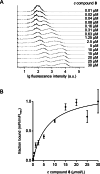
Compound 8 is a selective GluN2A labeling probe. (A) Concept of surface co-display of GluN1 and GluN2A ligand binding domains (LBDs) (Protein Data Bank (PDB) ID 5I56) via autodisplay. Full fusion proteins consist of (N- to C-terminal) signal peptide (SP), ligand binding domain S1 lobe (LBD S1), glycine-threonine dipeptide linker (GT), LBD S2, EhaA linker (Linker), and EhaA β-barrel (gray). Escherichia coli (E. coli) cells are co-transformed with both plasmids to facilitate co-display and dimerization of LBDs of both GluN1 (S1: red and S2: yellow) and GluN2A (S1: blue and S2: cyan) on the surface of the cells. (B) Structural formulas of GluN2A selective negative allosteric modulator of the N-methyl-d-aspartate receptor (NMDA) receptor TCN-201 and compound 8. (C) Flow cytometry-based binding assay with compound 8 binding to GluN1/GluN2A LBD co-displaying cells (colored) or control cells (black) displaying only one of the two types of LBDs. Median fluorescence intensity (MFI) for each sample is given in arbitrary units. Substantial binding was assumed when the MFI of a sample exceeded the MFI of cells with co-displayed LBDs, which were not incubated with any of the fluorescent compounds by a factor of two (MFI of cells with co-displayed LBDs without a fluorescent compound was 140, histogram not depicted). (D) Inhibitory effect of TCN-201, compounds 6a, 6b, and 8 evaluated by two-electrode voltage clamp (TEVC) at GluN1-1a/GluN2A expressing oocytes. Data of independent oocytes are presented as mean ± standard error of the mean (SEM) (n = 6–8). (E) Inhibitory effect of 1 μM TCN-201 compared to simultaneous application of 1 μM TCN-201 together with 9 μM compounds 6a, 6b, or 8 evaluated by TEVC at GluN1-1a/GluN2A expressing oocytes. Data of independent oocytes are presented as mean ± SEM (n = 4–6). (F) Overlay of docked (blue) and crystallized (cyan) binding pose of TCN-201 at the GluN1/GluN2A LBD interface. As seen in the overlay, applied docking parameters resulted in a reliable prediction of the binding pose of TCN-201 when compared to the reported crystallized binding pose (PDB ID 5I56). (G) Docking pose of compound 8 (blue). Notably, ring C is oriented into an opposite direction, when compared to TCN-201. The key residues for binding are supposed to be aromatic rings A and B, whereby both rings adopt a U-shaped π–π stack and fill a hydrophobic binding pocket. (H, I) Fluorescence microscopy of mouse L(tk−) cells expressing recombinant GluN1-1a/GluN2A (H) and GluN1-1a/GluN2B (I) subunits that were stained with compound 8. (J) Quantitative analysis of single cell fluorescence intensities after staining with compound 8 in mouse L(tk−) cells expressing recombinant GluN1-1a/GluN2A or GluN1-1a/GluN2B subunits. When indicated, GluN1-1a/GluN2A expressing mouse L(tk−) cells were additionally preincubated with TCN-201 before they were stained with compound 8. Data of single cells are presented as mean ± SEM (n = 7–16). (K) Immunocytochemical staining of two weeks old three-dimensional (3D) neural spheroids with compound 8 (green) and antibodies against neuronal marker Tuj1 (purple) and GluN2A (red). (L) Immunocytochemical staining of a neuro spheroid (as described in Fig. 1F) without overlay of anti Tuj1 staining. GluN2A staining depicted in green (compound 8) and red (anti-GluN2A antibody). (M) Immunocytochemical staining of a neuro spheroid (as described in Fig. 1F) without overlay of anti Tuj1 staining and the co-localization of compound 8 and anti-GluN2A antibody signals depicted in white. ∗∗∗P ≤ 0.001. ns: not significant.
figs1
figs1
figs2
figs2
figs3
figs3
figs4
figs4
figs5
figs5
figs6
figs6
figs7
figs7
figs8
figs8
figs9
figs9
figs10
figs10
figs11
figs11

References

    1. Hansen K.B., Wollmuth L.P., Bowie D., et al. Structure, function, and pharmacology of glutamate receptor ion channels. Pharmacol. Rev. 2021;73:298–487. - PMC - PubMed
    1. Furukawa H., Singh S.K., Mancusso R., et al. Subunit arrangement and function in NMDA receptors. Nature. 2005;438:185–192. - PubMed
    1. Jose J., Meyer T.F. The autodisplay story, from discovery to biotechnical and biomedical applications. Microbiol. Mol. Biol. Rev. 2007;71:600–619. - PMC - PubMed
    1. Hansen K.B., Ogden K.K., Traynelis S.F. Subunit-selective allosteric inhibition of glycine binding to NMDA receptors. J. Neurosci. 2012;32:6197–6208. - PMC - PubMed
    1. Yi F., Mou T.C., Dorsett K.N., et al. Structural basis for negative allosteric modulation of GluN2A-containing NMDA receptors. Neuron. 2016;91:1316–1329. - PMC - PubMed

LinkOut - more resources