. 2024 Jul 9:27:101152.
doi: 10.1016/j.mtbio.2024.101152. eCollection 2024 Aug.

Impact of uniaxial cyclic stretching on matrix-associated endothelial cell responses

Affiliations

Impact of uniaxial cyclic stretching on matrix-associated endothelial cell responses

Cuihong Ren et al. Mater Today Bio. .

Abstract

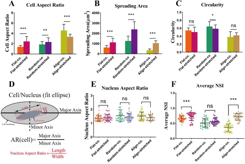
Uniaxial cyclic stretching plays a pivotal role in the fields of tissue engineering and regenerative medicine, influencing cell behaviors and functionality based on physical properties, including matrix morphology and mechanical stimuli. This study delves into the response of endothelial cells to uniaxial cyclic strain within the geometric constraints of micro-nano fibers. Various structural scaffold forms of poly(l-lactide-co-caprolactone) (PLCL), such as flat membranes, randomly oriented fiber membranes, and aligned fiber membranes, were fabricated through solvent casting and electrospinning methods. Our investigation focuses on the morphological variation of endothelial cells under diverse geometric constraints and the mechanical-dependent release of nitric oxide (NO) on oriented fibrous membranes. Our results indicate that while uniaxial cyclic stretching promotes endothelial cell spreading, the anisotropy of the matrix morphology remains the primary driving factor for cell alignment. Additionally, uniaxial cyclic stretching significantly enhances NO release, with a notably stronger effect correlated to the increasing strain amplitude. Importantly, this study reveals that uniaxial cyclic stretching enhances the mRNA expression of key proteins, including talin, vinculin, rac, and nitric oxide synthase (eNOS).

Keywords: Endothelial nitric oxide (NO) release; Matrix morphology; Uniaxial cyclic stretching.


Conflict of interest statement

The authors declare that they have no known competing financial interests or personal relationships that could have appeared to influence the work reported in this paper.

Figures

Image 1
Graphical abstract
Fig. 1
Fig. 1
Cyclic Stretching Device. a) Deformable polydimethylsiloxane (PDMS) culture chamber. b) Loading device. c) Ball screw reciprocating feeding system. d) Friction-enhancing grooves. e) Stepper motor.
Fig. 2
Fig. 2
Distribution of surface morphology. A) Scanning electron microscope images of ‘Flat’, ‘Random’ and ‘Align’. B) Atomic force microscope images of ‘Flat’, ‘Random’ and ‘Align’. The unit of Sq is um.
Fig. 3
Fig. 3
Statistical analysis of fiber diameters and the results of fast Fourier transform. A) fiber diameters images of Random and Align. B) fast Fourier transform (FFT) images of Random and Align.
Fig. 4
Fig. 4
Nanoindentation images A) Nanometer indentation displacement−load diagram B) Elastic modulus of different surface topography samples.
Fig. 5
Fig. 5
Stresses and strains curves of uniaxial Tensile Testing. A-C) 10 % strain. D-F) 15 %. G-I) 20 % strain.
Fig. 6
Fig. 6
Live−dead staining of HUVECs of different groups on day 1, day 3, day 4 (the scale bar is 100 μm), (Co. represents unstretched).
Fig. 7
Fig. 7
Immunofluorescence staining of HUVECs cultured in different groups under co. (unstretched) and stretched conditions (scale bar 20 μm). A-C) Stress fiber orientation of HUVECs cultured in different groups under co. (unstretched) and stretched conditions (scale bar 20 μm).
Fig. 8
Fig. 8
Statistical graphs of immunofluorescence (A, C) Cell polarization was evaluated by cell aspect ratio and circularity, respectively. (B) Spreading area of HUVECs. (D) AR definition diagram of cell and nucleus. (E, F) Nucleus aspect ratio and Nuclear shape index calculation. Mean ± standard deviation, n > 40, *p < 0.05, **p < 0.01, ***p < 0.001, ANOVA.
Fig. 9
Fig. 9
Immunofluorescence staining and Statistical analysis of NO release. A) DAF-FM staining. B) Laser intensity mean values of NO release under Co.(unstretched) and Stretched. C-D) Stretched/unstretched ratio of NO release in different Frequency and Strain (Reference to formula 5). E) mRNA expressions of the nitric oxide synthase (eNOS) genes related to cell adhesion quantified by q-PCR.
Fig. 10
Fig. 10
mRNA expressions of the genes related to dynamic actin rearrangement quantified by q-PCR under Co.(unstretched) and Stretched. (talin, vinculin, RhoA, Rac1,FAK, paxillin). Mean ± standard deviation, *p < 0.05, **p < 0.01, ***p < 0.001, t-test.
Fig. 11
Fig. 11
Cyclic stretching regulates the mechanism of EC mechanical conduction.

References

    1. Gordon E., Schimmel L., Frye M. The importance of mechanical forces for in vitro endothelial cell Biology. Front. Physiol. 2020;11 doi: 10.3389/fphys.2020.00684. 684–684. - DOI - PMC - PubMed
    1. Wang L., Wu S., Cao G., Fan Y., Dunne N., Li X. Biomechanical studies on biomaterial degradation and co-cultured cells: mechanisms, potential applications, challenges and prospects. J. Mater. Chem. B. 2019;7:7439–7459. doi: 10.1039/C9TB01539F. - DOI - PubMed
    1. Gupta P., Mandal B.B. Tissue-Engineered vascular grafts: emerging Trends and Technologies. Adv. Funct. Mater. 2021;31 doi: 10.1002/adfm.202100027. - DOI
    1. Shao Y., Mann J.M., Chen W., Fu J. Global architecture of the F-actin cytoskeleton regulates cell shape-dependent endothelial mechanotransduction. Integr Biol-Uk. 2014;6:300–311. doi: 10.1039/c3ib40223a. - DOI - PMC - PubMed
    1. Jufri N.F., Mohamedali A., Avolio A., Baker M.S. Mechanical stretch: physiological and pathological implications for human vascular endothelial cells. Vasc. Cell. 2015;7:1–12. doi: 10.1186/s13221-015-0033-z. - DOI - PMC - PubMed

LinkOut - more resources