

Wide-Awake Tenolysis for Tendon Entrapment in a Distal Radial Epiphyseal Separation (Volar Displacement Type): A Case Report
- PMID: 39104971
- PMCID: PMC11298766
- DOI: 10.7759/cureus.63837
Wide-Awake Tenolysis for Tendon Entrapment in a Distal Radial Epiphyseal Separation (Volar Displacement Type): A Case Report
Abstract
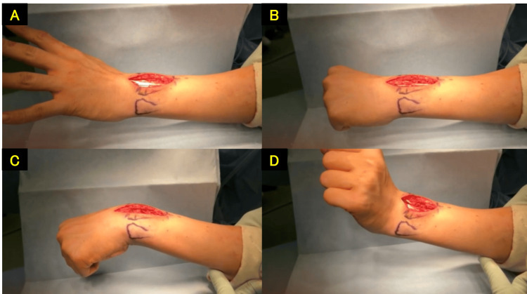
The patient was a 13-year-old male who fell while riding a bicycle and was initially diagnosed with a distal radial epiphyseal separation (volar displacement type) that was conservatively managed. Four months post-injury, he complained of limited movement in his left index finger and was referred to our hospital. Upon examination, the patient also complained of limited movement of the left index finger in wrist flexion. The wrist range of motion was 50° of volar flexion, 50° of dorsiflexion, 90° of pronation, and 90° of supination with the fingers extended. The X-ray revealed a radiolucent area in the distal radius. Ultrasound, computed tomography, and magnetic resonance imaging scans demonstrated entrapment of the extensor tendon within the medullary cavity of the radius. Five months post-injury, surgery was performed using the wide-awake local anesthesia no-tourniquet (WALANT) technique. A dorsal wrist approach was utilized, and the extensor digitorum communis tendon was found to be trapped within the medullary cavity of the radius. The tendon was released using an air drill, and sufficient improvement in the left index finger flexion was confirmed with active movement before concluding the surgery. At the 11-month postoperative follow-up, the patient showed excellent outcomes with a wrist range of motion of 75° of volar flexion, 85° of dorsiflexion, 90° of pronation, and 90° of supination. Tendon entrapment of the extensor tendons has been reported as a long-standing complication associated with distal radius fractures, particularly with volar displacement types. A benefit of the WALANT technique is the ability to communicate with the patient during surgery, allowing for active movements of the fingers and wrist. This is particularly useful in tendon surgeries for determining tendon tension. We report a case of successful tenolysis surgery using the WALANT technique for a patient with a conservatively managed distal radial epiphyseal separation (volar displacement type), who experienced a limited flexion of the index finger due to tendon entrapment.
Keywords: distal radial epiphyseal separation; extensor digitorum communis tendon; tendon entrapment; volar displacement type; walant technique.
Copyright © 2024, Mori et al.
Conflict of interest statement
Human subjects: Consent was obtained or waived by all participants in this study. Conflicts of interest: In compliance with the ICMJE uniform disclosure form, all authors declare the following: Payment/services info: All authors have declared that no financial support was received from any organization for the submitted work. Financial relationships: All authors have declared that they have no financial relationships at present or within the previous three years with any organizations that might have an interest in the submitted work. Other relationships: All authors have declared that there are no other relationships or activities that could appear to have influenced the submitted work.
Figures


References
-
- Reduction of Smith's fracture. TH FB. J Bone Joint Surg Br. 1957;39-B:463–470. - PubMed
-
- Minimally invasive anesthesia in wide awake hand surgery. Lalonde D. Hand Clin. 2014;30:1–6. - PubMed
-
- Dislocation of the extensor pollicis longus tendon in Smith's fracture of the radius. A case report. Hunt D. J Bone Joint Surg Am. 1969;51:991–994. - PubMed
-
- Traumatic entrapment of the extensor pollicis longus tendon in Smith’s fracture of the radius. Case report. Murakami Y, Todani K. J Hand Surg Am. 1981;6:238–240. - PubMed
Publication types
LinkOut - more resources
Full Text Sources