Review
. 2024 Jun 17;134(12):e179742.
doi: 10.1172/JCI179742.

Myasthenia gravis: the future is here

Affiliations
Review

Myasthenia gravis: the future is here

Henry J Kaminski et al. J Clin Invest. .

Abstract

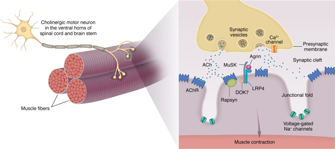
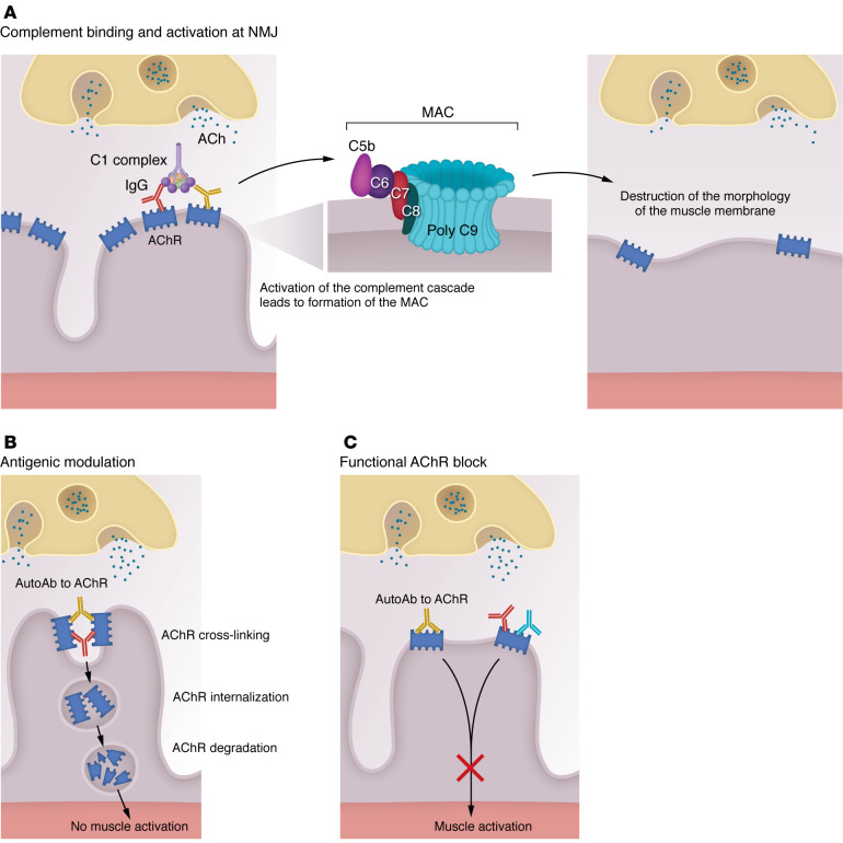
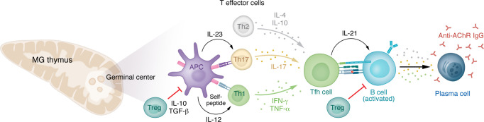
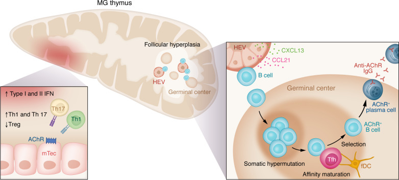
Myasthenia gravis (MG) stands as a prototypical antibody-mediated autoimmune disease: it is dependent on T cells and characterized by the presence of autoantibodies targeting proteins located on the postsynaptic surface of skeletal muscle, known as the neuromuscular junction. Patients with MG exhibit a spectrum of weakness, ranging from limited ocular muscle involvement to life-threatening respiratory failure. Recent decades have witnessed substantial progress in understanding the underlying pathophysiology, leading to the delineation of distinct subcategories within MG, including MG linked to AChR or MuSK antibodies as well as age-based distinction, thymoma-associated, and immune checkpoint inhibitor-induced MG. This heightened understanding has paved the way for the development of more precise and targeted therapeutic interventions. Notably, the FDA has recently approved therapeutic inhibitors of complement and the IgG receptor FcRn, a testament to our improved comprehension of autoantibody effector mechanisms in MG. In this Review, we delve into the various subgroups of MG, stratified by age, autoantibody type, and histology of the thymus with neoplasms. Furthermore, we explore both current and potential emerging therapeutic strategies, shedding light on the evolving landscape of MG treatment.


Conflict of interest statement

Conflict of interest: HJK is a consultant for Roche, Takeda, Cabaletta Bio, UCB Pharmaceuticals, Canopy Immunotherapeutics, EMD Serono, Ono Pharmaceuticals, ECoR1, Gilde Healthcare, and Admirix Inc. Argenix Inc. provides an unrestricted educational grant to George Washington University. HJK is an unpaid consultant for Care Constitution and has equity interest in MimiVax. LLK serves as a consultant for Alexion, GRO Biotechnology, CSL Berhing, Amplo Biotechnology, and Sanofi. LLK has equity interest in MimiVax.

Figures

Figure 1
Figure 1. Structure of the NMJ.
Each α–motor neuron axon divides into branches that innervate many individual muscle fibers. Each branch loses its myelin sheath and further subdivides into many presynaptic boutons, which face the surface of the postsynaptic surface of the muscle fiber and contain synaptic vesicles loaded with ACh. Between the synaptic bouton and the muscle surface lies the synaptic cleft, which contains acetylcholinesterase. The postsynaptic membrane has characteristic invaginations, with the AChRs densely packed at their tops. AChR density is influenced by both clustering and declustering signals, including ACh itself. Agrin, secreted by the nerve, binds to LRP-4 on the postsynaptic membrane, enhancing its binding with MuSK, which leads to MuSK autophosphorylation and ultimately the clustering of AChR. Rapsyn, a cytoplasmic protein, anchors AChR to the muscle cytoskeleton. When the nerve action potential reaches the synaptic bouton, voltage-gated Ca2+ channels are activated, leading to the fusion of synaptic vesicles with the nerve terminal membrane and release of ACh. ACh diffuses across the synaptic cleft, with some binding molecules the AChR. Binding triggers AChR ion channel opening, permitting influx of Na+ into the postsynaptic region. The resulting EPP activates voltage-gated Na+ channels at the bottom of the folds, leading to further Na+ influx and spreading of the action potential along the muscle fiber. Other proteins, including Rapsyn, MuSK, Dok-7, LRP-4, and agrin, which are involved in AChR clustering, are also present on the muscle membrane in close proximity to the AChR.
Figure 2
Figure 2. Effector mechanisms of AChR antibodies.
(A) Antibody binding to the AChR activates the complement cascade, resulting in the formation of membrane attack complex (MAC) and localized destruction of the postsynaptic NMJ membrane. This ultimately leads to a simplified, altered morphology of the postsynaptic membrane of the NMJ of patients with MG and EAMG animals. (B) Antibodies cross-link AChR molecules on the NMJ postsynaptic membrane, causing endocytosis of the cross-linked AChR molecules and their degradation (antigenic modulation). It is likely that antibodies attaching to different epitopes are required to produce modulation and complement activation. This ultimately leads to a reduced number of AChR molecules on the postsynaptic membrane. (C) Antibody binding of the ACh-binding sites of the AChR causes functional block of the AChR by interfering with binding of ACh released at the NMJ. It is important to appreciate that there may be overlap in the pathogenic mechanisms of individual AChR antibodies and these mechanisms may cooperate to induce disease.
Figure 3
Figure 3. Cytokine network and cells involved in the pathogenesis and immunoregulation of AChR antibody MG.
Th1 cytokines stimulate production of IgG subclasses that bind and activate complement effectively, whereas Th2 cytokines stimulate the production of Ig classes and IgG subclasses that do not. See text for details. AChRs are presented to naive T cells via antigen-presenting cells (APCs), leading to production of IL-23 and IL-17 that contributes to tissue inflammation in the MG thymus. Increased levels of Th1 cytokines (IFN-γ) promote the T follicular helper (Tfh) cell interaction with the recruited B cells. Th17 proinflammatory cytokine levels (IL-17) promote differentiation of B cells into antibody-secreting cells and production of complement-fixing antibodies. Tfh cells secrete IL-21, which promotes plasma cell differentiation. Tregs modulate proinflammatory responses by secreting antiinflammatory cytokines to suppress T cell and B cell responses. Dysfunction in circulating and thymic Tregs is associated with MG pathogenesis.
Figure 4
Figure 4. Thymic pathology associated early-onset MG.
The thymus is the organ of T cell maturation and establishment of central tolerance. Self-peptides are presented by medullary thymic epithelial cells (mTECs). Self-reactive T cells undergo apoptosis or are controlled by Tregs; however, suppressor functions of thymic Tregs are impaired in MG. Type I and II IFN induction in the thymus promotes expression of AChR, cytokines, and chemokines by thymic epithelial cells. Increased expression of IL-17 and IL-23 promotes expansion of Th1/Th17 cells. High endothelial venules (HEVs) and secretion of CCL21 and CXCL13 facilitate recruitment of B cells and ectopic germinal center formation associated with thymic hyperplasia. In the germinal center, B cells undergo somatic hypermutation, affinity maturation, and selection, processes that are implicated in development of AChR+ long-lived plasma cells. Anti-AChR–producing plasma cells exit the germinal center and migrate to the bone marrow. fDC, follicular DC.

References

    1. Nguyen-Cao TM, et al. Myasthenia gravis: Historical achievements and the “golden age” of clinical trials. J Neurol Sci. 2019;406:116428. doi: 10.1016/j.jns.2019.116428. - DOI - PubMed
    1. Kusner LL, Kaminski HJ. Editorial: Special issue on standardization of preclinical evaluation of animal models for myasthenia gravis. Exp Neurol. 2015;270:1–2. doi: 10.1016/j.expneurol.2015.05.001. - DOI - PubMed
    1. Smith VM, et al. A functional human-on-a-chip autoimmune disease model of myasthenia gravis for development of therapeutics. Front Cell Dev Biol. 2021;9:745897. doi: 10.3389/fcell.2021.745897. - DOI - PMC - PubMed
    1. Plomp JJ, et al. A bioassay for neuromuscular junction-restricted complement activation by myasthenia gravis acetylcholine receptor antibodies. J Neurosci Methods. 2022;373:109551. doi: 10.1016/j.jneumeth.2022.109551. - DOI - PubMed
    1. Hoch W, et al. Auto-antibodies to the receptor tyrosine kinase MuSK in patients with myasthenia gravis without acetylcholine receptor antibodies. Nat Med. 2001;7(3):365–368. doi: 10.1038/85520. - DOI - PubMed

MeSH terms