. 2024 Aug 21;481(16):1075-1096.
doi: 10.1042/BCJ20240110.

1-Deoxy-d-xylulose 5-phosphate reductoisomerase as target for anti Toxoplasma gondii agents: crystal structure, biochemical characterization and biological evaluation of inhibitors

Affiliations

1-Deoxy-d-xylulose 5-phosphate reductoisomerase as target for anti Toxoplasma gondii agents: crystal structure, biochemical characterization and biological evaluation of inhibitors

Flaminia Mazzone et al. Biochem J. .

Abstract

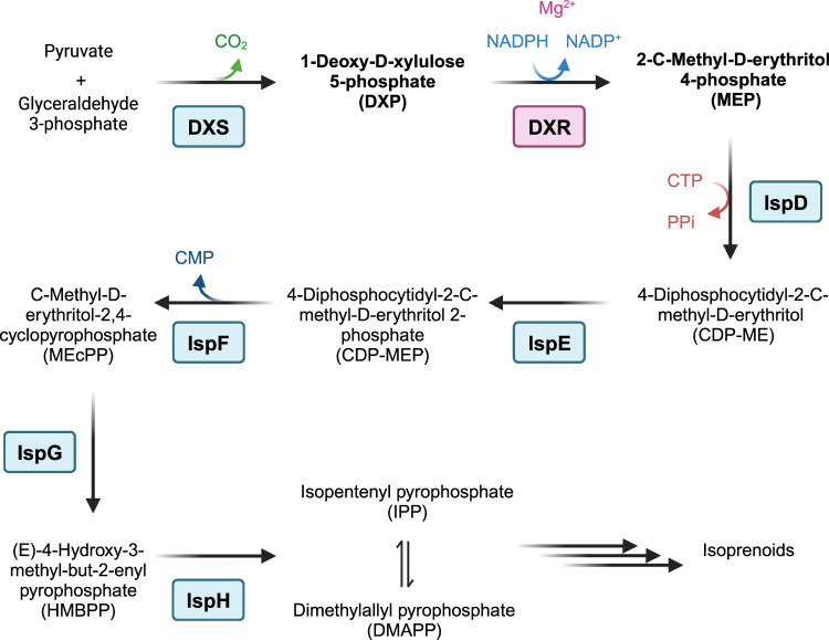
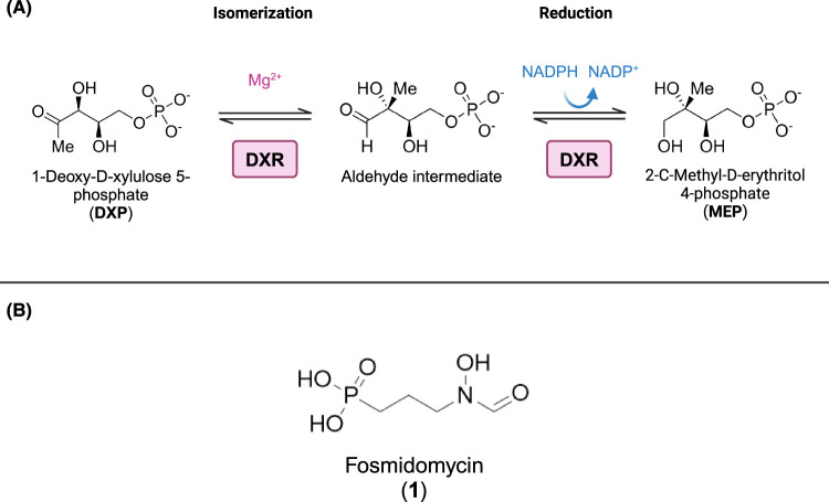
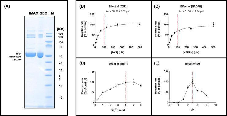
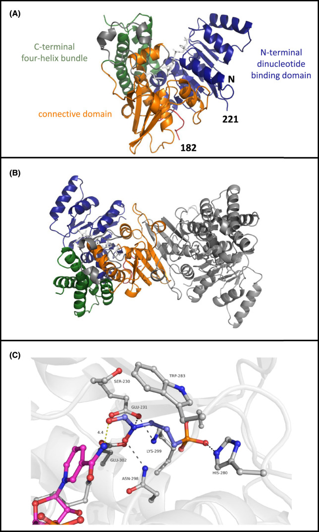
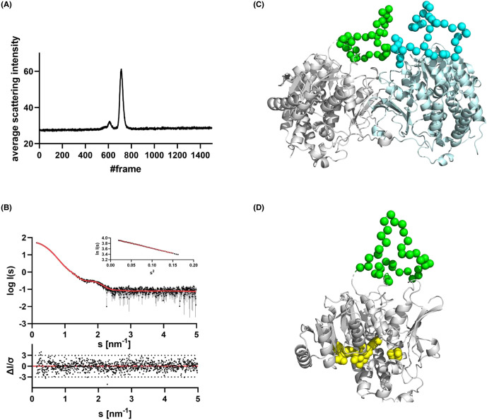
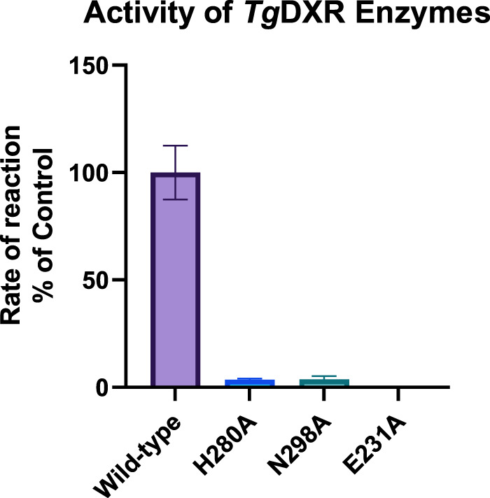
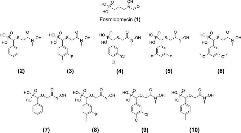
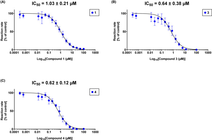
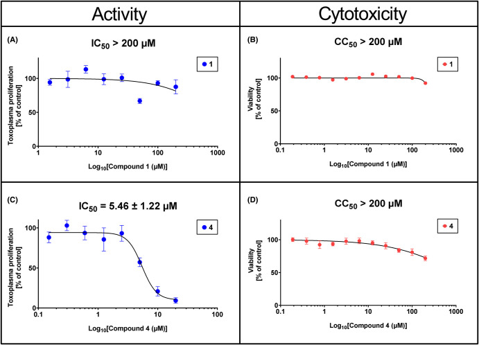
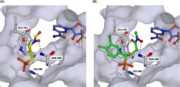
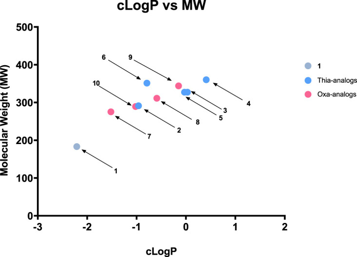
Toxoplasma gondii is a widely distributed apicomplexan parasite causing toxoplasmosis, a critical health issue for immunocompromised individuals and for congenitally infected foetuses. Current treatment options are limited in number and associated with severe side effects. Thus, novel anti-toxoplasma agents need to be identified and developed. 1-Deoxy-d-xylulose 5-phosphate reductoisomerase (DXR) is considered the rate-limiting enzyme in the non-mevalonate pathway for the biosynthesis of the isoprenoid precursors isopentenyl pyrophosphate and dimethylallyl pyrophosphate in the parasite, and has been previously investigated for its key role as a novel drug target in some species, encompassing Plasmodia, Mycobacteria and Escherichia coli. In this study, we present the first crystal structure of T. gondii DXR (TgDXR) in a tertiary complex with the inhibitor fosmidomycin and the cofactor NADPH in dimeric conformation at 2.5 Å resolution revealing the inhibitor binding mode. In addition, we biologically characterize reverse α-phenyl-β-thia and β-oxa fosmidomycin analogues and show that some derivatives are strong inhibitors of TgDXR which also, in contrast with fosmidomycin, inhibit the growth of T. gondii in vitro. Here, ((3,4-dichlorophenyl)((2-(hydroxy(methyl)amino)-2-oxoethyl)thio)methyl)phosphonic acid was identified as the most potent anti T. gondii compound. These findings will enable the future design and development of more potent anti-toxoplasma DXR inhibitors.

Keywords: Toxoplasma gondii; DXR; DXR inhibitors; SAXS; anti-infective; crystal structure; enzymatic assay; fosmidomycin; growth inhibition; parasite.


Conflict of interest statement

The authors declare that there are no competing interests associated with the manuscript.

Figures

Figure 1.
Figure 1.. The MEP pathway for the biosynthesis of isoprenoids.
Modified from Frank and Groll [86]. Created with BioRender.com.
Figure 2.
Figure 2.. Reaction catalyzed by 1-Deoxy-d-xylulose 5-phosphate reductoisomerase (DXR) and the inhibitor fosmidomycin.
(A) The DXR enzyme catalyzes the conversion of the substrate 1-Deoxy-d-xylulose 5-phosphate (DXP) into the product 2-C-Methyl-d-erythritol-4-phosphate (MEP). The catalysis occurs with dependency of a metallic dication (Mg2+) and NADPH as cofactors. One of the proposed mechanisms consists of a two-step reaction: first, the retro-aldol/aldol isomerization of DXP with the formation of the aldehyde intermediate [87]; then, a NADPH-depend reduction of the intermediate to MEP [88]. Modified from Kuzuyama et al. [43]. (B) Chemical structure of the DXR inhibitor fosmidomycin. Created with BioRender.com.
Figure 3.
Figure 3.. Purification of the His10-truncated TgDXR and its optimization and kinetic characterization.
(A) SDS–PAGE of samples taken during the purification of truncated His10-TgDXR after immobilized metal ion affinity chromatography (IMAC) and size exclusion chromatography (SEC). Marker (M): PageRuler™ Prestained Protein Ladder, Thermo Scientific™, Thermo Fisher Scientific, Waltham, MA, U.S.A., #26616, molecular mass in kDa are indicated. Running Buffer: 20× NuPAGE™ MOPS SDS Running buffer, Thermo Fisher Scientific, Waltham, MA, U.S.A., #NP000102. (B) Effects of [DXP] on TgDXR catalyzed reaction. Km values of DXP and NADPH are shown. (C) Effects of [NADPH] on the TgDXR catalyzed reaction. (D) Effects of [Mg2+] on the TgDXR catalyzed reaction. (E) Effects of pH on the TgDXR catalyzed reaction. See Materials and methods section for the experimental conditions. Optimal and selected concentrations or values are indicated (red line). Data shown are means of four independent experiments, each performed in duplicate (n = 8) ± S.D.
Figure 4.
Figure 4.. Structure of TgDXR A.
(A) Overall structure of TgDXR as observed in the crystal structure. Shown is chain A which is similar to chain B, as calculated by comparing the RMSD of both monomers after superimposing which is 0.3 Å overall. Three different domains are observed and colour coded being the N-terminal nucleotide binding domain (blue) the connective domain (orange) and the C-terminal four helix bundle (green). (B) The dimer of TgDXR is highlighted as observed in the asymmetric unit of the crystal. The dimer interface is mediated by the connective domain highlighted in orange in chain A. (C) The binding site of fosmidomycin is shown with in ball and stick representation. Fosmidomycin (blue) is bound via interaction with the sidechains of Glu231, His280, and Asn298. All distances are indicated with black line with a maximum distance of 3.6 Å. The distance of fosmidomycin to NADPH (purple) is 4.4 Å, which is too large to allow activity.
Figure 5.
Figure 5.. Small-angle X-ray scattering data from TgDXR and the most representative (62%) EOM model.
(A) Chromixs SEC SAXS elution profile. Each frame corresponds to 2 s. (B) Scattering data of TgDXR. Experimental data are shown in black dots, with grey error bars. The EOM ensemble model fit is shown as red line and below the residual plot of the data is given. The Guinier plot of TgDXR is added in the right corner. (C) The rigid body protomers of TgDXR from the crystal are shown in grey and cyan cartoon representation. The determined flexible loop is shown as green and blue spheres. (The other EOM models are part of the Supplementary Information). (D) The SAXS completed model of TgDXR is shown as monomer with the flexible loop shown in green spheres. The NADPH and fosmidomycin binding site is highlighted in yellow. It is clear that the flexible loop is remote of the active site and likely serves a more stabilizing role in the dimer of TgDXR.
Figure 6.
Figure 6.. Comparison of the activities of TgDXR wild-type and TgDXR mutants Glu321Ala, His280Ala and Asn298Ala.
Reaction rates of TgDXR wild-type (violet) and TgDXR mutants His280Ala (H280A, blue) and Asn298Ala (N298A, green). Glu321Ala (E321A) could not be determined. Data shown are from the means of three independent experiments each performed in duplicate (n = 6) ± S.D.
Figure 7.
Figure 7.. Chemical structures of DXR inhibitors investigated in this study.
Figure 8.
Figure 8.. In vitro inhibition of TgDXR by most potent DXR inhibitors.
The enzymatic inhibitory activity of 1 (A), 3 (B) and 4 (C) were determined by enzymatic assays in vitro. Assays were performed in 96 well plates at 30°C, containing 100 nM of purified TgDXR protein in dimeric state, 100 µM of NADPH and 4 mM of MgCl2 as cofactors, 100 µM of DXP as substrate in 50 mM HEPES buffer (pH 7.5) containing 50 µg/ml of bovine serum albumin (BSA). The investigated compounds were tested in a concentration range of 3.05 nM to 100 µM. Data shown are from the means of three independent experiments each performed in duplicate (n = 6) ± S.D. IC50 values of each compound are shown.
Figure 9.
Figure 9.. Anti-toxoplasma activity and cytotoxicity on human fibroblasts Hs27 of fosmidomycin and the 3,4-dichlorophenyl-thia analogue.
The antiprotozoal activity of 4 (A) was determined by the T. gondii inhibition assay via the amount of [3H]-uracil incorporation into the RNA of the parasite in vitro. Cytotoxicity of 4 (B) was measured by MTT assays on human fibroblasts Hs27. Data shown are the means of three independent experiments each performed in duplicate (n = 6) ± SEM. IC50 ± S.D. and CC50 values of compound 4 are shown.
Figure 10.
Figure 10.. Predicted binding modes of compound 4 in the X-ray crystal structure of TgDXR.
(A) The fosmidomycin binding site is illustrated, with fosmidomycin represented as yellow sticks, interacting with Glu231, Asn298, and His280. (B) Docking results for compound 4 reveal that the 3,4-dichlorophenyl substituent binds in a previously unoccupied pocket.
Figure 11.
Figure 11.. Plot of ClogP vs. MW for fosmidomycin and its thia- and oxa-analogs.
Fosmidomycin (1) is shown in grey, the thia-analogs in blue, and the oxa-analogs in pink. The investigated fosmidomycin derivatives exhibit increased lipophilicity compared with 1, with compounds 3 and 4, the most active compounds, being the most lipophilic. ClogP values have been predicted using the Marvin JS calculator.

References

    1. Levine, N.D. (2018) The Protozoan Phylum Apicomplexa: Volume 2, CRC Press, Boca Raton, FL
    1. Lourido, S. (2019) Toxoplasma gondii. Trends Parasitol. 35, 944–945 10.1016/j.pt.2019.07.001 - DOI - PubMed
    1. White, M.W. and Suvorova, E.S. (2018) Apicomplexa cell cycles: something old, borrowed, lost, and new. Trends Parasitol. 34, 759–771 10.1016/j.pt.2018.07.006 - DOI - PMC - PubMed
    1. Centers for Disease Control and Prevention. Parasites - toxoplasmosis (toxoplasma infection) - epidemiology & risk factors, https://www.cdc.gov/parasites/toxoplasmosis/epi.html
    1. Pleyer, U., Gross, U., Schlüter, D., Wilking, H. and Seeber, F. (2019) Toxoplasmosis in Germany: epidemiology, diagnosis, risk factors, and treatment. Dtsch. Ärztebl. Int. 116, 435 10.3238/arztebl.2019.0435 - DOI - PMC - PubMed

Publication types

MeSH terms

LinkOut - more resources