. 2024 Aug 20;18(33):22275-22297.
doi: 10.1021/acsnano.4c06286. Epub 2024 Aug 6.

Marginated Neutrophils in the Lungs Effectively Compete for Nanoparticles Targeted to the Endothelium, Serving as a Part of the Reticuloendothelial System

Affiliations

Marginated Neutrophils in the Lungs Effectively Compete for Nanoparticles Targeted to the Endothelium, Serving as a Part of the Reticuloendothelial System

Marco E Zamora et al. ACS Nano. .

Abstract

Nanomedicine has long pursued the goal of targeted delivery to specific organs and cell types but has yet to achieve this goal with the vast majority of targets. One rare example of success in this pursuit has been the 25+ years of studies targeting the lung endothelium using nanoparticles conjugated to antibodies against endothelial surface molecules. However, here we show that such "endothelial-targeted" nanocarriers also effectively target the lungs' numerous marginated neutrophils, which reside in the pulmonary capillaries and patrol for pathogens. We show that marginated neutrophils' uptake of many of these "endothelial-targeted" nanocarriers is on par with endothelial uptake. This generalizes across diverse nanomaterials and targeting moieties and was even found with physicochemical lung tropism (i.e., without targeting moieties). Further, we observed this in ex vivo human lungs and in vivo healthy mice, with an increase in marginated neutrophil uptake of nanoparticles caused by local or distant inflammation. These findings have implications for nanomedicine development for lung diseases. These data also suggest that marginated neutrophils, especially in the lungs, should be considered a major part of the reticuloendothelial system (RES), with a special role in clearing nanoparticles that adhere to the lumenal surfaces of blood vessels.

Keywords: lipid nanoparticles; lung targeting; lungs; marginated neutrophils; neutrophils; reticuloendothelial system.


Figures

Figure 1:
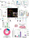
Figure 1:. Nanoparticles that have classically been labeled as “endothelial-targeted” also target pulmonary neutrophils equally well.
A. The classical model of lung uptake by nanoparticles conjugated to affinity ligands that bind to epitopes on endothelial cells, such as PECAM or ICAM-1. Although these epitopes are present at low concentrations on other cell types, the nanoparticles accumulate best in endothelial cells in the lungs due to the high capillary surface area, first-pass effect, and lack of competing cells in the vascular lumen. B. Biodistribution of radiolabeled nanoparticles (liposomes) conjugated to either anti-ICAM or isotype control antibodies, shown as percent injected dose per gram (%ID/g) 30 minutes after IV-injection into naive mice (n =3, p <0.05 by 2-way ANOVA). Inset: anti-ICAM increases the lung-to-blood concentration ratio by 72-fold compared to control (n=3, p<.05, by paired Students’ t-test. C-E. Flow cytometry reveals neutrophils as major cells of uptake for anti-ICAM liposomes. Mice were IV-injected with fluorescent nanoparticles similar to B, and, 30 minutes later, sacrificed to make a single-cell suspension of the lungs, which then underwent flow cytometry. C. Neutrophils had strong uptake of anti-ICAM (blue histogram in panel 1), but not isotype control (panel 2), liposomes; panel 4 shows similar results as a dot plot. D. Endothelial cells showed a similar trend as neutrophils, though with a lower % positive for liposome uptake. E. For each cell type, the positive fraction for liposome uptake showed neutrophils as the cells with the highest uptake percentage. Inset panel 1 shows the ratio of uptake in endothelial cells to neutrophils. Panel 2 shows the mean fluorescence intensity (MFI) of liposome-positive cells, which shows endothelial cells have only slightly higher MFIs than neutrophils. F. Mice treated as in C-E, but with lungs analyzed by fluorescent confocal microscopy. Red anti-ICAM liposomes are in many cell types, including endothelial cells (left panel) and neutrophils (right panel). G. Revised model of uptake of nanoparticles conjugated to endothelial epitopes. An overlooked population of cells, the marginated neutrophils, resides for long periods in the pulmonary capillaries, and they take up a large fraction of the nanoparticles that accumulate in the lungs. Endothelial cells also take up such nanoparticles, but they are far from the exclusive uptake cells of the classical model. Statistics. All figures were evaluated by 2-way ANOVA, n=3 mice, and error bars were the standard error of the mean (SEM). Fig 1B inset assessed by Welch’s t-test. ***= p<.001
Figure 2:
Figure 2:. Neutrophil uptake is generalizable to multiple targeting moieties classically described as targeting endothelial cells only.
A. Schema represents the known distribution of common epitopes to target the pulmonary endothelium. B. Biodistribution 30 minutes after IV injection of radiolabeled liposomes conjugated to antibodies that bind to putative endothelial targets: ICAM, PECAM, PLVAP, and isotype control. Displayed as percent injected dose per gram (%ID/g), the lungs are the dominant target organ). C. Nanoparticle biodistribution to the lungs is fast, peaking before 10 minutes and remaining stable up to 30 minutes. D. Cellular distribution of liposome uptake (measured by flow cytometry) among all targeting moieties. The two most commonly used moieties to target the pulmonary endothelium, anti-ICAM and -PECAM, both have strong neutrophil uptake. PLVAP, less commonly used for this purpose, has lower overall lung uptake due to lower access but a higher endothelial-to-neutrophil targeting ratio (inset). E. Representative histograms from flow cytometry performed in D, showing specific neutrophil uptake in all three targeting moieties but not isotype control. Statistics Fig 2D, 2-way ANOVA with Tukey post-hoc test **=p<0.01, ***=p<.0002, ****=p<.0001. Fig 2D inset, Brown-Forsythe and Welch ANOVA Test using Dunnet’s T3 post hoc test***=p<0.01
Figure 3.
Figure 3.. Most CAM-targeted liposomes are taken up by marginated neutrophils in the lungs.
Suppose a nanoparticle localizes to the lungs but is not internalized into cells. In that case, it could be taken up into cells after the mouse is sacrificed, especially during the single-cell-suspension step before flow cytometry. This could artificially inflate the fraction of a cell type taking up the particle. To quantify this potential artifact, we devised the following generalizable protocol (A): One mouse was given Alexa-594-labeled liposomes, and another had the same liposomes (and delivery route), labeled with Alexa-488. The left lungs of each mouse were combined into a single tube for processing for flow cytometry (the right lungs were treated similarly, as a replicate). B. Flow cytometry from an example mouse. Most endothelial cells were positive for only one color liposome (594 or 488, in the bottom-right or top-left quadrants, respectively). There were 6.85% of endothelial cells that were positive for both liposome colors, suggesting that a small fraction of liposomes can pass from one lung to the cells of the other while in the post-mortem single-cell suspension. This is similar to neutrophils from the same experiment, with about 9.81% of neutrophils being mixed C. We define the nanoparticle crossover index as listed in the figure, and it gives the fraction of liposome-positive cells that received a significant fraction of their liposomes post-mortem in the single-cell suspension. We show that this value for endothelial cells and neutrophils is very low. For comparison, an NCI =1 would indicate complete ex vivo transfer of our nanoparticle following flow cytometry preparation. D. We used high-resolution microscopy to visualize neutrophils in the capillary lumen, taking up our CAM-targeted liposomes. E. To quantify marginated neutrophils in each organ, we IV-injected radiolabeled anti-Ly6G, 5 minutes later sacrificed, and perfused. After normalizing each organ’s radioactivity to blood radioactivity (left panel), it is evident that marginated neutrophils are higher in the lung than the liver, especially when compared to control radiolabeled (non-avid) IgG. In the right panel, we normalized the anti-Ly6G signal by control IgG, which shows the lungs have ~4x higher concentration of marginated neutrophils than the liver. F. To determine if endothelial-targeted nanocarriers were leaking out of the endothelium and into the alveolar airspace, we IV-injected aPECAM-liposomes, sacrificed 30 minutes later, perfused the lungs, and then measured radioactivity in the airspace (via bronchoalveolar lavage [BAL]), and the residual lungs and blood. The lungs have by far the highest uptake, showing nearly no leak of nanoparticles into the airspace. G. We next developed a flow cytometry protocol to distinguish the different neutrophil compartments in the lung: neutrophils in the airspace, interstitial, and marginated neutrophils. Briefly, we inject liposomes labeled with AF594 and allow them to circulate for 30 min. 5 min before sacrifice, mice were injected with RED anti-Ly6G antibody. Mice were sacrificed, and blood and bronchoalveolar lavage (BAL) fluid samples were drawn for later analysis. Resultant single cell preparation from lung tissue is later post-stained with VIOLET anti-Ly6G antibody. This results in the following: blood samples representing circulating neutrophils stained only RED, marginated neutrophils stained with both RED and VIOLET, interstitial neutrophils stained only with VIOLET, and BAL samples stained only VIOLET. H. Gating on liposome-positive cells first, then identifying the percentage of each compartment, we find that in the lung, marginated neutrophils account for 84% of total liposome-positive neutrophils. I. MFI and histogram show that among neutrophils, marginated neutrophils in the lungs take up more nanoparticles than those in the blood, interstitium, or airspace. Statistics. n=3 per group unless otherwise stated. 1-way ANOVA with Tukey post hoc test *p<0.05, **p<0.01,***p<0.001, ****p<0.0001
Figure 4:
Figure 4:. Mechanisms underlying neutrophil uptake of lung-targeted nanoparticles.
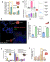
A. Kinetics of lung uptake of radiolabeled anti-ICAM liposomes, showing it is fast (< 10 minutes), with a slight decline by 1 hour. B. The ratio of liposome-positive neutrophils to liposome-positive endothelial cells also declines slightly over 1 hour. C. Expression of ICAM and PECAM proteins on various cells in the lungs (which had not received nanoparticles), measured by flow cytometry and presented as median fluorescence intensity (MFI) D. In the lungs of mice that received IV-injected nanocarriers targeted to the endothelium, neutrophil uptake of CAM-targeted liposomes increases with nanoparticle size. Further, cationic liposomes with known tropism for pulmonary endothelium have similar uptake into marginated neutrophils as CAM-antibody-targeted liposomes. E. Endothelial-to-neutrophil uptake ratio (derived from the experiment in D), showing larger-sized particles (200 nm) and cationic liposomes experience nearly co-equal uptake by neutrophils and endothelial cells. F. Neutrophil uptake of anti-ICAM nanoparticles is not specific to liposomes but generalizes to multiple particle types, here RNA-containing lipid nanoparticles (LNPs) and polystyrene nanoparticles. G. The endothelial-to-neutrophil nanoparticle uptake ratio does differ somewhat between particle types, but in no case is much above 1 (meaning endothelial cells never have significantly better uptake than neutrophils). H. Next, we measured the effect complement opsonization of nanoparticles has on neutrophil uptake of aPECAM liposomes. We injected 10 mg/kg AF594-labeled aPECAM liposomes into complement-deficient (C3 knockout) mice and wild-type control. We observe that knocking out complement increases the endothelial-to-neutrophil uptake ratio by >2-fold. I. Next, we tested whether the Fc region’s presentation on a liposome’s surface affected neutrophil recognition. We generated liposomes covalently conjugated with whole anti-ICAM antibody (YN1) or antibody fragments ICAM F(ab) generated from YN1 to test this. Removal of the Fc region increases the endothelial-to-neutrophil uptake ratio by 21x. This suggests that an Fc on nanoparticles is insufficient for neutrophil uptake (IgG2b-liposomes are not taken up). Still, when present with an antibody avid for a pulmonary epitope, the Fc promotes neutrophil uptake. J. Updating our schematic based on our findings, we propose two hypotheses for neutrophil uptake of endothelial CAM-targeted liposomes. First, direct binding of CAM-targeted liposomes to neutrophils, and second, after liposomes successfully associate with endothelial surfaces, marginated neutrophils in their surveillance role come and remove nanoparticles and prevent uptake by endothelial cells. Statistics. n=3 per group unless otherwise stated. Figs. 4D, F and H, 2-way ANOVA with Tukey post hoc test *p<0.05, **p<0.01,***p<0.001, ****p<0.0001. Fig 4E,F One-way ANOVA with Tukey post hoc test *p<0.05, **p<0.01, (Fig 4H and I), Students’ T-test, *p<0.05, ***p<.001.
Figure 5:
Figure 5:. Real-time imaging in breathing mice of marginated neutrophils taking up ‘pulmonary endothelial-targeted liposomes.
A. Representative still image from intravital microscopy videos shown in Supplemental Videos 2–4. B. Quantification of neutrophil movement into the frame of the video taken over time shows a steady increase in neutrophil accumulation over time after injection of aPECAM liposomes, with a reduction in the arrival rate after 20 minutes. C. The percentage of neutrophil-particle overlap in-frame over time gradually increases as neutrophils take up aPECAM liposomes in the lung. D. Quantifying the nanoparticle signal in neutrophils over time shows a delay from when nanoparticles land on an endothelial surface and engulfment by marginated neutrophils. E. Representative still frames of neutrophils taking up aPECAM liposomes depict the deposition of liposomes on the endothelial surface (Frame 1), migration of neutrophils towards the nanoparticles (Frame 2), engulfment by neutrophils (Frame 3), and clearance (Frame 4). F. Comparing the first and fourth from E, investigating liposomes taken up by marginated neutrophils (liposomes in white, neutrophils in green) versus liposomes on the endothelial surface (in red) shows the active internalization of liposomes by marginated neutrophils. G. A representative still from Supp Video 5. Here, liposomes on the endothelial surface colored in white, liposomes internalized neutrophils (neutrophils colored green, internalized liposomes in red) showing the direct uptake of liposomes from endothelial cells by neutrophils. H. Quantitative analysis of G assessing the percentage of nanoparticles found in neutrophils overlaid with the percentage of the neutrophil area containing nanoparticles (green box). This shows that over the time step of the recording, there is a delay before the uptake of nanoparticles by marginated neutrophils. This is similarly shown below (red box), where nanoparticle signal is observed in early time steps, however, the nanoparticle signal within neutrophils occurs only at later time steps, suggesting endothelial deposition.
Figure 6.
Figure 6.. Human lungs ex vivo also display neutrophil uptake of targeted liposomes.
A. Human lungs rejected for transplantation were oxygenated and endovascularly cannulated via the pulmonary artery for perfusion. Anti-ICAM conjugated liposomes were infused endovascularly, followed by washout with Steen’s solution and infusion of tissue dye to identify the perfused lung region for harvest. The perfused lung tissue was collected and processed into a single-cell suspension for flow cytometry. B. As in mice, anti-ICAM liposomes were taken up by endothelial cells and neutrophils. However, the endothelial-to-neutrophil uptake ratio was higher in humans than in mice. Histograms for neutrophils and endothelial cells are compared to liposome-negative samples, i.e., human lung samples that did not receive liposomes from the same donor.
Figure 7:
Figure 7:. During local and remote inflammation, marginated neutrophils in the lungs dominate the uptake of nanoparticles that bind to the vasculature.
A. Biodistribution of IV-injected I-125 radiolabeled anti-Ly6G antibody shows that 2 hours after CLP injury, neutrophil presence in the lung increases significantly B. CLP mice were IV-injected with fluorescent anti-PECAM liposomes and anti-Ly6G antibody (to label marginated neutrophils) via the same protocol as in Figure 3. This showed that marginated lung neutrophils take up nanoparticles to a greater extent than neutrophils in other lung compartments, blood, or spleen. C. Microscopy shows the marginated pool of neutrophils in the lungs colocalize well with aPECAM liposomes after CLP injury. However, in the spleen, while there is a presence of aPECAM liposome and neutrophils, there is less colocalization. D. In the CFA footpad inflammation model, flow cytometry reveals that ~60% of the marginated neutrophils in the lung take up fluorescently labeled aPECAM liposomes. In comparison, only ~20% of neutrophils take up nanoparticles in the injured footpad. Liposome MFI similarly shows that lung-marginated neutrophils more avidly take up aPECAM liposomes than neutrophils found in the footpad. E. Biodistribution of radiolabeled nanoparticles shows that ALI (nebulized LPS) decreases total lung uptake for ICAM- and PECAM-targeted nanoparticles but not PLVAP- or isotype control. F. Gating first to identify cell types, then identified the percentage of each cell type that takes up our CAM-targeted liposomes. Taking these percentages for endothelial cells and neutrophils, we get the endothelial-to-neutrophil uptake ratio and find that it is increased for all targeting moieties, but most for PLVAP, which was already the most endothelial-specific. Statistics. n=3 for all groups. Fig 7 A, E, and F, 2-way ANOVA with Tukey post hoc test **p<0.01,***p<0.001, ****p<0.0001. Fig 7B One-way ANOVA with Tukey post hoc test *p<0.05, **p<0.01, Fig7D Unpaired T-test with Welch’s correction, ****p<.0001, ***p<0.001.

References

    1. Glassman PM; Myerson JW; Ferguson LT; Kiseleva RY; Shuvaev VV; Brenner JS; Muzykantov VR Targeting Drug Delivery in the Vascular System: Focus on Endothelium. Adv. Drug Deliv. Rev. 2020, 157, 96–117. - PMC - PubMed
    1. Wilhelm S; Tavares AJ; Dai Q; Ohta S; Audet J; Dvorak HF; Chan WCW Analysis of Nanoparticle Delivery to Tumours. Nature Reviews Materials 2016, 1 (5), 1–12.
    1. Ouyang B; Poon W; Zhang Y-N; Lin ZP; Kingston BR; Tavares AJ; Zhang Y; Chen J; Valic MS; Syed AM; MacMillan P; Couture-Senécal J; Zheng G; Chan WCW The Dose Threshold for Nanoparticle Tumour Delivery. Nat. Mater. 2020, 19 (12), 1362–1371. - PubMed
    1. Bachmaier K; Stuart A; Singh A; Mukhopadhyay A; Chakraborty S; Hong Z; Wang L; Tsukasaki Y; Maienschein-Cline M; Ganesh BB; Kanteti P; Rehman J; Malik AB Albumin Nanoparticle Endocytosing Subset of Neutrophils for Precision Therapeutic Targeting of Inflammatory Tissue Injury. ACS Nano 2022, 16 (3), 4084–4101. - PMC - PubMed
    1. Chen S-J; Sanmiguel J; Lock M; McMenamin D; Draper C; Limberis MP; Kassim SH; Somanathan S; Bell P; Johnston JC; Rader DJ; Wilson JM Biodistribution of AAV8 Vectors Expressing Human Low-Density Lipoprotein Receptor in a Mouse Model of Homozygous Familial Hypercholesterolemia. Hum Gene Ther Clin Dev 2013, 24 (4), 154–160. - PMC - PubMed

LinkOut - more resources