. 2024 Aug 6;73(10):207.
doi: 10.1007/s00262-024-03791-6.

Unlocking the potential of HHLA2: identifying functional immune infiltrating cells in the tumor microenvironment and predicting clinical outcomes in laryngeal squamous cell carcinoma

Affiliations

Unlocking the potential of HHLA2: identifying functional immune infiltrating cells in the tumor microenvironment and predicting clinical outcomes in laryngeal squamous cell carcinoma

Wenjing Li et al. Cancer Immunol Immunother. .

Abstract

Background: HHLA2 (human endogenous retrovirus-H long terminal repeat-associating protein 2) represents a recently identified member of the B7 immune checkpoint family, characterized by limited expression in normal tissues but notable overexpression in various cancer types. Nevertheless, the precise function and interaction with immune cells remain poorly understood, particularly in laryngeal squamous cell carcinoma (LSCC). This investigation endeavored to elucidate the biological significance of HHLA2 within the tumor microenvironment of human LSCC tissues and delineate the clinical relevance and functional roles of HHLA2 in LSCC pathogenesis.

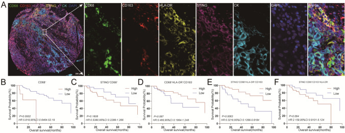
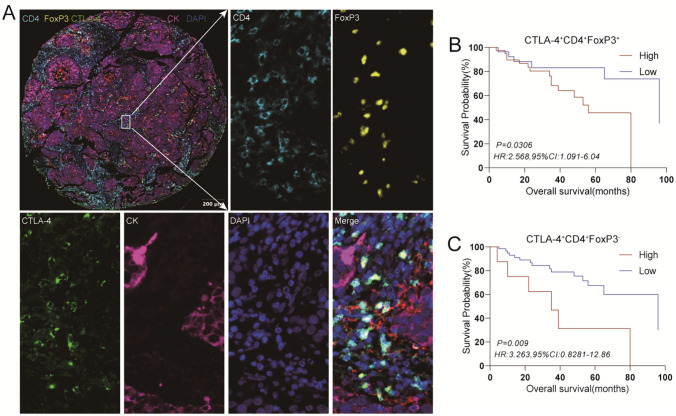
Methods: Through multiplexed immunohistochemistry analyses conducted on tissue microarrays sourced from LSCC patients (n = 72), the analysis was executed to assess the expression levels of HHLA2, density and spatial patterns of CD68+HLA-DR+CD163- (M1 macrophages), CTLA-4+CD4+FoxP3+ (CTLA-4+Treg cells), CTLA-4+CD4+FoxP3- (CTLA-4+Tcon cells), exhausted CD8+T cells, and terminally exhausted CD8+T cells in LSCC tissues. Survival analysis was conducted to evaluate the prognostic significance of HHLA2 and these immune checkpoints or immune cell populations, employing COX regression analysis to identify independent prognostic factors.

Results: Kaplan-Meier (K-M) survival curves revealed a significant association between HHLA2 expression and overall survival (OS) in LSCC. Elevated levels of HHLA2 were linked to reduced patient survival, indicating its potential as a prognostic marker (HR: 3.230, 95%CI 0.9205-11.34, P = 0.0067). Notably, increased infiltration of CD68+ cells (total macrophages), STING+CD68+HLA-DR+CD163- (STING+M1 macrophages), CTLA-4+CD4+FoxP3+, CTLA-4+CD4+FoxP3-, PD-1+LAG-3+CD8+T cells, and PD-1+LAG-3+TIM-3+CD8+T cells strongly linked to poorer survival outcomes (P < 0.05). A discernible trend was observed between the levels of these immune cell populations, STING+CD68+ (STING+ total macrophages), CD68+HLA-DR+CD163-, STING+CD68+CD163+HLA-DR- (STING+M2 macrophages), PD-1+LAG-3-CD8+T cells, PD-1+TIM-3+CD8+T cells, and PD-1+LAG-3+TIM-3-CD8+T cells and prognosis. Importantly, multivariate COX analysis identified HHLA2 as an independent predictive factor for OS in LSCC patients (HR = 3.86, 95% CI 1.08-13.80, P = 0.038). This underscored the potential of HHLA2 as a critical marker for predicting patient outcomes in LSCC.

Conclusions: HHLA2 emerged as a detrimental prognostic biomarker for assessing OS in LSCC patients. Relative to other immune checkpoints, HHLA2 exhibited heightened predictive efficacy for the prognosis of LSCC patients.

Keywords: HHLA2; Immune infiltrating cell; Laryngeal squamous cell carcinoma; Prognosis.


Conflict of interest statement

The authors declare no competing interests.

Figures

Fig. 1
Fig. 1
Characterization of the expression, localization, and prognostic values of HHLA2 and Ki67 in human LSCC tissues. An enlarged subsection of the core was highlighted, showing each of the individual markers in the composite image after spectral unmixing, together with the DAPI nuclear marker (pseudo-colored gray-blue) and the autofluorescence signal (pseudo-colored black). A. The expressions of HHLA2 (membrane, pseudo-colored green) and Ki67 (nuclear, pseudo-colored blue) in adjacent normal tissues of HCC were identified by CK (membrane, pseudo-colored purple), together with the DAPI nuclear marker (pseudo-colored gray-blue), The ratio of the main chart to the sub-chart is 1:11. B, C. Kaplan–Meier survival analysis was performed to predict the prognostic values of HHLA2+ and Ki67+ expression in human LSCC tissues. D. Kaplan–Meier survival analysis was performed to predict the prognostic value of HHLA2+ combined with Ki67+ in LSCC tissues. E. Correlation analysis was performed to predict the relationship between the expression of HHLA2 and Ki67. *P < 0.05, **P < 0.01, *** P < 0.001, ****P < 0.0001
Fig. 2
Fig. 2
Characterization of the expressions, localizations, and correlation of HHLA2 and immune checkpoint in human LSCC tissues. An enlarged subsection of the tissue core was highlighted, showing each of the individual markers in the composite image after spectral unmixing, together with the DAPI nuclear marker (pseudo-colored gray-blue) and the autofluorescence signal (pseudo-colored black). A. The expressions of HHLA2 (membrane, pseudo-colored green) and immune checkpoint in adjacent normal tissues of LSCC were identified by CK (membrane, pseudo-colored purple), together with the DAPI nuclear marker (pseudo-colored gray-blue). The ratio of the main chart to the sub-chart is 1:9. B, C. Correlation analysis was performed using the Spearman R to predict the relationship between HHLA2 expression and immune cells infiltration
Fig. 3
Fig. 3
Characterization of the expressions, localizations, and prognostic value of immune checkpoint of macrophages in human LSCC tissues. An enlarged subsection of the core was highlighted, showing each of the individual markers in the composite image after spectral unmixing, together with the DAPI nuclear marker (pseudo-colored gray-blue) and the autofluorescence signal (pseudo-colored black). A. The expressions of immune checkpoint in adjacent normal tissues of LSCC were identified by CK (membrane, pseudo-colored blue), together with the DAPI nuclear marker (pseudo-colored gray-blue), The ratio of the main chart to the sub-chart is 1:9. B-F. Kaplan–Meier survival analysis was performed to predict macrophages and subtypes of macrophages in human LSCC tissues
Fig. 4
Fig. 4
Characterization of the expressions, localizations, and prognostic value of immune checkpoint of CD4+ T cells in human LSCC tissues. An enlarged subsection of the core was highlighted, showing each of the individual markers in the composite image after spectral unmixing, together with the DAPI nuclear marker (pseudo-colored gray-blue) and the autofluorescence signal (pseudo-colored black), The ratio of the main chart to the sub-chart is 1:10. A. The expressions of immune checkpoint in adjacent normal tissues of LSCC were identified by CK (membrane, pseudo-colored purple), together with the DAPI nuclear marker (pseudo-colored gray-blue). B, C. Kaplan–Meier survival analysis was performed to predict the prognostic values of CD4+T cell subtypes in human LSCC tissues
Fig. 5
Fig. 5
Characterization of the expressions, localizations, and prognostic value of immune checkpoint of CD8+T cells in human LSCC tissues. An enlarged subsection of the core was highlighted, showing each of the individual markers in the composite image after spectral unmixing, together with the DAPI nuclear marker (pseudo-colored gray-blue) and the autofluorescence signal (pseudo-colored black). A. The expressions of immune checkpoints in adjacent normal tissues of LSCC were identified by CK (membrane, pseudo-colored purple), together with the DAPI nuclear marker (pseudo-colored gray-blue). The ratio of the main chart to the sub-chart is 1:11. B-F. Kaplan–Meier survival analysis was performed to predict the prognostic values of CD8+T cell subtype infiltration in human LSCC tissues
Fig. 6
Fig. 6
Kaplan–Meier survival analysis was performed to predict the prognostic values of HHLA2+ combined with immune infiltrating cells in human LSCC tissues. Patients were stratified according to the expression of HHLA2+ and density of immune infiltrating cells. A, B, and C. Cutoffs for expression or infiltration were defined with the Cutoff Finder method. *P < 0.05, **P < 0.01, *** P < 0.001, **** P < 0.0001

Similar articles

Cited by

References

    1. Wang X, Tian L, Li Y (2021) RBM15 facilitates laryngeal squamous cell carcinoma progression by regulating TMBIM6 stability through IGF2BP3 dependent. J Exp Clin Cancer Res 40(1):1–18. 10.1186/s13046-021-01871-4 10.1186/s13046-021-01871-4 - DOI - PMC - PubMed
    1. Cavaliere M, Bisogno A, Scarpa A et al (2021) Biomarkers of laryngeal squamous cell carcinoma: a review. Ann Diagn Pathol 54:151787. 10.1016/j.anndiagpath.2021.151787 10.1016/j.anndiagpath.2021.151787 - DOI - PubMed
    1. Cossu AM, Mosca L, Zappavigna S et al (2019) Long Non-coding RNAs as important biomarkers in laryngeal cancer and other head and neck tumours. Int J Mol Sci 20(14):3444. 10.3390/ijms20143444 10.3390/ijms20143444 - DOI - PMC - PubMed
    1. Koontongkaew S (2013) The tumor microenvironment contribution to development, growth, invasion and metastasis of head and neck squamous cell carcinomas. J Cancer 4(1):66–83. 10.7150/jca.5112 10.7150/jca.5112 - DOI - PMC - PubMed
    1. Horozoglu C, Sonmez D, Demirkol S et al (2021) Potential role of immune cell genetic variants associated with tumor microenvironment response in laryngeal squamous cell carcinoma (LSCC) in terms of clinicopathological features. Pathol Res Pract 228:153665. 10.1016/j.prp.2021.153665 10.1016/j.prp.2021.153665 - DOI - PubMed

MeSH terms

LinkOut - more resources