. 2024 Aug 13;121(33):e2405454121.
doi: 10.1073/pnas.2405454121. Epub 2024 Aug 6.

A bioactive supramolecular and covalent polymer scaffold for cartilage repair in a sheep model

Affiliations

A bioactive supramolecular and covalent polymer scaffold for cartilage repair in a sheep model

Jacob A Lewis et al. Proc Natl Acad Sci U S A. .

Abstract

Regeneration of hyaline cartilage in human-sized joints remains a clinical challenge, and it is a critical unmet need that would contribute to longer healthspans. Injectable scaffolds for cartilage repair that integrate both bioactivity and sufficiently robust physical properties to withstand joint stresses offer a promising strategy. We report here on a hybrid biomaterial that combines a bioactive peptide amphiphile supramolecular polymer that specifically binds the chondrogenic cytokine transforming growth factor β-1 (TGFβ-1) and crosslinked hyaluronic acid microgels that drive formation of filament bundles, a hierarchical motif common in natural musculoskeletal tissues. The scaffold is an injectable slurry that generates a porous rubbery material when exposed to calcium ions once placed in cartilage defects. The hybrid material was found to support in vitro chondrogenic differentiation of encapsulated stem cells in response to sustained delivery of TGFβ-1. Using a sheep model, we implanted the scaffold in shallow osteochondral defects and found it can remain localized in mechanically active joints. Evaluation of resected joints showed significantly improved repair of hyaline cartilage in osteochondral defects injected with the scaffold relative to defects injected with the growth factor alone, including implantation in the load-bearing femoral condyle. These results demonstrate the potential of the hybrid biomimetic scaffold as a niche to favor cartilage repair in mechanically active joints using a clinically relevant large-animal model.

Keywords: cartilage regeneration; peptide amphiphiles; self-assembly; supramolecular biomaterials.


Conflict of interest statement

Competing interests statement:J.A.L., N.A.S., M.T.M., and S.I.S. are inventors on patent application WO2023056433A1 filed by Northwestern University. N.A.S. and S.I.S. are stockholders of Amphix Bio, Inc.

Figures

Fig. 1.
Fig. 1.
Preparation and performance of supramolecular-covalent hybrid scaffold. (A) Chemical structure of the TGFβ-1 binding PA. (B) Chemical structure of the diluent PA. (C) Schematic showing the preparation and placement envisioned of the PA/HA hybrid material within a cartilage defect. (D) Photograph of the PA/HA hybrid material containing a 5-carboxytetramethylrhodamine (TAMRA)-dyed PA for color. (Scale bar, 5 mm.) (E) Storage modulus and loss modulus as a function of strain for gels containing PA only, PA added to 4 wt% unmodified HA, and PA added to 4 wt% crosslinked HA particles with flow strain indicated by dashed vertical lines (n = 3). (F) % swelling as a function of incubation time for PA-only gels and PA/HA gels with unmodified or crosslinked HA; statistical significance was calculated relative to PA-only gels (two-way ANOVA; n = 5). (G) Hyaluronidase catalyzed degradation of HA as a function of time for crosslinked HA alone, hybrid hydrogel of PA and unmodified HA, and hybrid hydrogel of PA and crosslinked HA determined using a carbazole assay (*, PA + HA vs. PA + Crosslinked HA; #, PA + HA vs. Crosslinked HA only; two-way ANOVA; n = 4) (*P ≤ 0.050; **P ≤ 0.010; ***P ≤ 0.001).
Fig. 2.
Fig. 2.
Self-assembled morphology of scaffold and chondrogenesis of encapsulated cells. (A) SEM micrographs of gels formed by 2 wt% PA only, 2 wt% PA added to crosslinked HA for a final concentration of 2 wt%, 4 wt%, and 10 wt%. (Scale bar, 1 μm.) (B) SAXS/MAXS/WAXS intensity as a function of the wave vector for PA only and PA/HA without CaCl2 added with d-spacings (in nm) associated with observed peaks indicated. (C) Immunocytochemistry confocal microscopy images showing hMSCs encapsulated by a PA-only gel and (D) a PA/HA gel; nuclei stained with Hoechst (blue), actin stained with phalloidin (green), and PA tagged with TAMRA (red). (Scale bar, 25 μm.) (E) SEM micrograph of an hMSC encapsulated in a PA/HA gel. (Scale bar, 2 μm.) (F) TGFβ-1 release from PA/HA gels as a function of time in which the PA component contained only diluent monomers or 90 mol% diluent and 10 mol% of the TGFβ-1-binding peptide sequence (two-way ANOVA; n = 4). (G) sGAG content normalized to DNA concentration 4 wk following encapsulation in PA-only and PA/HA gels without TGFβ-1, with 10 ng/mL TGFβ-1-supplemented medium, or with TGFβ-1 added to the PA solution prior to cell encapsulation such that the total TGFβ-1 in the gel equaled the total amount delivered in the supplemented medium group over the 4-wk experiment; significance calculated relative to control samples without TGFβ-1 for each gel (two-way ANOVA; n = 5; *P ≤ 0.050; ***P ≤ 0.001).
Fig. 3.
Fig. 3.
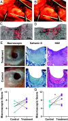
Osteochondral defect filling and scaffold integration. (A) Photographs of a 3 to 4 mm deep, 7 mm diameter osteochondral defect made by drilling into the medial condyle of a sheep stifle, and (B) the defect filled with the PA/HA hybrid. (C) Fluorescence imaging produced by TAMRA-labeled PA filaments (red) in the hybrid in medial condyle histological sections for slung and (D) contralateral nonslung stifles obtained 7 d following implantation. (E) Photographs showing a representative macroscopic appearance of the defects, Safranin O and H&E-stained histological sections along with (F) blinded ICRS scoring of macroscopic appearance of medial condyle defects and (G) trochlear groove defects treated with the hybrid material delivering TGFβ-1 and contralateral controls where TGFβ-1 was applied in saline solution (Wilcoxon signed-rank test; n = 6). (Scale bar, 1 mm.) (*P ≤ 0.050)
Fig. 4.
Fig. 4.
Repair of hyaline-like tissue in supramolecular-covalent hybrid treated cartilage defects. (A) Representative macroscopic appearance, Safranin O-stained sections, H&E-stained sections, and immunohistology staining of collagen II images for trochlea groove defects 12 wk postsurgery. (B) Comparison of subject-matched histological scores for control and treated trochlear groove and (C) femoral condyle defects 12 wk postsurgery (Paired Student’s t test; n = 13). (D) Representative macroscopic appearance, Safranin O-stained sections, H&E-stained sections, and immunohistology staining of collagen II images for femoral condyle defects 24 wk postimplantation. (E) Comparison of subject-matched histological scores for control and treated femoral condyle defects 24 wk postsurgery (Paired Student’s t test; n = 8). (Scale bar, 1 mm.) (*P ≤ 0.050).

References

    1. Huey D. J., Hu J. C., Athanasiou K. A., Unlike bone, cartilage regeneration remains elusive. Science 338, 917–921 (2012), 10.1126/science.1222454. - DOI - PMC - PubMed
    1. Malda J., Groll J., van Weeren P. R., Rethinking articular cartilage regeneration based on a 250-year-old statement. Nat. Rev. Rheumatol. 15, 571–572 (2019), 10.1038/s41584-019-0278-7. - DOI - PubMed
    1. Kurtz S. M., et al. , International survey of primary and revision total knee replacement. Int. Orthop. 35, 1783–1789 (2011). - PMC - PubMed
    1. Price A. J., et al. , Knee replacement. Lancet 392, 1672–1682 (2018), 10.1016/S0140-6736(18)32344-4. - DOI - PubMed
    1. Armiento A. R., Stoddart M. J., Alini M., Eglin D., Biomaterials for articular cartilage tissue engineering: Learning from biology. Acta Biomater. 65, 1–20 (2018), 10.1016/j.actbio.2017.11.021. - DOI - PubMed

MeSH terms

LinkOut - more resources