. 2024 Oct:48:102074.
doi: 10.1016/j.tranon.2024.102074. Epub 2024 Aug 5.

Combination of radiotherapy and PD-L1 blockade induces abscopal responses in EGFR-mutated lung cancer through activating CD8+ T cells

Affiliations

Combination of radiotherapy and PD-L1 blockade induces abscopal responses in EGFR-mutated lung cancer through activating CD8+ T cells

Wu-Yan Xia et al. Transl Oncol. 2024 Oct.

Abstract

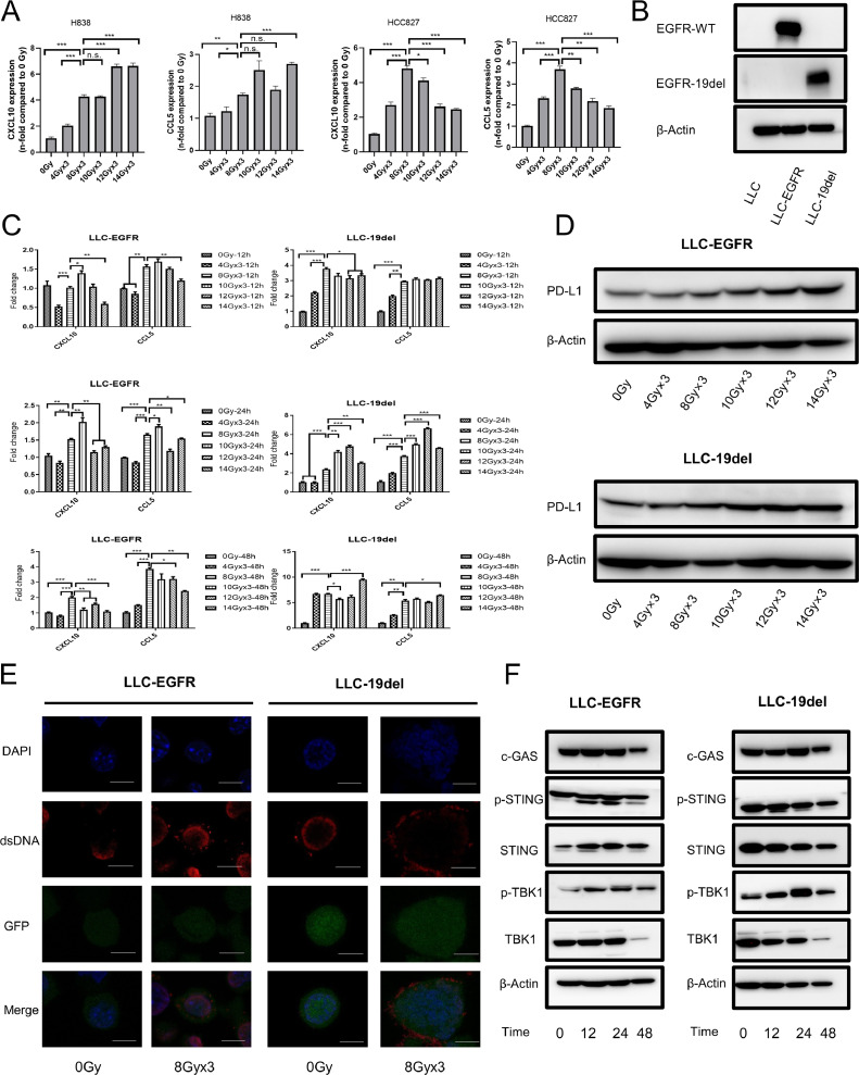
Patients with EGFR-mutated non-small cell lung cancer (NSCLC) respond poorly to immune checkpoint inhibitors (ICIs). It has been reported that the number of CD8+T cells is reduced in EGFR-mutated NSCLC. However, the extent of heterogeneity and effector function of distinct populations of CD8+T cells has not been investigated intensively. In addition, studies investigating whether a combination of radiotherapy and ICIs can improve the efficacy of ICIs in EGFR-mutated lung cancer are lacking. Single-cell RNA sequencing (scRNA-seq) was used to investigate the heterogeneity of CD8+T cell populations in EGFR-mutated NSCLC. The STING pathway was explored after hypofractionated radiation of EGFR-mutated and wild-type cells. Mice bearing LLC-19del and LLC-EGFR tumors were treated with radiotherapy plus anti-PD-L1. The scRNA-seq data showed the percentage of progenitor exhausted CD8+T cells was lower in EGFR-mutated NSCLC. In addition, CD8+T cells in EGFR-mutated NSCLC were enriched in oxidative phosphorylation. In EGFR-mutated and wild-type cells, 8 Gy × 3 increased the expression of chemokines that recruit T cells and activate the cGAS-STING pathway. In the LLC-19del and LLC-EGFR mouse model, the combination of radiation and anti-PD-L1 significantly inhibited the growth of abscopal tumors. The enhanced abscopal effect was associated with systemic CD8+T cell infiltration. This study provided an intensive understanding of the heterogeneity and effector functions of CD8+T cells in EGFR-mutated NSCLC. We showed that the combination of hypofractionated radiation and anti-PD-L1 significantly enhanced the abscopal responses in both EGFR-mutated and wild-type lung cancer by activating CD8+T cells in mice.

Keywords: Abscopal effect; Anti-PD-L1; Epidermal growth factor receptor; Radiotherapy; Single-cell RNA sequencing.


Conflict of interest statement

Declaration of competing interest The authors declare that they have no known competing financial interests or personal relationships that could have appeared to influence the work reported in this paper.

Figures

Fig 1
Fig. 1
The response to ICIs and percentage of lymphocytes in NSCLC patients with EGFR mutation. (A) The frequency of best overall response to ICIs is shown in the pie chart. (B) The PFS curve for patients. (C) Summary of the percentage of lymphocytes subsets (CD3+, CD4+, CD8+, CD19+, CD16+CD56+ cells) and the CD4+/CD8+ ratio in peripheral blood from EGFR-19del and EGFR wild-type LUADs. ICI, immune checkpoint inhibitor; NSCLC, non-small cell lung cancer; EGFR, epidermal growth factor receptor; MT, mutated; n.s.: not significant.
Fig 2
Fig. 2
Profiling of CD8+T cells in LUADs with different EGFR mutation status from single-cell RNAseq samples. (A) The UMAP projection of 5989 CD8+T cells, visualizing the formation of 11 main subtypes. (B) Canonical cell markers used to identify CD8+T cell subtypes. (C) Heatmap of functional markers used to annotate distinct CD8+T subtypes. (D) Average proportion of each CD8+T cell subtype in EGFR-19del and EGFR-WT group. (E) The average expression level of activation (left), cytotoxic (middle), and exhausted (right) marker genes of CD8+T cell differing in EGFR status. P value was calculated by unpaired Wilcoxon rank-sum test. (F) Boxplots showing the progenitor exhausted and terminal exhausted gene signature score derived from Tpex versus Tex-term CD8+T cells. P value was calculated by unpaired Wilcoxon rank-sum test. (G) Bubble plot of the immune checkpoint receptor expression among CD8+T cell clusters from different groups. n.s.: not significant, ****P < 0.001.
Fig 3
Fig. 3
Gene set enrichment analysis, metabolic analysis and SCENIC analysis for CD8+T cells differs by EGFR mutation status. (A) Top enriched pathways of CD8+T cells isolated from EGFR-19del (left) or EGFR-WT (right) group as determined by GSEA using the hallmark gene sets. (B) SCENIC analysis of TRM (left), Tpex (right) and Tex-term (bottom) cells from EGFR-19del and EGFR-WT group. (C) Heatmap showing differences in metabolic pathways between CD8+T cells in EGFR-19del and EGFR-WT group.
Fig 4
Fig. 4
Chemokines expression and STING pathway activation in the EGFR wild-type and EGFR-19del cells treated with different radiation doses. (A) RT-qPCR measurement of chemokines CXCL10 and CCL5 in H838 and HCC827 cells treated with different radiation doses at 24 h post irradiation. (B) Human EGFR wild-type or mutant (exon 19 deletion)-transfected LLC mouse cell line was established, and the expression of wild-type EGFR and exon 19-deleted EGFR were detected by Western blotting. (C) RT-qPCR analysis of CXCL10 and CCL5 expression in LLC-EGFR and LLC-19del cells treated with various radiation doses at 12, 24, 48 h post irradiation. (D) PD-L1 expression in LLC-EGFR and LLC-19del cells treated with various radiation doses at 24 h post irradiation. (E) Representative micrographs of cytoplasmic dsDNA in LLC-EGFR and LLC-19del cells treated with three 8 Gy radiation doses. Representative micrographs show DAPI-stained nuclei (blue), cytoplasmic dsDNA (red), cytoplasmic GFP (green), and the three channels combined. Magnification: × 400; white bars, 10 μm. (F) Representative immunoblotting of cGAS, STING, p-STING, TBK1 and p-TBK1 in LLC-EGFR and LLC-19del cells treated with three 8 Gy radiation doses at 12, 24, 48 h post irradiation. All data are mean±s.e.m. *P < 0.05, **P < 0.01, ***P < 0.001.
Fig 5
Fig. 5
Systemic CD8+T-cell responses after different treatments in mice bearing LLC-EGFR or LLC-19del tumors. (A) Scheme for tumor model and treatment schedule. (B and C) Growth of the irradiated and abscopal tumors in mice bearing either LLC-EGFR or LLC-19del tumor (n = 7–10mice /group, two-way analysis of variance) treated with PBS(control), α-PD-L1, 8 Gy × 3 and 8 Gy × 3 + α-PD-L1. On day 14 after treatment start, the frequency of CD8+T cells and IFN-γ producing CD8+T cells were determined in blood by flow cytometry. (D) Representative contour plots depicting CD8+T cells that produce IFN-γ in blood. (E) Quantitation of the percentage of CD8+T cells and IFN-γ producing CD8+T cells. The frequency of CD8+T cells that produce IFN-γ and TNF-α was detected in spleen. (F) Representative contour plots depicting IFN-γ+CD8+T cells and TNF-α+ CD8+T cells. (G) Quantitation of the percentage of IFN-γ+CD8+T cells and IFN-γ+ TNF-α+ CD8+T cells. *P < 0.05, **P < 0.01, ***P < 0.001, n.s.: not significant.
Fig 6
Fig. 6
Combination of radiotherapy and PD-L1 blockade induced abscopal responses in CD8+T cells-dependent manner in mice bearing LLC-EGFR or LLC-19del tumors. Mice were inoculated with LLC-EGFR or LLC-19del without treatment. On day 14, tumor-infiltrating CD8+T cells were analyzed. (A) Left: Representative flow cytometry staining of tumor-infiltrating CD8+T cells. Right: Quantitation of the percentage of CD8+T cells. (B and C) The abscopal tumor was analyzed on day 14 after treatment start for infiltrating total, IFN-γ producing, and IFN-γ and TNF-α coproducing tumor-infiltrating CD8+T cells. (D and E) Growth of the primary and secondary tumors in mice bearing either LLC-EGFR or LLC-19del tumors treated with 8 Gy × 3 plus PD-L1 blockade with or without CD8 depleting antibody. All data are mean±s.e.m. *P < 0.05, **P < 0.01, ***P < 0.001.

References

    1. Shi Y., Au J.S., Thongprasert S., Srinivasan S., Tsai C.M., Khoa M.T., Heeroma K., Itoh Y., Cornelio G., Yang P.C. A prospective, molecular epidemiology study of EGFR mutations in Asian patients with advanced non-small-cell lung cancer of adenocarcinoma histology (PIONEER) J. Thorac. Oncol. 2014;9(2):154–162. doi: 10.1097/JTO.0000000000000033. - DOI - PMC - PubMed
    1. Camidge D.R., Pao W., Sequist L.V. Acquired resistance to TKIs in solid tumours: learning from lung cancer. Nat. Rev. Clin. Oncol. 2014;11(8):473–481. doi: 10.1038/nrclinonc.2014.104. - DOI - PubMed
    1. Reck M., Rodriguez-Abreu D., Robinson A.G., Hui R., Csoszi T., Fulop A., Gottfried M., Peled N., Tafreshi A., Cuffe S., O'Brien M., Rao S., Hotta K., Leiby M.A., Lubiniecki G.M., Shentu Y., Rangwala R., Brahmer J.R., Investigators K. Pembrolizumab versus chemotherapy for PD-L1-positive non-small-cell lung cancer. N. Engl. J. Med. 2016;375(19):1823–1833. doi: 10.1056/NEJMoa1606774. - DOI - PubMed
    1. Lee C.K., Man J., Lord S., Cooper W., Links M., Gebski V., Herbst R.S., Gralla R.J., Mok T., Yang J.C. Clinical and molecular characteristics associated with survival among patients treated with checkpoint inhibitors for advanced non-small cell lung carcinoma: a systematic review and meta-analysis. JAMA Oncol. 2018;4(2):210–216. doi: 10.1001/jamaoncol.2017.4427. - DOI - PMC - PubMed
    1. Lee C.K., Man J., Lord S., Links M., Gebski V., Mok T., Yang J.C. Checkpoint inhibitors in metastatic EGFR-mutated non-small cell lung cancer-a meta-analysis. J. Thorac. Oncol. 2017;12(2):403–407. doi: 10.1016/j.jtho.2016.10.007. - DOI - PubMed