. 2024 Sep 5;111(9):1970-1993.
doi: 10.1016/j.ajhg.2024.07.008. Epub 2024 Aug 5.

Expanding the genetic and phenotypic landscape of replication factor C complex-related disorders: RFC4 deficiency is linked to a multisystemic disorder

Affiliations

Expanding the genetic and phenotypic landscape of replication factor C complex-related disorders: RFC4 deficiency is linked to a multisystemic disorder

Marie Morimoto et al. Am J Hum Genet. .

Abstract

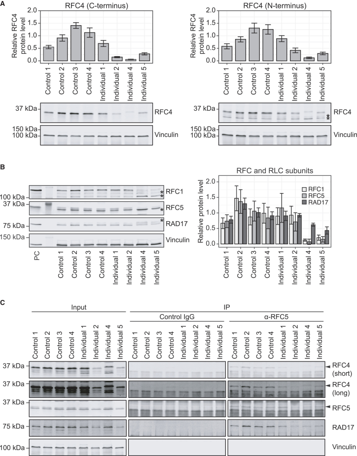
The precise regulation of DNA replication is vital for cellular division and genomic integrity. Central to this process is the replication factor C (RFC) complex, encompassing five subunits, which loads proliferating cell nuclear antigen onto DNA to facilitate the recruitment of replication and repair proteins and enhance DNA polymerase processivity. While RFC1's role in cerebellar ataxia, neuropathy, and vestibular areflexia syndrome (CANVAS) is known, the contributions of RFC2-5 subunits on human Mendelian disorders is largely unexplored. Our research links bi-allelic variants in RFC4, encoding a core RFC complex subunit, to an undiagnosed disorder characterized by incoordination and muscle weakness, hearing impairment, and decreased body weight. We discovered across nine affected individuals rare, conserved, predicted pathogenic variants in RFC4, all likely to disrupt the C-terminal domain indispensable for RFC complex formation. Analysis of a previously determined cryo-EM structure of RFC bound to proliferating cell nuclear antigen suggested that the variants disrupt interactions within RFC4 and/or destabilize the RFC complex. Cellular studies using RFC4-deficient HeLa cells and primary fibroblasts demonstrated decreased RFC4 protein, compromised stability of the other RFC complex subunits, and perturbed RFC complex formation. Additionally, functional studies of the RFC4 variants affirmed diminished RFC complex formation, and cell cycle studies suggested perturbation of DNA replication and cell cycle progression. Our integrated approach of combining in silico, structural, cellular, and functional analyses establishes compelling evidence that bi-allelic loss-of-function RFC4 variants contribute to the pathogenesis of this multisystemic disorder. These insights broaden our understanding of the RFC complex and its role in human health and disease.

Keywords: DNA replication; Mendelian disorder; gene discovery; rare disease; replication factor C complex; translational research.


Conflict of interest statement

Declaration of interests The authors declare no competing interests.

Figures

None
Graphical abstract
Figure 1
Figure 1
Bi-allelic variants in RFC4 segregate with a multisystemic disorder in nine individuals from eight unrelated families (A) The pedigrees of eight unrelated families with at least one affected individual presenting with a clinical spectrum of incoordination, muscle weakness, hearing impairment, and decreased body weight demonstrate segregation of bi-allelic RFC4 variants in an autosomal-recessive mode of inheritance. (B) Brain magnetic resonance imaging (MRI) of affected individuals with bi-allelic variants in RFC4. Individuals 1, 2, 5, and 6 showed evidence of cerebellar atrophy (arrowhead, left panel). Individuals 1, 4, and 5 were also noted to have a hypoplastic pituitary gland. Neuroimaging of individual 1 showed severe vermis cerebellar atrophy (arrowhead, left panel) and cerebral atrophy in the frontotemporal/perisylvian regions (asterisks, right panel) at 47 years of age. Neuroimaging of individual 2 showed severe cerebellar atrophy (arrowhead, left panel), mild cerebral atrophy, and enlarged ventricles and subarachnoid/cerebral spinal fluid spaces at 28 years of age. Neuroimaging of individual 4 showed abnormal T2 hyperintensity in the basal ganglia (arrowheads, right panel) at 6 months of age. Neuroimaging of individual 5 showed cerebellar atrophy (arrowhead, left panel). Neuroimaging was unremarkable for individual 3 at 17 months of age and individual 7 at 12 months of age. (C) Available anthropometry data showing the Z scores of birth OFC, length, and weight as well as postnatal OFC, height, and weight for the nine affected individuals. The median value is represented by the red horizontal bar. OFC, occipital frontal circumference.
Figure 2
Figure 2
The RFC4 variants in the affected individuals alter conserved residues and cluster in the C-terminal domain required for RFC complex formation (A) RFC4 schematic highlighting variant positions (asterisks). The two transcript variants of RFC4, GenBank: NM_002916 and NM_181573, differ in the 5′ UTR and encode the same isoform; GenBank: NM_002916 is represented here. The relative exonic or intronic locations of the variants are indicated by asterisks above or below the schematic, respectively. (B) RFC4 schematic illustrating the AAA+ domain (medium blue, residues 70–202) encompassing the Walker A (residues 78–85) and Walker B (residues 146–151) motifs, and the arginine finger (residue Arg 193) (dark blue), which is essential for ATP binding. The C-terminal domain (medium blue, residues 270–353), critical for interactions with the other subunits of RFC, is also depicted. Amino acid alterations are indicated by asterisks. The c.996+2dup variant was shown to have multiple consequences by cDNA Sanger sequencing, exon 9 skipping leading to p.Val268_Lys294del, exon 10 skipping leading to p.Asp295_Ala332del, or a combined exon 9 and 10 skipping leading to p.Val268_Ala332del were most frequently observed. (C) Cross-species amino acid alignment for RFC4, comparing human (Homo sapiens) sequences with those from chimpanzee (Pan troglodytes), rat (Rattus norvegicus), mouse (Mus musculus), dog (Canis familiaris), chicken (Gallus gallus), frog (Xenopus tropicalis), zebrafish (Danio rerio), fruit fly (Drosophila melanogaster), nematode (Caenorhabditis elegans), and budding yeast (Saccharomyces cerevisiae). The amino acid residue of interest is bolded and highlighted by a black box. The level of conservation is indicated by the color where light blue represents modest conservation, medium blue represents moderate conservation, and dark blue represents high conservation. aa, amino acid.
Figure 3
Figure 3
RFC4 variants and neighboring residues, as located within a cryogenic electron-microscopy structure of RFC bound to PCNA. (A) Structure of the 5-subunit RFC complex bound to the homotrimer DNA clamp PCNA, as determined by cryogenic electron microscopy (cryo-EM) (PDB: 6VVO). The top view (center) was obtained from the side view (left) by a 90-degree rotation. The side chains of Asp 275, Cys 281, Ala 303, Ile 327, and Glu 344 in the C-terminal domain of RFC4 are shown as space-filling (right). (B) Stereo views of RFC4 variant sites within the cryo-EM structure (PDB: 6VVO). The view at right was obtained by a 90-degree rotation about the vertical axis. The alpha-carbon trace of RFC4 residues 267 to 361 is shown (green). The side chains of Ala 303, Cys 281, and Glu 344 are shown as space filling. Although the effective resolution of the cryo-EM structure is limited (3.4 Å), the structure suggests that Cys 281 forms hydrogen bonds with Val 277 and Cys 352 (shown as sticks). Selected side chains near Glu 344 are also shown as sticks (Asp 343 of RFC4; Asp 138, Ser 167, Ser 169, Lys 170 of RFC3 with carbon atoms light blue).
Figure 4
Figure 4
Analysis of relative RFC4 mRNA expression and RFC4 protein levels, RFC/RLC subunit stability, and RFC complex formation in RFC4-deficient HeLa cell lines (A) RFC4-deficient HeLa cell lines were generated by CRISPR-Cas9 gene editing using two guide RNAs targeting exons 8 and 10 in a Cas9-expressing HeLa cell line. Guide RNA sequences (blue), protospacer adjacent motifs (red), and cleavage sites (arrowheads) are mapped on the gene schematic. PCR primer locations for screening clones are also depicted. (B) The protein schematic highlights the cleavage sites (red arrowhead), predicting partial loss of the C-terminal domain of RFC4 essential for RFC complex interactions. (C) PCR of the targeted region demonstrates genomic deletions in RFC4 for RFC4-deficient clone 1 and clone 2, in contrast to the intact 3,240 bp product (denoted by an arrowhead) in the control HeLa cell line. A 331-bp positive control PCR amplifying a region downstream of the target region confirms the presence of genomic DNA. (D) Quantitative PCR was used to measure relative RFC4 mRNA expression in a control and two RFC4-deficient cell lines (clone 1 and clone 2) with TaqMan assays spanning exon junctions 3–4 and 10–11. Data are presented as the mean of three technical replicates relative to the control HeLa cell line equal to 1. Two independent experiments were performed, and representative data from a single independent experiment are presented. Expression of HPRT1 and TBP were used as internal controls to normalize gene expression; error bars represent one standard deviation. (E and F) Immunoblot analysis quantifying relative RFC4 (E) and RFC1, RFC5, and RAD17 (F) protein. Primary antibodies recognizing the N terminus (1–50 aa) or C terminus (181–363 aa) of human RFC4 were used. Data are presented as the mean of three technical replicates relative to the control HeLa cell line equal to 1. Two independent experiments were performed for quantifying relative RFC4 protein, and one experiment was performed for quantifying relative RFC1, RFC5, and RAD17 protein; representative blots of each target are shown. Vinculin was used as a loading control; error bars represent one standard deviation. Asterisks () denote non-specific bands. (G) Endogenous RFC5 protein was immunoprecipitated from the control or RFC4-deficient cell lines (clone 1 and clone 2) using a polyclonal RFC5 antibody and Protein A Sepharose beads. Normal rabbit IgG was used as an isotype control antibody. RFC complex formation in the RFC4-deficient cell lines was assessed by analyzing co-immunoprecipitated RFC1, RFC4, and RAD17 by immunoblot. One experiment was performed for assessing RFC complex formation. Asterisks () denote non-specific bands. aa, amino acid; IP, immunoprecipitation; NTC, no template control; PAM, protospacer adjacent motif; PC, positive control.
Figure 5
Figure 5
Analysis of relative RFC4 protein abundance, RFC/RLC subunit stability, and RFC complex formation in available cultured primary fibroblasts from the affected individuals with bi-allelic RFC4 variants (A and B) Relative RFC4 (A) and RFC1, RFC5, and RAD17 (B) protein abundance were quantified by immunoblot in the cultured primary fibroblasts of four unaffected controls and individuals 1, 2, 4, and 5. Primary antibodies recognizing the C terminus (181–363 aa) or N terminus (1–50 aa) of human RFC4 were used. Data are presented as the mean of four technical replicates relative to the average of all unaffected controls equal to 1. Two independent experiments were performed for quantifying relative RFC4 protein, and one experiment was performed for quantifying relative RFC1, RFC5, and RAD17 protein; representative blots of each target are shown. Vinculin was used as a loading control; error bars represent one standard deviation. HeLa cell lysate was used as a positive control (PC). Asterisks () denote non-specific bands. (C) Endogenous RFC5 protein from cultured primary fibroblasts of four unaffected controls and individuals 1, 2, 4, and 5 was immunoprecipitated using a polyclonal RFC5 antibody and Protein A Sepharose beads. Normal rabbit IgG was used as an isotype control antibody. RFC complex formation in the cultured primary fibroblasts was assessed by analyzing co-immunoprecipitated RFC4 and RAD17 by immunoblot. One experiment was performed for assessing RFC complex formation. Asterisks () denote non-specific bands. For RFC4 and RFC5, arrowheads denote the band of interest while the bands below represent IgG light chain from the IP. IP, immunoprecipitation; n.s., not significant; PC, positive control; RLC, RFC-like complex.
Figure 6
Figure 6
Analysis of RFC complex formation and PCNA loading activity of the RFC4 variants (A) The replication factor C (RFC) complex, critical for DNA replication, consists of five subunits: the large RFC1 subunit and the four small subunits, RFCs 2–5. The RFC complex loads proliferating cell nuclear antigen (PCNA) onto DNA to recruit essential proteins and enhance DNA polymerase processivity. In addition to RFC, three RFC-like complexes (RLCs) share the four small subunits but feature alternative large subunits—ATAD5, RAD17, and CTF18—each facilitating unique and shared processes. (B) RFC4 variants fused to Strep-tag II were transiently expressed in HEK293T cells. These fusion proteins were affinity purified using Strep-Tactin Sepharose beads. Immunoblot analysis of co-precipitated proteins indicated that all RFC4 variants exhibited impaired interactions with the small subunit RFC5 and large subunits RFC1, ATAD5, and CTF18. (C) To assess PCNA loading activity, wild-type or mutant RFC complex and PCNA were incubated with a magnetic bead-coupled DNA substrate (upper panel). To visualize and quantify the PCNA that had been loaded onto the DNA substrate by RFC complex containing wild-type or mutant RFC4, the magnetic bead-coupled DNA was washed to remove unloaded PCNA, and the DNA-loaded PCNA was released from the magnetic beads by DNase I treatment and analyzed via immunoblotting. Data are presented as the mean of four technical replicates relative to RFC complex containing wild-type RFC4 (12.5 nM) equal to 1. Two independent experiments were performed, and representative data from a single independent experiment is presented. FLAG (TALE) was used as a loading control; error bars represent one standard deviation. PCNA, proliferating cell nuclear antigen; Pol, polymerase; RFC, replication factor C; RLC, RFC-like complex; TALE, transcription activator-like effector.

References

    1. Guilliam T.A., Yeeles J.T.P. An updated perspective on the polymerase division of labor during eukaryotic DNA replication. Crit. Rev. Biochem. Mol. Biol. 2020;55:469–481. - PubMed
    1. Bernad A., Blanco L., Lazaro J.M., Martin G., Salas M. A conserved 3'→5' exonuclease active site in prokaryotic and eukaryotic DNA polymerases. Cell. 1989;59:219–228. - PubMed
    1. Shevelev I.V., Hubscher U. The 3'–5' exonucleases. Nat. Rev. Mol. Cell Biol. 2002;3:364–376. - PubMed
    1. Bellelli R., Boulton S.J. Spotlight on the Replisome: aetiology of DNA replication-associated genetic diseases. Trends Genet. 2021;37:317–336. - PubMed
    1. Schmidt S.L., Pautz A.L., Burgers P.M. ATP utilization by yeast replication factor C. IV. RFC ATP-binding mutants show defects in DNA replication, DNA repair, and checkpoint regulation. J. Biol. Chem. 2001;276:34792–34800. - PubMed

Substances