Development of international consensus on a standardised image acquisition protocol for diagnostic evaluation of the sacroiliac joints by MRI: an ASAS-SPARTAN collaboration
- PMID: 39107080
- PMCID: PMC11671998
- DOI: 10.1136/ard-2024-225882
Development of international consensus on a standardised image acquisition protocol for diagnostic evaluation of the sacroiliac joints by MRI: an ASAS-SPARTAN collaboration
Abstract
Background: A range of sacroiliac joint (SIJ) MRI protocols are used in clinical practice but not all were specifically designed for diagnostic ascertainment. This can be confusing and no standard diagnostic SIJ MRI protocol is currently accepted worldwide.
Objective: To develop a standardised MRI image acquisition protocol (IAP) for diagnostic ascertainment of sacroiliitis.
Methods: 13 radiologist members of Assessment of SpondyloArthritis International Society (ASAS) and the SpondyloArthritis Research and Treatment Network (SPARTAN) plus two rheumatologists participated in a consensus exercise. A draft IAP was circulated with background information and online examples. Feedback on all issues was tabulated and recirculated. The remaining points of contention were resolved and the revised IAP was presented to the entire ASAS membership.
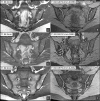
Results: A minimum four-sequence IAP is recommended for diagnostic ascertainment of sacroiliitis and its differential diagnoses meeting the following requirements. Three semicoronal sequences, parallel to the dorsal cortex of the S2 vertebral body, should include sequences sensitive for detection of (1) changes in fat signal and structural damage with T1-weighting; (2) active inflammation, being T2-weighted with fat suppression; (3) bone erosion optimally depicting the bone-cartilage interface of the articular surface and (4) a semiaxial sequence sensitive for detection of inflammation. The IAP was approved at the 2022 ASAS annual meeting with 91% of the membership in favour.
Conclusion: A standardised IAP for SIJ MRI for diagnostic ascertainment of sacroiliitis is recommended and should be composed of at least four sequences that include imaging in two planes and optimally visualise inflammation, structural damage and the bone-cartilage interface.
Keywords: Arthritis; Axial Spondyloarthritis; Magnetic Resonance Imaging.
© Author(s) (or their employer(s)) 2024. Re-use permitted under CC BY-NC. No commercial re-use. See rights and permissions. Published by BMJ on behalf of EULAR.
Conflict of interest statement
Competing interests: RGWL: consulting fees from CARE Arthritis and Image Analysis Group. XB: contract with Novartis; Consulting fees from Amgen, Bristol-Myers Squibb, Chugai, Eli Lilly, Galapagos, Janssen, Merck Sharp & Dohme, Novartis, Pfizer, Roche, Sandoz, Sanofi, and UCB; Payment or honoraria from Amgen, Bristol-Myers Squibb, Chugai, Eli Lilly, Galapagos, Janssen, Merck Sharp & Dohme, Novartis, Pfizer, Roche, Sandoz, Sanofi, and UCB; Meeting support from Eli Lilly, Janssen, Novartis, Pfizer and UCB; Participation on a Data Safety Monitoring Board or Advisory Board: Amgen, Bristol-Myers Squibb, Eli Lilly, Galapagos, Janssen, Merck Sharp & Dohme, Novartis, Pfizer, Roche, Sandoz, Sanofi and UCB; Leadership role: Editorial Board Member of Annals of Rheumatic Diseases, ASAS President, EULAR President Elect. SAB: Royalties from Elsevier. JAC: Consulting fees from AstraZeneca and Covera Health; Participation on a Data Safety Monitoring Board or Advisory Board: Carestream, Image Analysis Group, Image Biopsy Lab; Leadership role: RSNA, ACR, IAOAI. TD: Grants or contracts from Berlin Institute of Health (BIH); Payment or honoraria from Berlinflame, Canon Medical Systems, Janssen, MSD, Novartis, UCB. IE: Payment or honoraria from Lilly, Novartis. KGH: Payment or honoraria from MSD, AbbVie Novartis; Cofounder of BerlinFlame. NH: None declared. JJ: Stock in Exo. LJ: None declared. AGJ: None declared. JMDO'N:– None declared. MR: ISS Seed Grant; Consultant for ASAS. MJT: Consulting fees from GE HealthCare; Meeting support from International Skeletal Society; Leadership role—President-elect International Skeletal Society. WPM: Grants or contracts from Abbvie, BMS, Eli-Lilly, Pfizer, UCB; Consulting fees from Abbvie, Celgene, BMS, Eli-Lilly, Galapagos, Pfizer, UCB; Payment or honoraria from Abbvie, Janssen, Pfizer, Novartis; Leadership role—SPARTAN Board of Directors; Chief Medical Officer, CARE Arthritis.
Figures


References
-
- Docherty P, Mitchell MJ, MacMillan L, et al. Magnetic resonance imaging in the detection of sacroiliitis. J Rheumatol. 1992;19:393–401. - PubMed
Publication types
MeSH terms
LinkOut - more resources
Full Text Sources
Medical
Research Materials

