. 2024 Jun 24;32(3):101289.
doi: 10.1016/j.omtm.2024.101289. eCollection 2024 Sep 12.

Lipid nanoparticle-encapsulated DOCK11-siRNA efficiently reduces hepatitis B virus cccDNA level in infected mice

Affiliations

Lipid nanoparticle-encapsulated DOCK11-siRNA efficiently reduces hepatitis B virus cccDNA level in infected mice

Hikari Okada et al. Mol Ther Methods Clin Dev. .

Abstract

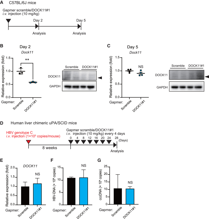
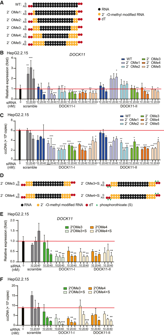
The hepatitis B virus (HBV) infects many people worldwide. As HBV infection frequently leads to liver fibrosis and carcinogenesis, developing anti-HBV therapeutic drugs is urgent. Therapeutic drugs for preventing covalently closed circular DNA (cccDNA) production, which can eliminate HBV infection, are unavailable. The host factor dedicator of cytokinesis 11 (DOCK11) is involved in the synthesis and maintenance of HBV cccDNA in vitro. However, the effectiveness of DOCK11 as a target for the in vivo elimination of HBV cccDNA remains unclear. In this study, we assess whether DOCK11 inhibitors suppress HBV cccDNA production in mouse models of HBV infection. The tocopherol-conjugate hetero- gapmer, a DNA/RNA duplex of gapmer/complementary RNA targeting the DOCK11 sequence, partially reduces the expression of DOCK11, but not that of HBV cccDNA, in the livers of HBV-infected human hepatocyte chimeric mice, along with weight loss and decreased serum human albumin levels. Lipid nanoparticle-encapsulated chemically modified siRNAs specific for DOCK11 suppress DOCK11 expression and decrease HBV cccDNA levels without adverse effects in the mice. Therefore, nucleic acid-based drugs targeting DOCK11 in hepatocytes are potentially effective anti-HBV therapeutics that can reduce HBV cccDNA levels in vivo.

Keywords: DOCK11; HBV; LNP-siRNA; cccDNA; gapmer; hetero gapmer; nucleic acid medicine.


Conflict of interest statement

The authors declare no competing interests.

Figures

None
Graphical abstract
Figure 1
Figure 1
DOCK11-targeting gapmers suppress DOCK11 expression and HBV cccDNA in vitro (A) Schematic illustration of the full-length human DOCK11 and the gapmer constructs. (B and C) DOCK11 mRNA expression in Huh7 cells (B) and mouse primary hepatocytes (C) 48 h after gapmer treatment. (D) Schematic illustration of 13, 15, and 17 bp DOCK11#1 gapmers. (E) DOCK11 mRNA expression in Huh7 cells transfected with gapmer scramble and DOCK11#1. (F and G) DOCK11 mRNA expression (F) and HBV cccDNA copy number (G) in Hep2.2.15 cells transfected with gapmer scramble and DOCK11#1. In (B), (C), and (E–G), data are presented as the mean (SD) (N = 3) and analyzed using the one-way ANOVA. ∗p < 0.05, ∗∗p < 0.01, ∗∗∗p < 0.001; NS, not significant.
Figure 2
Figure 2
The DOCK11-targeting gapmer transiently suppresses the expression of DOCK11 but is insufficient to eliminate HBV cccDNA in vivo (A) The schedule for gapmer administration in animals. (B and C) Expression levels of DOCK11 mRNA (N = 3) and protein (N = 2) in the liver at 2 days (B) and 5 days (N = 4) (C) after injecting gapmers. (D) Experimental schedule for administration of gapmer scramble and DOCK11#1 in human liver chimeric mice with HBV chronic infections. (E–G) DOCK11 mRNA expression (E), and copy numbers of HBV DNA (F) and cccDNA (G) in the liver of HBV-infected human liver chimeric mice treated with gapmers (N = 2). In (B), (C), and (E–G), data are presented as the mean (SD) and analyzed using the Mann-Whitney U test. ∗∗p < 0.01; NS, not significant.
Figure 3
Figure 3
The DOCK11-targeting hetero-gapmer exhibits stable DOCK11-suppressive effects and decreases HBV cccDNA levels in vivo (A) Schematic presentation demonstrating hetero-gapmer constructs. (B) Schedule for nucleic acid administration in animals. (C) Expression of Dock11 mRNA (N = 4) and protein in the liver of C57BL6/J mice 5 days after administration of hetero-gapmer. (D) Schedule for the administration of hetero-gapmers in AAV8-HBV1.3mer virus-infected C57BL/6J mice (early-phase and chronic-phase models). (E and F) Expression of Dock11 mRNA, protein (E) and cccDNA copy number (F) in the liver of hetero-gapmer-treated mice on days 15 and 30 (N = 5). In (C), (E), and (F), data are presented as the mean (SD) and analyzed using the Mann-Whitney U test. ∗p < 0.05, ∗∗p < 0.01.
Figure 4
Figure 4
Hetero-gapmers are hepatotoxic in HBV-infected human liver chimeric mice (A) Schedule for the administration of hetero-gapmers in HBV-infected human liver chimeric mice. (B–E) Expression of hepatic DOCK11 mRNA, protein (B), hepatic HBV cccDNA (C), serum HBV-DNA (D), and serum HBsAg (E) in human liver chimeric mouse models of HBV chronic infection, after hetero-gapmer administration. (F–I) Changes in body weights (F and G) and h-Alb levels (H and I) of HBV-infected human liver chimeric mouse after administration of the hetero- gapmer scramble (F and H) and DOCK11#1 (G and I). In (B)–(I), data are presented as the mean (SD) (N = 3) and analyzed using the two-sided unpaired t test with Welch’s correction. ∗p < 0.05, ∗∗p < 0.01, ∗∗∗p < 0.001; NS, not significant.
Figure 5
Figure 5
LNP-encapsulated siRNA targeting DOCK11 reduces HBV cccDNA in vivo (A) Construction of siRNA against DOCK11. (B and C) The relative expression levels of DOCK11 mRNA (B) and HBV cccDNA copy number (C) in Hep2.2.15 cells 48 h after siRNA transfection; data are presented as the mean (SD) (N = 3) and were analyzed using the one-way ANOVA. ∗p < 0.05, ∗∗p < 0.01, ∗∗∗p < 0.001. (D) Schedule for the administration of LNP-siRNA in AAV8-HBV1.3mer-infected C57BL6/J mice. (E and F) The relative expression levels of DOCK11 mRNA, protein (E) and HBV cccDNA copy number (F) in the livers of LNP-siRNA-treated mice. Data are presented as the mean (SD) (N = 4) and analyzed using the Mann-Whitney U test. ∗p < 0.05.
Figure 6
Figure 6
Chemically modified DOCK11-targeting siRNA exhibits enhanced efficacy for gene suppression and HBV cccDNA elimination in vitro (A) Schematic representation of the chemically modified siRNAs indicating positions of 2′-O-methylation (2′OMe). (B and C) The relative expression levels of DOCK11 mRNA (B) and HBV cccDNA copy number (C) in Hep2.2.15 cells after transfection of 2′-OMe siRNA (N = 3). (D) Schematic representation of the chemically modified siRNAs indicating positions of 2′OMe and phosphorothioate (S) modifications. (E and F) The relative expression levels of DOCK11 mRNA (E) and HBV cccDNA copy number (F) in HepG2.2.15 cells 72 h after transfection of siRNA modified with 2′OME and phosphorothioate. The red line, indicating the data obtained from the untreated group, is considered for comparative analyses. In (B), (C), (E), and (F), data are presented as the mean (SD) (N = 3) and analyzed using the one-way ANOVA. ∗p < 0.05, ∗∗p < 0.01, ∗∗∗p < 0.001.
Figure 7
Figure 7
LNP chemically modified siRNA suppresses HBV cccDNA in AAV8-HBV1.3mer chronic infection model mice and HBV relapse model of HBV infection post-ETV discontinuation (A) Experimental schedule for administering LNP-siRNA DOCK11-I (2′OMe3+S) to AAV8-HBV1.3mer chronically infected mice. (B and C) The relative expression of DOCK11 mRNA and protein (B) and copy numbers of HBV cccDNA (C) in the livers of mice treated with saline, LNP-gapmer DOCK11#1, LNP-siRNA DOCK11 (non-modified), and LNP-siRNA DOCK11 (2′OMe3+S). (D) Schedule for establishing the HBV relapse model with entecavir and administration of LNP-siRNA DOCK11 (2′OMe3+S). (E–H) The relative expressions of DOCK11 mRNA, protein (E), HBV DNA (F), and HBV cccDNA (G) in the liver of relapse mouse model of AAV8-HBV1.3mer HBV infection 7 days after a single administration of LNP-siRNA DOCK11-I (2′ OMe3+S). Data are presented as the mean (SD) (N = 3–4) and analyzed using the two-sided unpaired t test with Welch’s correction. ∗p < 0.05, ∗∗p < 0.01.
Figure 8
Figure 8
LNP-encapsulated chemically modified DOCK11-targeting siRNA reduces HBV replication and cccDNA in HBV-infected human hepatocyte chimeric mice (A) Experimental schedule for administration of LNP-siRNA DOCK11-I (2′OMe3+S) in HBV-infected human liver chimeric mice. (B–G) The relative expression levels of DOCK11 mRNA and protein in the liver (B), the copy number of blood HBV-DNA (C), serum HBsAG (D), and HBeAG (E) levels, and the copy numbers of HBV cccDNA (F) and HBV-DNA (G) in the liver of HBV-infected human liver chimeric mice after administering LNP-siRNA DOCK11-I (2′OMe3+S). (H) The amounts of rcDNA, linearDNA, and cccDNA in the liver tissues of human liver chimeric mice were analyzed by Southern blotting. (I) Immunohistochemical staining analyses of HBsAg and HBcAg in the liver of human liver chimeric mice. Scale bar, 100 μm. Data are presented as the mean (SD) (N = 3) and analyzed using the two-sided unpaired t test with Welch’s correction. ∗p < 0.05, ∗∗p < 0.01, ∗∗∗p < 0.001.

References

    1. Guvenir M., Arikan A. Hepatitis B Virus: From Diagnosis to Treatment. Pol. J. Microbiol. 2020;69:391–399. doi: 10.33073/pjm-2020-044. - DOI - PMC - PubMed
    1. Mak L.Y., Wong D.K.H., Pollicino T., Raimondo G., Hollinger F.B., Yuen M.F. Occult hepatitis B infection and hepatocellular carcinoma: Epidemiology, virology, hepatocarcinogenesis and clinical significance. J. Hepatol. 2020;73:952–964. doi: 10.1016/j.jhep.2020.05.042. - DOI - PubMed
    1. Bray F., Ferlay J., Soerjomataram I., Siegel R.L., Torre L.A., Jemal A. Global cancer statistics 2018: GLOBOCAN estimates of incidence and mortality worldwide for 36 cancers in 185 countries. CA Cancer J. Clin. 2018;68:394–424. doi: 10.3322/caac.21492. - DOI - PubMed
    1. Revill P.A., Locarnini S.A. New perspectives on the hepatitis B virus life cycle in the human liver. J. Clin. Invest. 2016;126:833–836. doi: 10.1172/JCI86650. - DOI - PMC - PubMed
    1. El-Sayed M.H., Feld J.J. Vaccination at the forefront of the fight against hepatitis B and C. Nat. Rev. Gastroenterol. Hepatol. 2022;19:87–88. doi: 10.1038/s41575-021-00570-x. - DOI - PMC - PubMed

LinkOut - more resources