Review
. 2024 Aug;25(8):726-741.
doi: 10.3348/kjr.2024.0105.

Amyloid-Related Imaging Abnormalities in the Era of Anti-Amyloid Beta Monoclonal Antibodies for Alzheimer's Disease: Recent Updates on Clinical and Imaging Features and MRI Monitoring

Affiliations
Review

Amyloid-Related Imaging Abnormalities in the Era of Anti-Amyloid Beta Monoclonal Antibodies for Alzheimer's Disease: Recent Updates on Clinical and Imaging Features and MRI Monitoring

So Yeong Jeong et al. Korean J Radiol. 2024 Aug.

Abstract

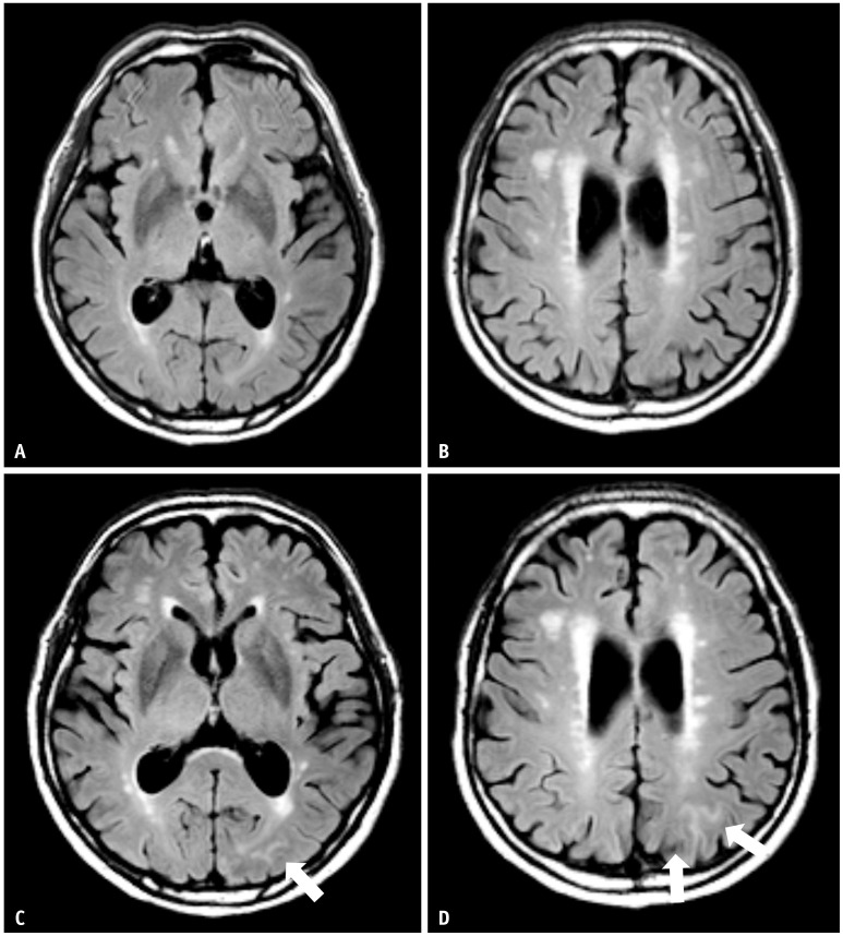
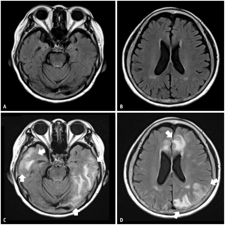
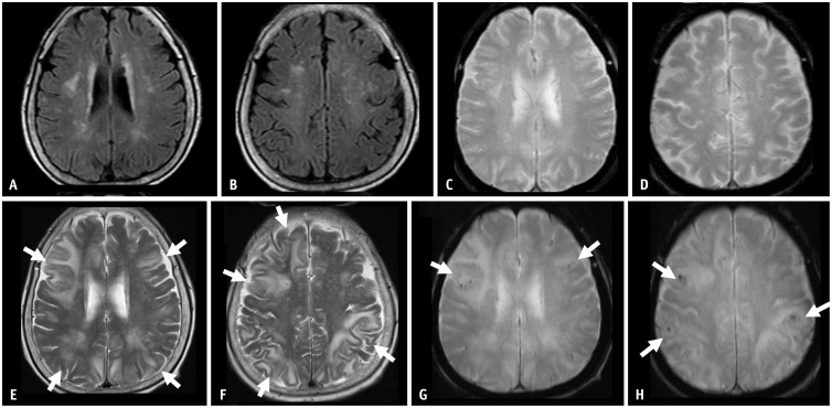
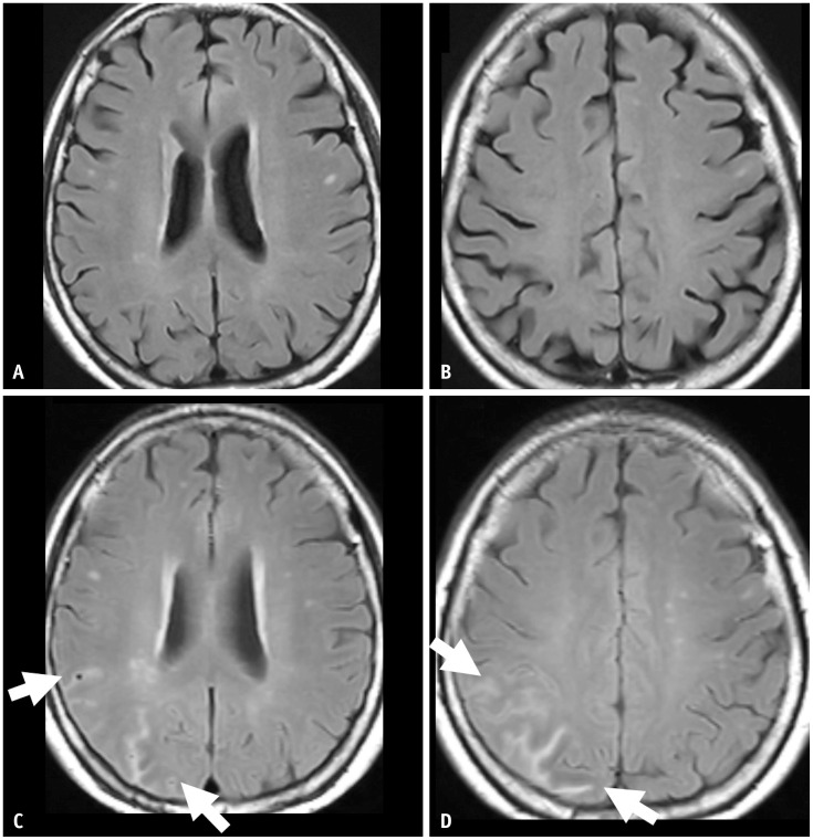
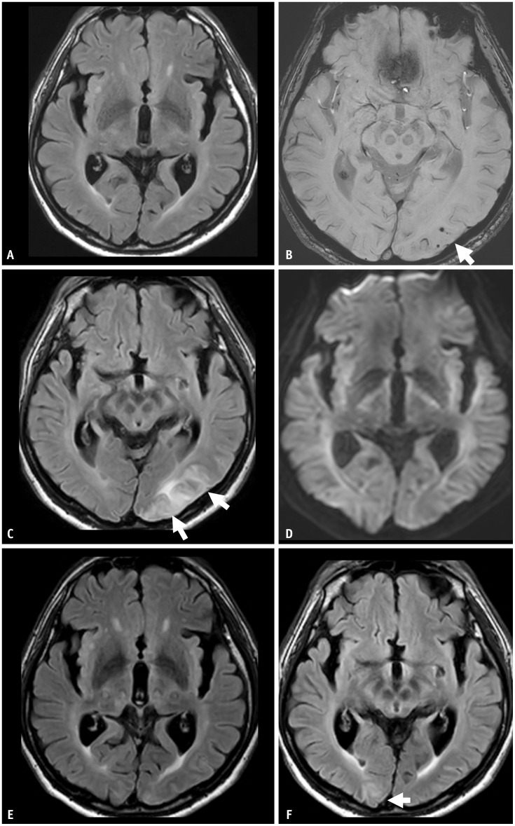
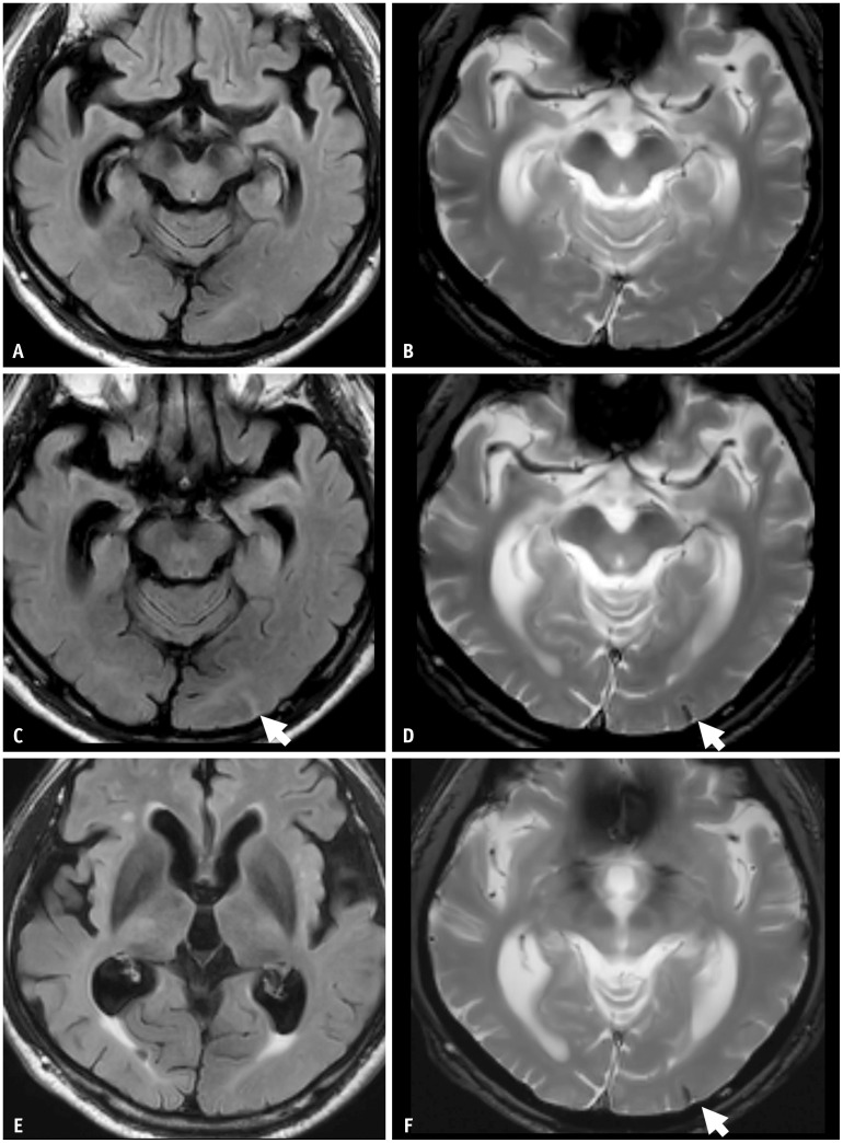
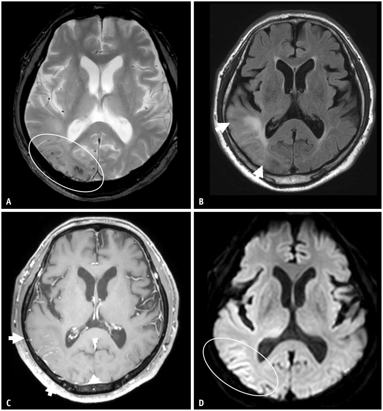
Recent advancements in Alzheimer's disease treatment have focused on the elimination of amyloid-beta (Aβ) plaque, a hallmark of the disease. Monoclonal antibodies such as lecanemab and donanemab can alter disease progression by binding to different forms of Aβ aggregates. However, these treatments raise concerns about adverse effects, particularly amyloid-related imaging abnormalities (ARIA). Careful assessment of safety, especially regarding ARIA, is crucial. ARIA results from treatment-related disruption of vascular integrity and increased vascular permeability, leading to the leakage of proteinaceous fluid (ARIA-E) and heme products (ARIA-H). ARIA-E indicates treatment-induced edema or sulcal effusion, while ARIA-H indicates treatment-induced microhemorrhage or superficial siderosis. The minimum recommended magnetic resonance imaging sequences for ARIA assessment are T2-FLAIR, T2* gradient echo (GRE), and diffusion-weighted imaging (DWI). T2-FLAIR and T2* GRE are necessary to detect ARIA-E and ARIA-H, respectively. DWI plays a role in differentiating ARIA-E from acute to subacute infarcts. Physicians, including radiologists, must be familiar with the imaging features of ARIA, the appropriate imaging protocol for the ARIA workup, and the reporting of findings in clinical practice. This review aims to describe the clinical and imaging features of ARIA and suggest points for the timely detection and monitoring of ARIA in clinical practice.

Keywords: Amyloid-related imaging abnormalities; Magnetic resonance imaging; Monoclonal antibodies.


Conflict of interest statement

Chong Hyun Suh, who holds the respective position as Assistant to the Editor of the Korean Journal of Radiology, was not involved in the editorial evaluation or decision to publish this article. The remaining author has declared no conflicts of interest.

Figures

Fig. 1
Fig. 1. A 75-year-old male patient with mild cognitive impairment presented with transient ARIA-E during gantenerumab therapy. A, B: His baseline MRI shows a moderate degree of white matter hyperintensity. C, D: Nine weeks later, a surveillance MRI showed sulcal FLAIR with high signal intensity in the left parietal area, suggesting ARIA-E (arrows). The patient was asymptomatic. Since a single region and extent measuring less than 5 cm would be classified as mild, this was an asymptomatic and mild case, and the gantenerumab was continuously taken. At follow-up surveillance MRI after initial ARIA-E detection, it was completely resolved.
Fig. 2
Fig. 2. A 74-year-old female patient with mild cognitive impairment presented with severe ARIA-E during lecanemab (BAN-2401) therapy. A, B: Her baseline MRI shows a mild degree of white matter hyperintensity. C, D: Eight weeks later, she had a headache and confusion. MRI shows multifocal FLAIR high signal intensity parenchymal edema with swelling in the bilateral cerebral hemispheres, suggestive of ARIA-E (arrows). Multiple lesions with an extent measuring more than 10 cm were present and classified as severe. The lecanemab therapy was discontinued. The extent of ARIA-E gradually decreased on follow-up MRI and completely resolved at the follow-up surveillance MRI after the initial ARIA-E detection.
Fig. 3
Fig. 3. An 80-year-old female patient with mild cognitive impairment concurrently presented with both ARIA-E and ARIA-H during aducanumab therapy. She was an APOE-ε4 homozygote carrier. A-D: Her baseline MRI shows moderate white matter hyperintensity but no microbleeds. Five months later, she complained of a headache and confusion. E-H: Multifocal FLAIR high signal intensity edema in both cerebral hemispheres showed multiple microbleeds, suggestive of ARIA-E and ARIA-H on follow-up MRI. The MRI showed multifocal FLAIR high signal intensity edema (arrows in E, F) and multiple microbleeds (arrows in G, H) in both cerebral hemispheres, suggestive of ARIA-E and ARIA-H. She was discontinued from the lecanemab therapy, and her symptoms gradually resolved. Three months after the infusion suspension, ARIA-E was resolved, and ARIA-H was stabilized on a follow-up MRI.
Fig. 4
Fig. 4. A 69-year-old female patient presented with mild cognitive impairment and was treated with gantenerumab therapy. A, B: Her baseline MRI shows a mild degree of white matter hyperintensity. C, D: Seven months after the initiation of gantenerumab therapy, the patient had an episode of syncope, and the MRI showed multifocal sulcal FLAIR high-signal intensity in the right parietal lobe, suggesting ARIA-E (arrows). The case had two regions (arrows) with an extent measuring between 5 cm and 10 cm, classified as moderate. As a symptomatic and moderate case, the gantenerumab therapy was suspended. ARIA-E was resolved after suspending treatment for 1 month.
Fig. 5
Fig. 5. A 67-year-old male patient presented with mild cognitive impairment and was treated with lecanemab therapy. He was an APOE-ε4 homozygote carrier. A, B: His baseline MRI shows a mild degree of white matter hyperintensity and three microbleeds (arrow) in the left occipital lobe. Eleven months after initiation of the lecanemab therapy, the patient complained of a headache. C, D: MRI shows sulcal and parenchymal FLAIR high-signal intensity without diffusion restriction in the left occipital lobe, suggesting ARIA-E (arrows). A single region with an extent measuring between 5 cm and 10 cm was classified as moderate. As a symptomatic and moderate case, the lecanemab therapy was suspended. E: On follow-up MRI after suspending treatment for 2 months, ARIA-E was resolved. After the resolution of ARIA-E, the lecanemab treatment was resumed. F: Two months after the resumption of the therapy, a new sulcal and parenchymal FLAIR high-signal intensity (arrow) appeared in the right occipital lobe, and the patient permanently suspended the treatment.
Fig. 6
Fig. 6. A 68-year-old male patient presented with mild cognitive impairment and was treated with lecanemab therapy. He was an APOE-ε3/ε4 heterozygote carrier. A, B: His baseline MRI shows no abnormalities except bilateral medial temporal lobe atrophy. C, D: On the surveillance MRI performed 6 months after the lecanemab therapy, there was focal sulcal FLAIR high-signal intensity effusion and superficial siderosis on gradient echo sequences in the left occipital lobe, suggestive of ARIA-E and ARIA-H (arrows). Both the sulcal effusion and superficial siderosis were single lesions with an extent less than 5 cm, classified as mild ARIA-E and ARIA-H. As an asymptomatic mild ARIA case, lecanemab therapy was continued. E, F: On a 1-month follow-up MRI after the ARIA event, the ARIA-E was resolved, and the ARIA-H (arrow) remained stable.
Fig. 7
Fig. 7. A 70-year-old male patient presented with confusion. A: On T2* gradient echo sequence, there were multiple lobar distributed cortical and subcortical microbleeds in the right temporooccipital lobes. B: On FLAIR, there was confluent parenchymal high signal intensity (arrows). C: Postcontrast axial T1 images demonstrated marked gyral enhancement in the same area (arrows), suggestive of prominent leptomeningeal vessels. D: On the diffusion-weighted image, there was subtle high signal intensity along the cortex. These images are very similar to amyloid-related imaging abnormalities; however, the patient had not undergone anti-amyloid-beta immunotherapy. The patient was diagnosed with cerebral amyloid angiopathy-related inflammation.

References

    1. Karran E, De Strooper B. The amyloid hypothesis in Alzheimer disease: new insights from new therapeutics. Nat Rev Drug Discov. 2022;21:306–318. - PubMed
    1. Decourt B, Boumelhem F, Pope ED, 3rd, Shi J, Mari Z, Sabbagh MN. Critical appraisal of amyloid lowering agents in AD. Curr Neurol Neurosci Rep. 2021;21:39. - PMC - PubMed
    1. Jeong SY, Suh CH, Shim WH, Lim JS, Lee JH, Kim SJ. Incidence of amyloid-related imaging abnormalities in patients with Alzheimer disease treated with anti-β-amyloid immunotherapy: a meta-analysis. Neurology. 2022;99:e2092–e2101. - PubMed
    1. Barakos J, Purcell D, Suhy J, Chalkias S, Burkett P, Marsica Grassi C, et al. Detection and management of amyloid-related imaging abnormalities in patients with Alzheimer’s disease treated with anti-amyloid beta therapy. J Prev Alzheimers Dis. 2022;9:211–220. - PubMed
    1. Arndt JW, Qian F, Smith BA, Quan C, Kilambi KP, Bush MW, et al. Structural and kinetic basis for the selectivity of aducanumab for aggregated forms of amyloid-β. Sci Rep. 2018;8:6412. - PMC - PubMed

Publication types

MeSH terms

Substances