Histopathological Patterns of Otosclerosis Progression: Exploring Otic Capsule and Round Window Involvement
- PMID: 39109802
- PMCID: PMC11635137
- DOI: 10.1002/lary.31680
Histopathological Patterns of Otosclerosis Progression: Exploring Otic Capsule and Round Window Involvement
Abstract
Objectives: Obliteration of the round window (RW) in cases of otosclerosis presents a significant clinical challenge due to its association with more severe hearing loss and a poorer prognosis for functional recovery after stapes surgery. The objective is to assess and characterize the occurrence of RW involvement in otosclerosis cases and to identify patterns of disease progression that may indicate a potential for RW obliteration.
Methods: We selected archival temporal bones from donors with otosclerosis. We evaluated the degree of RW obliteration using a semi-quantitative scale and the location of the foci within the temporal bone, and whether the foci were continuous or isolated.
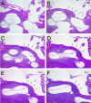
Results: Most of the foci were located anteriorly to the oval window (89.2%), while RW area involvement was seen in 26.9% of the ears. In cases with fenestral foci, 68.1% directly involved and/or fixed the footplate. Among donors with bilateral otosclerosis, foci affected both ears in a similar pattern in 64.2%. Among donors with RW involvement, ones with continuous, large lesions that extended from the oval window associated with complete RW obliteration, while ones with smaller degrees of obliteration had solitary foci scattered within the otic capsule.
Conclusion: Our results demonstrate a high rate of RW involvement in cases of otosclerosis. Ears with continuous lesions extending from the oval window region to the RW area were more likely to present with complete RW obliteration. These results provide insights that could lead to better prognostic assessment of patients with otosclerosis in the future.
Level of evidence: NA Laryngoscope, 135:324-330, 2025.
Keywords: otopathology; otosclerosis; oval window; round window.
© 2024 The Author(s). The Laryngoscope published by Wiley Periodicals LLC on behalf of The American Laryngological, Rhinological and Otological Society, Inc.
Figures


References
-
- National Institute on Deafness and Other Communication Disorders. Otosclerosis. 2022. Accessed May 3, 2024. https://www.nidcd.nih.gov/health/otosclerosis/
-
- American Hearing Research Foundation. Otosclerosis. 2024. Accessed May 3, 2024. https://www.american-hearing.org/disease/otosclerosis/
-
- Guild SR. Histologic otosclerosis. Ann Otol Rhino Laryng. 1944;53:246.
MeSH terms
Grants and funding
- CAPES- Finance Code:001/Coordenação de Aperfeiçoamento de Pessoal de Nível Superior - Brasil
- International Hearing Foundation
- U24 DC020851/DC/NIDCD NIH HHS/United States
- Technological Research Council of Türkiye (TUBITAK)
- U24 DC020851-01/National Institutes of Health / National Institute on Deafness and Other Communication Disorders
LinkOut - more resources
Full Text Sources
Research Materials

