. 2024 Oct;33(5):513-525.
doi: 10.1007/s11248-024-00402-z. Epub 2024 Aug 7.

A Mouse Model for Conditional Expression of Activated β-Catenin in Epidermal Keratinocytes

Affiliations

A Mouse Model for Conditional Expression of Activated β-Catenin in Epidermal Keratinocytes

Vineet K Maurya et al. Transgenic Res. 2024 Oct.

Abstract

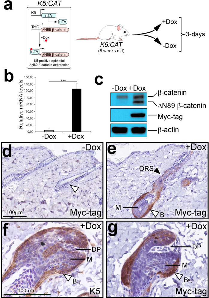
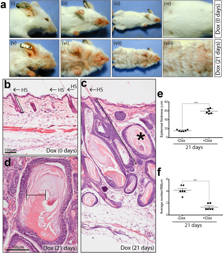
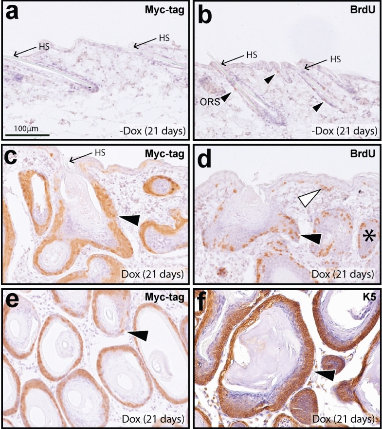
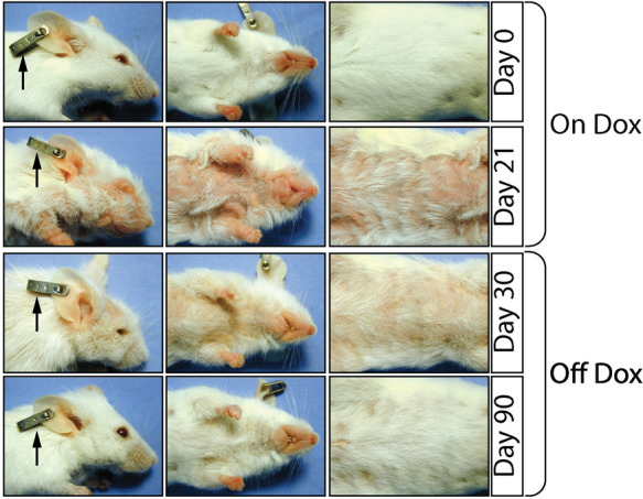
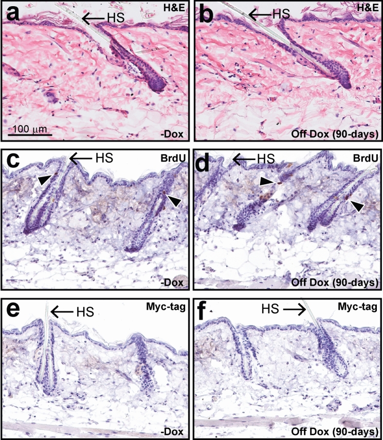
We report the generation and characterization of the K5: CAT bigenic mouse in which the constitutively activated form of β-catenin (ΔN89 β-catenin) is conditionally expressed in cytokeratin-5 (K5) positive epidermal keratinocytes. Following short-term doxycycline intake during the telogen resting phase, the adult K5: CAT bigenic develops enlarged pilosebaceous units that expand deep into the dermis, an expansion usually observed during the anagen growth phase. Prolonged doxycycline treatment results in significant thickening and folding of the K5: CAT epidermis. During this persistent induction period, there is clear evidence of increased keratinocyte proliferation, particularly in the epidermal basal cell layer and the outer root sheath of the hair follicle. This unscheduled increase in cellular proliferation likely explains the decrease in hair density observed in the K5: CAT mouse following persistent doxycycline intake. Numerous hyperplastic endometrioid cysts, which display cornification toward their lumens, are also observed during this treatment period. Remarkably, de-induction of ΔN89 β-catenin expression through doxycycline withdrawal results in a marked reversal of the skin phenotype, suggesting that these morphological changes are dependent on continued signaling by β-catenin and/or its downstream molecular mediators. Joining a small group of mouse models for conditional β-catenin signaling, our K5: CAT mouse model will be particularly useful in identifying those molecular mediators of β-catenin that are responsible for initiating and maintaining these phenotypic responses in the K5: CAT skin. Such studies are predicted to shed more light on β-catenin signaling in epidermal epithelial morphogenesis, hair follicle cycling, and hair growth pathologies.

Keywords: Cytokeratin-5; Doxycycline; Keratinocytes; Mouse; Transgenic; β-catenin.


Conflict of interest statement

Declarations. Conflict of interests: The authors declare no competing interests.

Figures

Fig. 1
Fig. 1
Doxycycline induction of ΔN89 β-catenin expression in the K5: CAT epidermis. a Left schematic summarizes the doxycycline induction of ΔN89 β-catenin protein in K5: CAT keratinocytes. The right schematic shows the experimental design in which K5: CAT adult (8 weeks old) mice were either treated with doxycycline (+ Dox) or left untreated (-Dox) for 72 h before molecular and histological analyses. For all experiments, mice (+ Dox or –Dox) were siblings or age-matched. b Compared with age-matched untreated K5: CAT mice (-Dox), qRT-PCR analysis shows a significant induction for β-catenin transcripts in the skin of K5: CAT mice when treated with doxycycline (+ Dox) for 3 days (n = 3 per treatment group). c In addition to detection of endogenous β-catenin, Western analysis shows a significant induction of transgene-derived ΔN89 β-catenin protein in the K5: CAT skin in response to 3 days of doxycycline administration (+ Dox), which is confirmed by the induction of transgene-derived myc epitope-tagged protein (+ Dox); β-actin serves as a loading control. Note: skin protein extracts from four mice per treatment group (-Dox and + Dox) were pooled per lane of the polyacrylamide gel. d A parasagittal skin section from an untreated K5: CAT mouse (-Dox (representative of N = 11 mice)); white arrowhead indicates the follicle bulb. Although the tissue section was immunohistochemically stained for the myc epitope-tag, immunopositivity was not observed, as expected. e A parasagittal section of skin tissue derived from a K5: CAT mouse treated with doxycycline (+ Dox) for 3 days (representative of N = 8 mice). Note the significant immunopositivity for the myc epitope-tag in the outer keratinocytes of the follicle bulb while the inner cells of the matrix (M) are negative for the myc epitope-tag. Along the outer root sheath (ORS (black arrowhead)), there is sparse immunopositivity for the myc epitope-tag in the basal keratinocyte layer (black arrowhead). f Higher power magnification of the bulb region in the 3-day doxycycline-treated K5: CAT skin shows the location of K5 positive cells in the outer layer of the follicle bulb (B); the inner matrix (M) cells are negative for K5 positivity. g The doxycycline-treated K5: CAT follicle bulb immunostained for the myc epitope-tag shows immunopositivity for the epitope-tag in the outer keratinocytes of the follicle bulb, with no immunopositivity detected in the matrix (M) cells; DP denotes dermal papilla. Scale bar in panels (d) and (f) apply to panels (e) and (g) respectively. Note: the significant difference in follicular bulb size between the + Dox and –Dox treated K5: CAT mouse groups is shown in Fig. S2
Fig. 2
Fig. 2
Hyperkeratosis induced by doxycycline in the K5: CAT epidermis. a Top left panel (i) shows the side view of the K5: CAT mouse at day 0 of doxycycline treatment. The top second and third panels (ii and iii respectively) show ventral views of the mouse whereas the fourth top panel (iv) represents a higher power magnification image of the third panel (iii). At this treatment time, the K5: CAT mouse is a phenocopy of wild type (data not shown). The bottom panels (v-viii) are the same view perspectives as the top panels (i-iv) of the same mouse which has been treated with doxycycline for 21 days. Note the clear thickening of the epidermis, which is accompanied by numerous thick ridges and a reduction in hair density. b Hematoxylin & eosin (H&E) stained parasagittal section of K5: CAT back skin on day 0 of doxycycline treatment. Note the presence of hair follicles with hair shaft (HS); the interfollicular interval region, the epidermis and the lower white adipocyte cell layer are indicated. c A parasagittal section of H&E stained skin tissue from a K5: CAT mouse treated with doxycycline for 21 days. Note the striking enlargement of the epidermis accompanied by the presence of numerous enlarged pilosebaceous units (bracketed) that extend deep into the underlying dermis; horn pearl-like structures of multi concentric layers of squamous cell layers with increasing keratinization toward the center are also evident (asterisk). Scale bar in panel (b) applies to panel (c). (d) A higher power magnification of a horn pearl-like structure that features prominently in the epidermis of the K5: CAT mouse treated with doxycycline for 21 days. Note the extensive internal cornification of keratinocytes (bracket) that is reminiscent of the cornification of the stratum corneum. The histological data are representative of six age-matched K5: CAT mice per treatment condition. Quantification of epidermal thickness (μm) and the number of hair fibers per 500 μm are displayed in panels (e) and (f) respectively
Fig. 3
Fig. 3
A hyperplastic epidermis is induced in the K5: CAT mouse by doxycycline administration for 21 days. a A parasagittal skin section from an adult K5: CAT mouse without doxycycline induction stained for the transgene-derived myc epitope-tag. Note the absence of immunopositivity for the myc epitope-tag and the presence of normally spaced hair follicles (black arrowheads); hair shafts (HS) are indicated. b A parasagittal skin section from a K5: CAT mouse without doxycycline intake stained for BrdU incorporation. Note the presence of a few keratinocytes scoring positive for BrdU incorporation, mainly along the outer root sheath (ORS (black arrowhead)). c A parasagittal skin section from a K5: CAT mouse treated with doxycycline for 21 days. Immunohistochemical staining for the myc epitope-tag shows a significant induction of immunopositivity for the myc epitope-tag (black arrowhead). d Immunohistochemical detection of BrdU incorporation in parasagittal skin sections from the K5: CAT treated with doxycycline for 21 days shows a marked increase in the number of BrdU positive keratinocytes both in the basal cell layer of the epidermis (white arrowhead) and in the outer keratinocyte layer of large pilosebaceous units (black arrowhead); multilayered pearls of squamous cell layers with cornified keratin in their lumens is also observed (asterisk). Quantitation of the average number BrdU positive epithelial cells in K5: CAT skin per treatment (-Dox or + Dox) at day 0 and day 21 are shown in Fig. S4. e Immunohistochemistry reveals that transgene-derived myc tag-epitope expression is present in similar basal keratinocyte layers that display a hyperproliferative response phenotype shown in panel ((d) black arrow). f Immunohistochemical detection reveals that endogenous K5 protein is expressed in similarly located keratinocytes that score positive for myc epitope-tag positivity as shown in panel (e). Scale bar in panel (a) applies to all panels. The immunohistochemical data are representative of six age-matched K5: CAT mice per group
Fig. 4
Fig. 4
The skin histopathology of the K5: CAT mouse treated with 21 days of doxycycline is reversible. Top three horizontal panels show both the side and ventral views of a K5: CAT mouse at day 0 of doxycycline (Dox) treatment. The second three horizontal panels show the same mouse after 21 days of doxycycline treatment; at the gross level, note the characteristic thickening of skin elicited by induction of β-catenin signaling. The third set of horizontal panels shows the same mouse that is 30 days off doxycycline. Note the decrease in the severity of the skin phenotype compared with that shown in the second set of horizontal panels. The bottom set of panels shows the same mouse following 90 days off doxycycline. At this stage of doxycycline de-induction of β-catenin signaling, the skin phenotype is nearly nonexistent. Black arrow in the left panels indicates the same ear tag (#319) throughout the study. The result is representative of similar doxycycline de-induction assays applied to four different mice (see also supplementary video file folder). The histological data are representative of six age-match mice per group
Fig. 5
Fig. 5
Myc epitope-tag expression and hyperkeratosis are not observed in the K5: CAT epidermis following a 21 days ON followed by a 90 days OFF doxycycline treatment regimen. a A H&E stained parasagittal section of skin from a K5: CAT mouse that was not treated with doxycycline (-Dox) showing a typical hair follicle, with hair shaft (HS). b A H&E stained parasagittal skin section from a K5: CAT mouse, which was previously treated with doxycycline for 21 days and subsequently left without doxycycline for 90 days; hair shaft is denoted by HS. Note the absence of the typical skin histopathology in the skin section following 3-months of de-induction of the transgene. c Immunohistochemical detection of BrdU incorporation shows only a small number of keratinocytes along the ORS that are positive for BrdU staining in the K5: CAT skin in the absence of doxycycline treatment. d Immunohistochemical staining for BrdU positivity in parasagittal skin sections from the K5: CAT mouse induced with doxycycline for 21 days and then de-induced following 90 days off doxycycline (Off Dox) shows a histomorphology that is indistinguishable from that shown for skin sections obtained from K5: CAT mice that were not treated with doxycycline (panel (c) -Dox). Quantitation of the number of BrdU positive cells per mouse group is shown in Fig. S6. e Immunohistochemistry shows the typical absence of expression of the myc epitope-tag in the skin of the K5: CAT mouse that is not treated with doxycycline (-Dox). f The expression of the transgene-derived myc epitope-tag in the skin of the K5: CAT mouse, which is induced with doxycycline for 21 days and then left without doxycycline for 90 days, is not present. Scale bar in panel (a) applies to all panels. The immunohistochemical data are representative of six age-matched K5: CAT mice per treatment group

Similar articles

References

    1. Aberle H, Bauer A, Stappert J, Kispert A, Kemler R (1997) beta-catenin is a target for the ubiquitin-proteasome pathway. EMBO J 16:3797–3804. 10.1093/emboj/16.13.3797 - PMC - PubMed
    1. Baker CM, Verstuyf A, Jensen KB, Watt FM (2010) Differential sensitivity of epidermal cell subpopulations to beta-catenin-induced ectopic hair follicle formation. Dev Biol 343:40–50. 10.1016/j.ydbio.2010.04.005 - PMC - PubMed
    1. Behrens J, Jerchow BA, Wurtele M, Grimm J, Asbrand C, Wirtz R, Kuhl M, Wedlich D, Birchmeier W (1998) Functional interaction of an axin homolog, conductin, with beta-catenin, APC, and GSK3beta. Science 280:596–599. 10.1126/science.280.5363.596 - PubMed
    1. Clevers H (2006) Wnt/beta-catenin signaling in development and disease. Cell 127:469–480. 10.1016/j.cell.2006.10.018 - PubMed
    1. Clevers H, van de Wetering M (1997) TCF/LEF factor earn their wings. Trends Genet 13:485–489. 10.1016/s0168-9525(97)01305-x - PubMed

LinkOut - more resources