

Optic nerve sheath measurement to monitor disease activity in giant cell arteritis: a pilot study
- PMID: 39110327
- PMCID: PMC11442530
- DOI: 10.1007/s10067-024-07095-z
Optic nerve sheath measurement to monitor disease activity in giant cell arteritis: a pilot study
Abstract
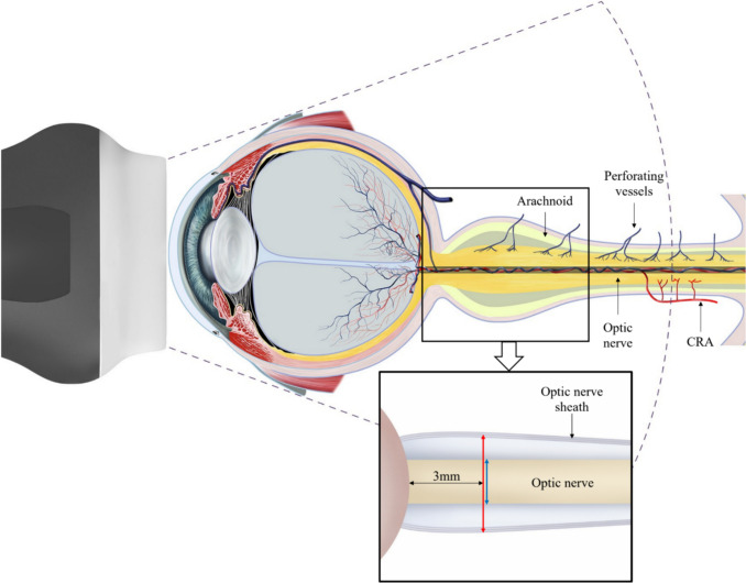
Introduction/objectives: Optic nerve sheath (ONS) enhancement using magnetic resonance imaging of the orbits was observed in patients with giant cell arteritis (GCA). We previously showed that ONS diameter (ONSD) by bedside ultrasound is increased in patient with active GCA. This study aims to assess whether ONSD decreases with clinical remission in patients with GCA.
Methods: A prospective cohort study was conducted from June 2022 to January 2023. Patients who had an optic nerve ultrasound at GCA diagnosis as part of a previous crosssectional study were eligible. Optic nerve ultrasound was performed by the same investigator at diagnosis and month 3. ONSD (includes the optic nerve and its sheath) and optic nerve diameter (OND) were measured. Descriptive statistics for baseline characteristics and paired sample t-test were performed to assess the mean difference in OND and ONSD between diagnosis and month 3.
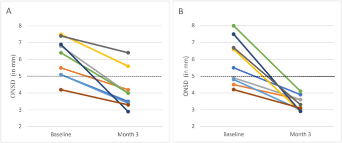
Results: Nine patients with GCA were included. The median age at disease onset was 79 years (interquartile range (IQR) of 79-82 years), and 7 patients were males. All patients were in clinical remission at month 3 on prednisone (median dose of 15 mg/day, IQR of 10-25 mg). The mean ONSD was lower at month 3 (3.76 mm) compared to baseline (5.98 mm), with a paired mean difference of 2.22 mm (95% CI 1.41-3.03 mm, p < 0.001). As anticipated, OND measurements did not vary between diagnosis and month 3.
Conclusion: ONSD on ultrasound improves after 3 months of therapy in patients with GCA. A longer prospective study is required to determine if ONSD is useful to assess disease activity in GCA. Key Points • ONS ultrasound can identify patients with active GCA. • The ONSD on ultrasound is dynamic and improved after 3 months of GCA therapy. • ONS ultrasound may be useful to monitor disease activity in GCA.
Keywords: Biomarkers; Diagnosis; Giant cell arteritis; Optic nerve; Ultrasonography.
© 2024. The Author(s).
Conflict of interest statement
Dr. Ross has received consultation fees from Otsuka® outside the submitted work. Dr. Ducharme-Bénard has received presentation and focus group fees from Novartis® outside the submitted work. Dr. Hussein has received presentation fees from Janssen® and is an investigator in clinical trials funded by Astra-Zeneca outside the submitted work. Dr. Pagnoux has received consultation or presentation fees from Roche®, Otsuka®, GlaxoSmithKline®, Astra-Zeneca®, Sanofi ®, BMS®, and ChemoCentryx® and has received research funds from CanVasc, the Vasculitis Clinical Research Consortium (VCRC), GSK®, Astra-Zeneca®, and Otsuka® outside the submitted work. Dr. Makhzoum has received consultation or presentation fees from Roche®, Otsuka®, GlaxoSmithKline®, Astra-Zeneca®, and Sanofi ® and is an investigator in clinical trials funded by Health Canada, the Vasculitis Clinical Research Consortium (VCRC), Janssen®, and Novartis® outside the submitted work. All other authors had no conflicts of interests to declare.
Figures
References
MeSH terms
Substances
LinkOut - more resources
Full Text Sources
Medical
Research Materials
