. 2024 Sep 1;132(3):953-967.
doi: 10.1152/jn.00155.2024. Epub 2024 Aug 7.

Neural pathways associated with reduced rigidity during pallidal deep brain stimulation for Parkinson's disease

Affiliations

Neural pathways associated with reduced rigidity during pallidal deep brain stimulation for Parkinson's disease

Emily Lecy et al. J Neurophysiol. .

Abstract

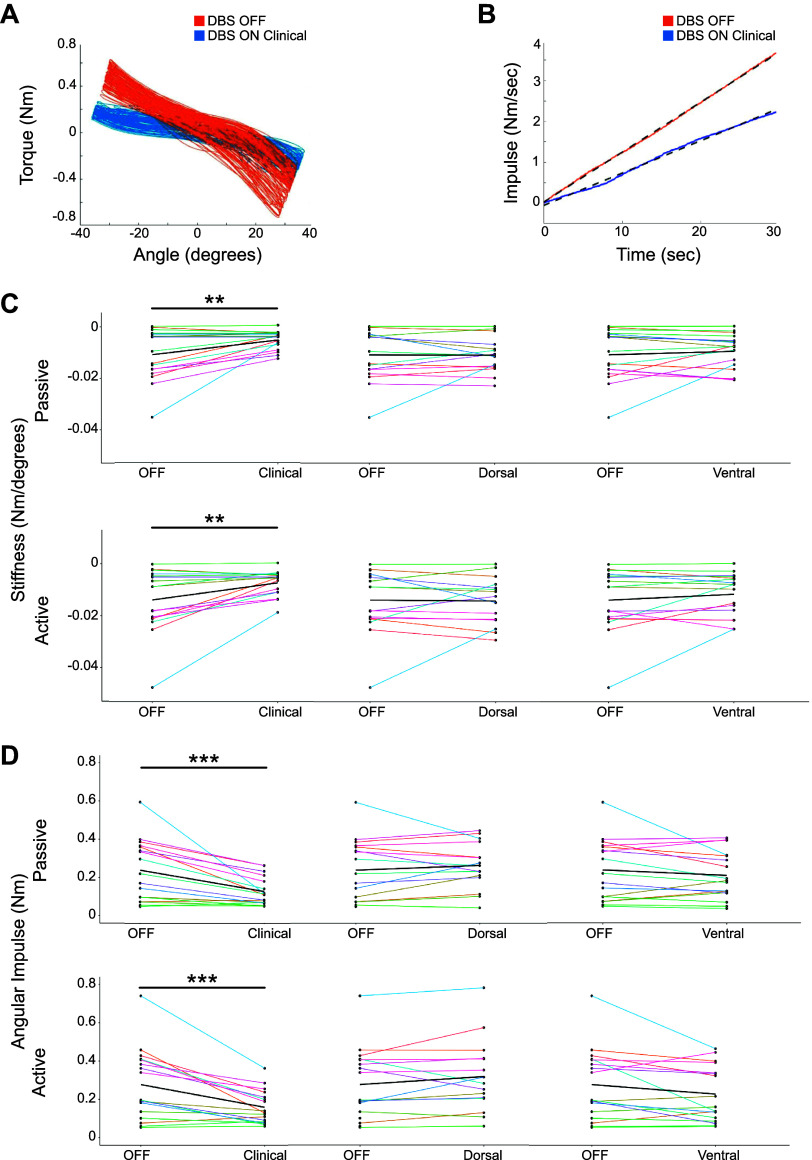
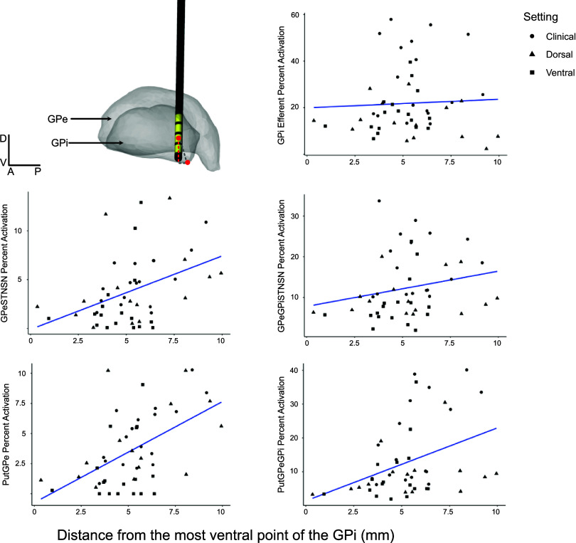
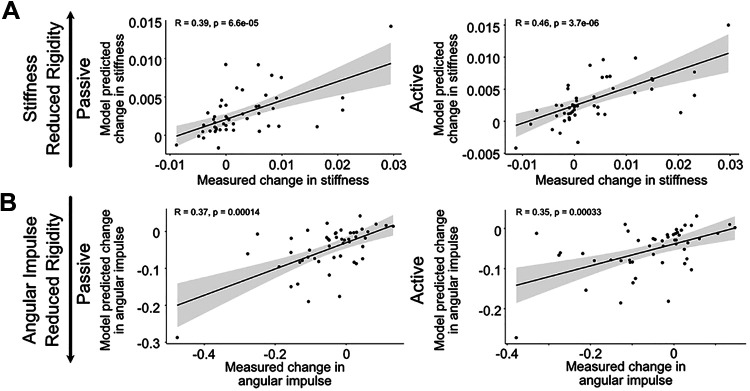
Deep brain stimulation (DBS) of the internal segment of the globus pallidus (GPi) can markedly reduce muscle rigidity in people with Parkinson's disease (PD); however, the mechanisms mediating this effect are poorly understood. Computational modeling of DBS provides a method to estimate the relative contributions of neural pathway activations to changes in outcomes. In this study, we generated subject-specific biophysical models of GPi DBS (derived from individual 7-T MRI), including pallidal efferent, putamenal efferent, and internal capsule pathways, to investigate how activation of neural pathways contributed to changes in forearm rigidity in PD. Ten individuals (17 arms) were tested off medication under four conditions: off stimulation, on clinically optimized stimulation, and on stimulation specifically targeting the dorsal GPi or ventral GPi. Quantitative measures of forearm rigidity, with and without a contralateral activation maneuver, were obtained with a robotic manipulandum. Clinically optimized GPi DBS settings significantly reduced forearm rigidity (P < 0.001), which aligned with GPi efferent fiber activation. The model demonstrated that GPi efferent axons could be activated at any location along the GPi dorsal-ventral axis. These results provide evidence that rigidity reduction produced by GPi DBS is mediated by preferential activation of GPi efferents to the thalamus, likely leading to a reduction in excitability of the muscle stretch reflex via overdriving pallidofugal output.NEW & NOTEWORTHY Subject-specific computational models of pallidal deep brain stimulation, in conjunction with quantitative measures of forearm rigidity, were used to examine the neural pathways mediating stimulation-induced changes in rigidity in people with Parkinson's disease. The model uniquely included internal, efferent and adjacent pathways of the basal ganglia. The results demonstrate that reductions in rigidity evoked by deep brain stimulation were principally mediated by the activation of globus pallidus internus efferent pathways.

Keywords: Parkinson’s disease; computational model; deep brain stimulation; globus pallidus; rigidity.


Conflict of interest statement

No conflicts of interest, financial or otherwise, are declared by the authors.

Figures

None
Graphical abstract
Figure 1.
Figure 1.
Subject-specific internal segment of the globus pallidus (GPi) deep brain stimulation (DBS) computational modeling pipeline. A: finite element models were data-driven from preoperative MRI and postoperative computed tomography (CT) scans. B: the combination of the finite element model and biophysical axon models enabled determination of the stimulation threshold for eliciting action potentials within each axon within a pathway. C: model-predicted axonal activation is shown in orange [external segment of globus pallidus (GPe) axons passing through the GPi to the subthalamic nucleus (STN) (GPe-GPi-STN)] and blue (GPi efferents).
Figure 2.
Figure 2.
Reconstructions of axons passing through or adjacent to the internal segment of the globus pallidus (GPi). A: axons traversing through both external (GPe) and internal (GPi) segments of the globus pallidus. Axons were reconstructed with curvatures based on nonhuman primate single-axon histological reconstructions (–43). B: corticospinal tract axon examples from 2 viewpoints, representative of 3 subtracts, that project from motor, premotor, and supplementary motor area cortical regions. Images are oriented anatomically: anterior (A), posterior (P), medial (M), lateral (L), dorsal (D), and ventral (V). STN, subthalamic nucleus.
Figure 3.
Figure 3.
Axon breakdown for all internal pathways showing different types of axon curvature that could be randomly created in each pathway for internal segment of the globus pallidus (GPi) efferents (A), external segment of the globus pallidus (GPe) efferents (B), and striatofugal fibers (C). All fiber curvature is based on nonhuman primate single-axon tracing studies (–43). Put, putamen; SN, substantia nigra; STN, subthalamic nucleus.
Figure 4.
Figure 4.
Change in rigidity measures with internal segment of the globus pallidus (GPi) deep brain stimulation (DBS). A: trace of position and torque taken from an upper extremity robotic manipulandum in both DBS OFF and DBS ON settings from a representative participant. B: traces of impulse (integrated torque) in both DBS OFF and DBS ON settings from a representative participant. The dashed line shows the line of best fit for the impulse, the slope of which is defined as angular impulse. C and D: changes in stiffness (C) and angular impulse (D) across DBS settings for all participants. Top: passive measures. Bottom: trials in which an activation maneuver was completed. The thick black line indicates the average change in rigidity scores between OFF and the tested setting, and thin colored lines are individual responses. Paired Wilcoxon signed-rank tests compared changes in rigidity differences between OFF and ON-DBS settings. **P ≤ 0.005, ***P ≤ 0.0005.
Figure 5.
Figure 5.
Violin plots showing pathway activation distributions across clinically optimized stimulation (A) and low-amplitude dorsal and ventral settings (B) in all participants. Dots are individual data points (N = 17). Solid colored horizontal lines represent means; open circles represent the median. All colored dots are individual data points. GPe, external segment of the globus pallidus; GPi, internal segment of the globus pallidus; M1, primary motor cortex; PM, premotor cortex; Put, putamen; SMA, supplementary motor cortex; STN, subthalamic nucleus.
Figure 6.
Figure 6.
Electrode localization within the internal segment of the globus pallidus (GPi) for all leads. Viewpoints are from superior to the GPi, with direction labeled in the key. Images are oriented anatomically: anterior (A), posterior (P), medial (M), lateral (L), dorsal (D), ventral (V).
Figure 7.
Figure 7.
Relationship between the distance from the most ventral point of the internal segment of the globus pallidus (GPi) and neural pathway activation of all internal fibers of passage. Top left quadrant shows how distance was calculated, using an example participant: by taking the distance along the dorsal-ventral plane (dashed red line) between the most ventral point of the GPi and the center of the active electrode. Five scatterplots show the relationship between the distance from the ventral GPi and pathway activation across internal axonal pathways. Trend lines were created using a general linear model of pathway activation by distance. Stimulation settings are shown in shapes according to the key. GPe, external segment of the globus pallidus; Put, putamen; STN, subthalamic nucleus; SN, substantia nigra.
Figure 8.
Figure 8.
Model predicted change in rigidity scores (y-axes) compared to measured change in rigidity scores (x-axes) for stiffness (A) and angular impulse (B) in passive (left) and active (right) conditions. Linear mixed effects model (LME) intercept and pathway slopes were fed into an equation that used actual pathway activation to predict change in rigidity scores. The relationship between the measured change in rigidity scores and the LME predicted scores was assessed with a Kendall’s rank correlation coefficient (R and P values shown on each plot). The regression line is shown in blue, with the 95% confidence interval indicated by the gray shading.
Figure 9.
Figure 9.
Scatterplots showing the relationship between stiffness (A) and angular impulse (B) and pathway activation differences in both passive (top) and active (bottom) conditions. The x-axis represents the difference in the number of axons activated between external segment of the globus pallidus globus pallidus (GPe) efferent (GPeSTNSN, GPeGPiSTNSN) and internal segment of the globus pallidus (GPi) efferent pathways by GPi deep brain stimulation (DBS) (GPe efferents − GPi efferents). Motor output measures are shown as changes, calculated by subtracting the measured outcome between DBS-ON and DBS-OFF conditions. Settings are represented in shapes according to the key. STN, subthalamic nucleus; SN, substantia nigra.

Similar articles

References

    1. Martin WE, Loewenson RB, Resch JA, Baker AB. Parkinson’s disease. Clinical analysis of 100 patients. Neurology 23: 783–790, 1973. doi:10.1212/WNL.23.8.783. - DOI - PubMed
    1. Mutch WJ, Strudwick A, Roy SK, Downie AW. Parkinson’s disease: disability, review, and management. Br Med J (Clin Res Ed) 293: 675–677, 1986. doi:10.1136/bmj.293.6548.675. - DOI - PMC - PubMed
    1. Ruonala V, Pekkonen E, Airaksinen O, Kankaanpää M, Karjalainen PA, Rissanen SM. Levodopa-induced changes in electromyographic patterns in patients with advanced Parkinson’s disease. Front Neurol 9: 35, 2018. doi:10.3389/fneur.2018.00035. - DOI - PMC - PubMed
    1. Shapiro MB, Vaillancourt DE, Sturman MM, Metman LV, Bakay RA, Corcos DM. Effects of STN DBS on rigidity in Parkinson’s disease. IEEE Trans Neural Syst Rehabil Eng 15: 173–181, 2007. doi:10.1109/TNSRE.2007.896997. - DOI - PMC - PubMed
    1. Fasano A, Romito LM, Daniele A, Piano C, Zinno M, Bentivoglio AR, Albanese A. Motor and cognitive outcome in patients with Parkinson’s disease 8 years after subthalamic implants. Brain 133: 2664–2676, 2010. doi:10.1093/brain/awq221. - DOI - PubMed

Publication types

LinkOut - more resources