

Fabry Disease Rat Model Develops Age- and Sex-Dependent Anterior Segment Ocular Abnormalities
- PMID: 39110587
- PMCID: PMC11314710
- DOI: 10.1167/iovs.65.10.14
Fabry Disease Rat Model Develops Age- and Sex-Dependent Anterior Segment Ocular Abnormalities
Abstract
Purpose: Fabry disease is an X-linked lysosomal storage disorder that results in multi-systemic renal, cardiovascular, and neuropathological damage, including in the eyes. We evaluated anterior segment ocular abnormalities based on age, sex (male and female), and genotype (wild-type, knockout [KO] male, heterozygous [HET] female, and KO female) in a rat model of Fabry disease.
Methods: The α-Gal A KO and WT rats were divided into young (6-24 weeks), adult (25-60 weeks), and aged (61+ weeks) groups. Intraocular pressure (IOP) was measured. Eyes were clinically scored for corneal and lens opacity as well as evaluated for corneal epithelial integrity and tear break-up time (TBUT). Anterior chamber depth (ACD) and central corneal thickness (CCT) using anterior segment-optical coherence tomography (AS-OCT).
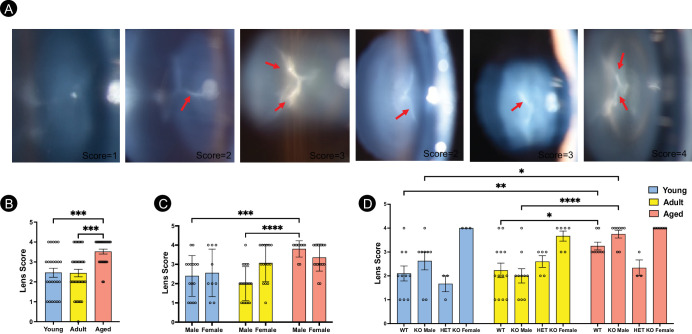
Results: The Fabry rats showed an age-dependent increase in IOP, predominantly in the male genotype. TBUT was decreased in both male and female groups with aging. Epithelial integrity was defective in KO males and HET females with age. However, it was highly compromised in KO females irrespective of age. Corneal and lens opacities were severely affected irrespective of sex or genotype in the aging Fabry rats. AS-OCT quantification of CCT and ACD also demonstrated age-dependent increases but were more pronounced in Fabry versus WT genotypes.
Conclusions: Epithelial integrity, corneal, and lens opacities worsened in Fabry rats, whereas IOP and TBUT changes were age-dependent. Similarly, CCT and ACD were age-related but more pronounced in Fabry rats, providing newer insights into the anterior segment ocular abnormalities with age, sex, and genotype in a rat model of Fabry disease.
Conflict of interest statement
Disclosure:
Figures


References
-
- Zarate YA, Hopkin RJ.. Fabry's disease. Lancet. 2008; 372: 1427–1435. - PubMed
-
- Kint JA. The enzyme defect in Fabry's disease. Nature. 1970; 227: 1173–1173. - PubMed
-
- Brady RO, Gal AE, Bradley RM, Martensson E, Warshaw AL, Laster L.. Enzymatic defect in Fabry's disease. N Engl J Med. 1967; 276: 1163–1167. - PubMed
-
- Sweeley CC, Klionsky B.. Fabry's disease: classification as a sphingolipidosis and partial characterization of a novel glycolipid. J Biol Chem. 1963; 238: PC3148–PC3150. - PubMed
MeSH terms
Substances
Grants and funding
LinkOut - more resources
Full Text Sources
Research Materials
