. 2024 Oct 7;228(2):iyae128.
doi: 10.1093/genetics/iyae128.

Fdo1, Fkh1, Fkh2, and the Swi6-Mbp1 MBF complex regulate Mcd1 levels to impact eco1 rad61 cell growth in Saccharomyces cerevisiae

Affiliations

Fdo1, Fkh1, Fkh2, and the Swi6-Mbp1 MBF complex regulate Mcd1 levels to impact eco1 rad61 cell growth in Saccharomyces cerevisiae

Gurvir Singh et al. Genetics. .

Abstract

Cohesins promote proper chromosome segregation, gene transcription, genomic architecture, DNA condensation, and DNA damage repair. Mutations in either cohesin subunits or regulatory genes can give rise to severe developmental abnormalities (such as Robert Syndrome and Cornelia de Lange Syndrome) and also are highly correlated with cancer. Despite this, little is known about cohesin regulation. Eco1 (ESCO2/EFO2 in humans) and Rad61 (WAPL in humans) represent two such regulators but perform opposing roles. Eco1 acetylation of cohesin during S phase, for instance, stabilizes cohesin-DNA binding to promote sister chromatid cohesion. On the other hand, Rad61 promotes the dissociation of cohesin from DNA. While Eco1 is essential, ECO1 and RAD61 co-deletion results in yeast cell viability, but only within a limited temperature range. Here, we report that eco1rad61 cell lethality is due to reduced levels of the cohesin subunit Mcd1. Results from a suppressor screen further reveals that FDO1 deletion rescues the temperature-sensitive (ts) growth defects exhibited by eco1rad61 double mutant cells by increasing Mcd1 levels. Regulation of MCD1 expression, however, appears more complex. Elevated expression of MBP1, which encodes a subunit of the MBF transcription complex, also rescues eco1rad61 cell growth defects. Elevated expression of SWI6, however, which encodes the Mbp1-binding partner of MBF, exacerbates eco1rad61 cell growth and also abrogates the Mpb1-dependent rescue. Finally, we identify two additional transcription factors, Fkh1 and Fkh2, that impact MCD1 expression. In combination, these findings provide new insights into the nuanced and multi-faceted transcriptional pathways that impact MCD1 expression.

Keywords: Cornelia de Lange Syndrome (CdLS); ECO1/ESCO2; Fdo1; Fkh1; Fkh2; MBF (Mbp1 and Swi6); Mcd1/Scc1/RAD21; Rad61/WAPL; Roberts Syndrome (RBS); cohesins.


Conflict of interest statement

Conflicts of interest The author(s) declare no conflict of interest.

Figures
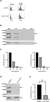
Fig. 1.
Fig. 1.
eco1 Δ rad61Δ double mutant cells contain reduced Mcd1 levels. a) Flow cytometry data of DNA contents for wildtype (YBS255) and eco1Δ rad61Δ double mutant cells (YMM828). b) Representative Western Blot of Mcd1 (top panel) and Pgk1 (lower panel) protein levels obtained from serially diluted (100%, 50%, 25%, 12.5%) extracts of HU-synchronized wildtype cells indicated in (A). c) Quantification of Mcd1 (left panel) and Pgk1 (right panel) of the serially diluted (100%, 50%, 25%, 12.5%) sample in (B). d) Representative Western Blot of Mcd1 (top panel) and Pgk1 (lower panel) protein levels of 50% diluted wildtype and eco1rad61 null cell extracts obtained from HU-synchronized cells indicated in (A). e) Quantification of Mcd1, normalized to Pgk1 loading controls. Statistical analysis was performed using a two-tailed t-test. Statistical differences (**) are based on a P < 0.01 obtained across three experiments (n = 3). P = 0.0031 for eco1Δ rad61Δ compared to wildtype cells. Error bars indicate the standard deviation.
Fig. 2.
Fig. 2.
MCD1 overexpression rescues eco1rad61 double mutant cell ts-growth. a) Streak test of three independent isolates of an mcd1-1 temperature-sensitive strain (YMM396) transformed with vector alone, compared to three independent isolates of mcd1-1 ts cells transformed with vector overexpressing MCD1. Temperature growth conditions are indicated. b) Growth of 10-fold serial dilutions of wildtype cells overexpressing vector alone (YGS26) or overexpressing MCD1 (YGS27), and eco1Δ rad61Δ double mutant cells overexpressing vector alone (YGS28), or overexpressing MCD1 (2 isolates shown; YGS29, YGS30). Temperatures and days of growth are indicated.
Fig. 3.
Fig. 3.
Identification of FDO1 deletion as a suppressor of eco1Δ rad61Δ cell temperature-sensitive growth defects. a) Growth of 10-fold serial dilutions of wildtype (YBS255), eco1Δ rad61Δ double mutant cells (YMM828) and three independent isolates of eco1Δ rad61Δ fdo1Δ triple mutant cells (YGS4, YGS36, YGS37). Temperatures and days of growth are indicated. b) Growth of 10-fold serial dilutions of eco1Δ rad61Δ double mutant cells (YMM828), two independent isolates of eco1Δ rad61Δ fdo1Δ triple mutant cells (YGS3, YGS1), and two independent isolates of eco1Δ rad61Δ fdo11–82Δ (YGS5, YGS6) triple mutant cells. Temperature and days of growth are indicated.
Fig. 4.
Fig. 4.
Deletion of FDO1 elevates Mcd1 levels in eco1Δ rad61Δ double mutant cells. a) Flow cytometry data of DNA content for three independent isolates of eco1Δ rad61Δ fdo1Δ triple mutant cells (YGS3, YGS4, YGS31). Log phase and early S phase synchronized (HU, hydroxyurea) DNA profiles are shown for each strain. b) Representative Western Blot of Mcd1 (top panel) and Pgk1 (lower panel) protein levels of the serially diluted (100%, 50%, 25%, 12.5%) eco1Δ rad61Δ fdo1Δ triple mutant cell extracts obtained from HU-synchronized cells indicated in (a). c) Quantifications of Mcd1 (left panel) and Pgk1 (right panel) of the serially diluted (100%, 50%, 25%, 12.5%) sample in (b). d) Representative Western Blot of Mcd1 (top panel) and Pgk1 (lower panel) protein levels of a 50% diluted eco1Δ rad61Δ fdo1Δ triple mutant and eco1Δ rad61Δ fdo1Δ double mutant cell extracts obtained from HU-synchronized cells indicated in (a). e) Quantification of Mcd1, normalized to Pgk1 loading controls. Statistical analysis was performed using a two-tailed t-test. Statistical differences (*) are based on a P < 0.05 obtained across three experiments (n = 3). P = 0.0133 for eco1Δ rad61Δ fdo1Δ compared to eco1Δ rad61Δ cells. Error bars indicate the standard deviation.
Fig. 5.
Fig. 5.
Increased Mcd1 levels partially rescue the growth defect caused by elevated Fdo1 levels. a) Top panel: Growth of 10-fold serial dilutions of wildtype cells overexpressing vector alone (YGS26) and three independent isolates of wildtype cells overexpressing FDO1 (YGS98, YGS100, YGS102). Temperatures and days of growth are indicated. Bottom panel: Growth of 10-fold serial dilutions of wildtype cells overexpressing vector alone (YGS26), eco1Δ rad61Δ double mutant cells overexpressing vector alone (YGS28), and three independent isolates of eco1Δ rad61Δ double mutant cells overexpressing FDO1 (YGS99, YGS101, and YGS103). Temperatures and days of growth are indicated. b) Growth of 10-fold serial dilutions of eco1Δ rad61Δ double mutant cells (YMM828) overexpressing MCD1 (YGS179), overexpressing FDO1 (YGS193), and two independent isolates of eco1Δ rad61Δ co-overexpressing both FDO1 and MCD1 (YGS195, YGS196). Temperature and days of growth are indicated.
Fig. 6.
Fig. 6.
FKH2 deletion exacerbates eco1Δ rad61Δ cell ts-growth. a) Growth of 10-fold serial dilutions of eco1Δ rad61Δ double mutant cells (YMM828) and two independent isolates of eco1Δ rad61Δ fkh1Δ triple mutant cells (YGS 11, YGS12). Temperatures and days of growth are indicated. b) Growth of 10-fold serial dilutions of eco1Δ rad61Δ double mutant cells (YMM828) and two independent isolates of eco1Δ rad61Δ fkh2Δ triple mutant cells (YGS13, YGS14). Temperatures and days of growth are indicated.
Fig. 7.
Fig. 7.
MCD1 overexpression rescues the deleterious effect of FKH2 deletion from eco1rad61 null cells. Growth of 10-fold serial dilutions of eco1Δ rad61Δ doble mutant cells (YMM828) overexpressing MCD1 (YGS29), eco1Δ rad61Δ fkh2Δ triple mutant cells overexpressing vector alone (YGS176), and two independent isolates of eco1Δ rad61Δ fkh2Δ overexpressing MCD1 (YGS177, YGS178). Temperature and days of growth are indicated.
Fig. 8.
Fig. 8.
Overexpression of FKH1 and FKH2 each are detrimental to the growth of the eco1Δ rad61Δ double mutant cells. a) Left panel: Growth of 10-fold serial dilutions of wildtype cells overexpressing vector alone (YGS26), and two independent isolates of wildtype cells overexpressing FKH2 (YGS126, YGS127). Temperatures and days of growth are indicated. Right panel: Growth of 10-fold serial dilutions of eco1Δ rad61Δ double mutant cells overexpressing vector alone (YGS28), and two independent isolates of eco1Δ rad61Δ double mutant cells overexpressing FKH2 (YGS53, YGS55). Temperatures and days of growth are indicated. b) Left panel: Growth of 10-fold serial dilutions of wildtype cells overexpressing vector alone (YGS26), and two independent isolates of wildtype cells overexpressing FKH1 (YGS79, YGS83). Temperatures and days of growth are indicated. Right panel: Growth of 10-fold serial dilutions of wildtype cells overexpressing vector alone (YGS26), eco1Δ rad61Δ double mutant cells overexpressing vector alone (YGS28), and two independent isolates of eco1Δ rad61Δ double mutant cells overexpressing FKH1 (YGS80, YGS84). Temperatures and days of growth are indicated.
Fig. 9.
Fig. 9.
MCD1 overexpression rescues the deleterious growth effect that result from elevated FKH1 and FKH2 levels in eco1rad61 null cells. a) Growth of 10-fold serial dilutions of eco1Δ rad61Δ doble mutant cells (YMM828) overexpressing MCD1 (YGS179), eco1Δ rad61Δ double mutant cells overexpressing FKH1 (YGS180), and two independent isolates of eco1Δ rad61Δ co-overexpressing FKH1 and MCD1 (YGS181, YGS182). Temperature and days of growth are indicated. b) Growth of 10-fold serial dilutions of eco1Δ rad61Δ doble mutant cells (YMM828) overexpressing MCD1 (YGS179), eco1Δ rad61Δ double mutant cells overexpressing FKH2 (YGS183), and two independent isolates of eco1Δ rad61Δ co-overexpressing FKH2 and MCD1 (YGS184, YGS185). Temperature and days of growth are indicated.
Fig. 10.
Fig. 10.
SWI6 overexpression reduces eco1rad61 cell growth, whereas MBP1 overexpression partially restores eco1rad61 cell growth at 37°C. a) Left panel: Growth of 10-fold serial dilutions of wildtype cells overexpressing vector alone (YGS26), and three independent isolates of wildtype cells overexpressing SWI6 (YGS104, YGS106, YGS108). Temperatures and days of growth are indicated in this and all subsequent panels. Right panel: Growth of 10-fold serial dilutions (dilution series used in all subsequent panels) of wildtype cells overexpressing vector alone (YGS26), eco1Δ rad61Δ double mutant cells overexpressing vector alone (YGS28), and three independent isolates of eco1Δ rad61Δ double mutant cells overexpressing SWI6 (YGS105, YGS107, YGS109). b) Left panel: Serial dilutions of wildtype cells overexpressing vector alone (YGS158), and three independent isolates of wildtype cells overexpressing MBP1 (YGS162, YGS164, YGS166) are shown. Right panel: Growth of serial dilutions of wildtype cells overexpressing vector alone (YGS158), eco1Δ rad61Δ double mutant cells overexpressing vector alone (YGS160), and three independent isolates of eco1Δ rad61Δ double mutant cells overexpressing MBP1 (YGS163, YGS165, YGS167). Temperatures and days of growth are indicated. c) Left panel: Serial dilutions of wildtype cells overexpressing two empty vectors (YGS168), and three independent isolates of wildtype cells overexpressing SWI6 and MBP1 (YGS170, YGS171, YGS172) are shown. Right panel: Serial dilutions of wildtype cells overexpressing two empty vectors (YGS168), eco1Δ rad61Δ double mutant cells overexpressing two empty vectors (YGS130), and three independent isolates of eco1Δ rad61Δ double mutant cells overexpressing SWI6 and MBP1 (YGS134, YGS138, YGS142).
Fig. 11.
Fig. 11.
Swi6 negatively regulates Mbp1-based transcription of MCD1. Growth of 10-fold serial dilutions of eco1Δ rad61Δ double mutant cells (YMM828) overexpressing MCD1 (YGS179), overexpressing SWI6 (YGS197), and two independent isolates co-overexpressing SWI6 and MCD1 (YGS199, YGS200). Temperature and days of growth are indicated.
Fig. 12.
Fig. 12.
Schematic highlights integrated mechanisms of MCD1 regulation. MBF complex (Swi6 and Mbp1) and Forkhead (Fkh1 and Fkh2) transcription factors bind non-overlapping DNA sequences upstream of MCD1. Swi6 and Mbp1 play antagonistic roles in MCD1 expression: Swi6 inhibits the positive role that Mpb1 otherwise performs in MCD1 transcription. Of the two forkhead transcription factors in yeast, Fkh2 appears to play a positive role in MCD1 expression, although elevated expression of either FKH1 or FKH2 (in bold with up arrow) is detrimental to eco1Δ rad61Δ cell growth. Fdo1, a co-repressor that binds Fkh1, inhibits MCD1 expression. Notably, elevated expression of MCD1 rescues (or partially suppresses) the negative effects produced by alterations in any one of these three (MBF, FKHs, and Fdo1) pathways. Further analysis is required to clarify the dual role of FKHs in regulating MCD1 expression, as well the molecular mechanisms through which Cln2-CDK may impact the transcriptional pathways defined here to regulate Mcd1 levels.

Similar articles

References

    1. Alomer RM, Da Silva EML, Chen J, Piekarz KM, McDonald K, Sansam CG, Sansam CL, Rankin S. 2017. Esco1 and Esco2 regulate distinct cohesin functions during cell cycle progression. Proc Natl Acad Sci. 114(37):9906–9911. doi:10.1073/pnas.1708291114. - DOI - PMC - PubMed
    1. Antony J, Chin CV, Horsfield JA. 2021. Cohesin mutations in cancer: emerging therapeutic targets. Int J Mol Sci. 22(13):6788. doi:10.3390/ijms22136788. - DOI - PMC - PubMed
    1. Aref R, Sanad MNME, Schüller H-J. 2021. Forkhead transcription factor Fkh1: insights into functional regulatory domains crucial for recruitment of Sin3 histone deacetylase complex. Curr Genet. 67(3):487–499. doi:10.1007/s00294-021-01158-3. - DOI - PMC - PubMed
    1. Arumugam P, Gruber S, Tanaka K, Haering CH, Mechtler K, Nasmyth K. 2003. ATP hydrolysis is required for Cohesin's association with chromosomes. Curr Biol. 13(22):1941–1953. doi:10.1016/j.cub.2003.10.036. - DOI - PubMed
    1. Bean JM, Siggia ED, Cross FR. 2005. High functional overlap between mlui cell-cycle box binding factor and swi4/6 cell-cycle box binding factor in the G1/S transcriptional program in Saccharomyces cerevisiae. Genetics. 171(1):49–61. doi:10.1534/genetics.105.044560. - DOI - PMC - PubMed

MeSH terms