. 2024 Oct;634(8033):424-431.
doi: 10.1038/s41586-024-07874-3. Epub 2024 Aug 7.

A virally encoded tRNA neutralizes the PARIS antiviral defence system

Affiliations

A virally encoded tRNA neutralizes the PARIS antiviral defence system

Nathaniel Burman et al. Nature. 2024 Oct.

Erratum in

  • Author Correction: A virally encoded tRNA neutralizes the PARIS antiviral defence system.
    Burman N, Belukhina S, Depardieu F, Wilkinson RA, Skutel M, Santiago-Frangos A, Graham AB, Livenskyi A, Chechenina A, Morozova N, Zahl T, Henriques WS, Buyukyoruk M, Rouillon C, Saudemont B, Shyrokova L, Kurata T, Hauryliuk V, Severinov K, Groseille J, Thierry A, Koszul R, Tesson F, Bernheim A, Bikard D, Wiedenheft B, Isaev A. Burman N, et al. Nature. 2024 Dec;636(8043):E8. doi: 10.1038/s41586-024-08427-4. Nature. 2024. PMID: 39633059 Free PMC article. No abstract available.

Abstract

Viruses compete with each other for limited cellular resources, and some deliver defence mechanisms that protect the host from competing genetic parasites1. The phage antirestriction induced system (PARIS) is a defence system, often encoded in viral genomes, that is composed of a 55 kDa ABC ATPase (AriA) and a 35 kDa TOPRIM nuclease (AriB)2. However, the mechanism by which AriA and AriB function in phage defence is unknown. Here we show that AriA and AriB assemble into a 425 kDa supramolecular immune complex. We use cryo-electron microscopy to determine the structure of this complex, thereby explaining how six molecules of AriA assemble into a propeller-shaped scaffold that coordinates three subunits of AriB. ATP-dependent detection of foreign proteins triggers the release of AriB, which assembles into a homodimeric nuclease that blocks infection by cleaving host lysine transfer RNA. Phage T5 subverts PARIS immunity through expression of a lysine transfer RNA variant that is not cleaved by PARIS, thereby restoring viral infection. Collectively, these data explain how AriA functions as an ATP-dependent sensor that detects viral proteins and activates the AriB toxin. PARIS is one of an emerging set of immune systems that form macromolecular complexes for the recognition of foreign proteins, rather than foreign nucleic acids3.


Conflict of interest statement

The authors declare no competing interests.

Figures

Fig. 1
Fig. 1. PARIS is a two-component system that assembles into a propeller-shaped supramolecular complex.
a, Schematic of the integrated P4 satellite in the E. coli genome. Grey arrows labelled sid (size determination), delta (δ transcriptional activator), psu (polarity suppression) and int (integrase) are P4 genes flanking AriA and AriB genes. b, PARIS genes coloured according to domain organization. AriA is an ABC ATPase whereas AriB is a DUF4435 that includes a TOPRIM nuclease. IS1 is composed of three helices flanked by the ABC ATPase domain, whereas IS2 adds two additional helices to the coiled-coil resulting in a helical bundle. c, AlphaFold2-predicted structures of AriA and AriB as homodimers coloured according to the structural features shown in b. d, Size exclusion chromatography showing that AriA and AriB copurify as a complex of roughly 430 kDa. The size exclusion chromatography profile is representative of three independent biological experiments. e, Experimentally determined density maps of the PARIS complex showing a homohexameric assembly of AriA with D3 symmetry. The AriA hexamer is decorated by AriB subunits that attach to AriA homodimers in one of two potential orientations: a ‘cis’ arrangement where the three AriB subunits are facing the same direction, or a ‘trans’ arrangement where one AriB subunit is rotated by 180° relative to the other two.
Fig. 2
Fig. 2. Interaction between AriA ATPase and AriB is essential for PARIS-mediated defence.
a, Each asymmetric unit of the PARIS complex contains an AriA homodimer with two NBDs. A 90° rotation of the asymmetric unit, showing 1–53 N-terminal residues (yellow) of AriB positioned over the ATPase active sites of AriA. b, AriB attaches to AriA through electrostatic interactions near helix α2 of AriB. TOPRIM active site residues predicted to participate in metal binding (grey spheres) and catalysis are shown as sticks. c, Close-up of NBD1 showing ATPγS bound in an open conformation. d, Close-up of NBD2 showing the bound ligand trapped in a closed precleavage state. e, Plaque assays were performed in duplicate with two independent clones of each mutant, using a serial dilution of phage T7 on E. coli MG1655 carrying the WT or mutated PARIS system.
Fig. 3
Fig. 3. AriA interacts with Ocr in an ATPase-dependent manner, leading to the release of AriB from the PARIS complex.
a, Ocr-Strep pulls down AriA, but not AriB. A mutation in the ATPase active site of AriA (K39A) blocks binding to Ocr-Strep. b, Ocr releases AriB (E26A) from WT AriA (lane 3), but Ocr does not trigger AriB release from an ATP-binding-deficient variant of AriA (K39A) (lane 5). c, A 4.4-Å-resolution reconstruction of AriA that was purified using Ocr-Strep as a bait. The structure shows three radial pores symmetrically positioned around a central pore. The pores contain disordered loops with several positively charged residues. Ocr (PDB: 1S7Z) is shown as an electrostatic surface. d, Mutations in the central pore (R116A or R119A) reduce the efficiency of phage protection. e, Ocr-Strep binds WT AriA and ejects AriB (E26A), and the charge swap mutations in the central pore of AriA (R116E/R119E) limit interaction with Ocr-Strep. f, AriB-Strep (E26A) pulls down WT AriA but not AriA (R116E/R119E). g, ATP and ADP were separated using thin-layer chromatography (Extended Data Fig. 7), and the rates of ATP hydrolysis for PARIS, PARIS mixed with tenfold excess Ocr and PARIS with an ATPase mutation in AriA (E393A) were measured. Experiments were performed in triplicate, and error bars show ±s.e.m. Two-sided statistical analysis performed using a post hoc Dunnett’s test, ****P < 0.0001. h, Size exclusion chromatography of AriB-Strep (around 36 kDa) following Ocr-mediated release from AriA. The column was calibrated using molecular weight standards (grey lines). AriB elutes in a single peak with an estimated molecular weight of 81 kDa (Extended Data Fig. 5a). i, Glutaraldehyde (GA) cross-linking assay with activated AriB (E26A). j, Mutations predicted to block AriB dimerization (E285R and F137A) prevent PARIS-mediated defence (Extended Data Fig. 1g). Plaque assays were performed in duplicate with two independent clones of each mutant.
Fig. 4
Fig. 4. Activation of PARIS leads to cell death and inhibition of translation.
a, Expression of Ocr from the pBAD vector in PARIS+ culture resulted in cell toxicity, whereas the empty vector (EV) had no impact. b, Ocr expression in PARIS+ cells induced cell death (propidium iodide staining, red) and DNA compactization (DAPI staining, blue). Arrows indicate zoomed-in images of nucleoid structures from PARIS+/Ocr (top) and PARIS/Ocr (bottom). Cells were imaged 1 h following induction of Ocr expression. c, Metabolic labelling experiments show a drop in 3H-methionine incorporation following induction of Ocr, indicative of translation inhibition following PARIS activation. d, In vitro translation in the presence of activated AriB (WT) or active site mutant (E26A). Translation of firefly luciferase mRNA was monitored by luminescence. In the presence of activated AriB, luciferase signal was reduced markedly, further indicating that PARIS functions through a mechanism of translational inhibition. c,d, Mean and s.d. were calculated for each time point for three independent experiments. Scale bars, 10 µm. a.u., Arbitrary units; 3H-t, 3H-thymidine. Source Data
Fig. 5
Fig. 5. PARIS cleaves E. coli tRNALys and T5 encodes a non-cleavable tRNALys.
a, Plaque assays conducted using cells with (+) or without (−) PARIS against mutants T5WT, T5Mos and T5Mos (T5Mut1–Mut4). b, Graphical map of the WT T5 genome. ORFs are represented by grey arrows and tRNAs by red ovals. T5Mos is missing an 8 kb fragment (red), with T5123 having a smaller 3 kb deletion (blue). c, T5Mos phages acquire mutations in ORF094 (Ptr1) and ORF103 (Ptr2) (black arrows) to escape PARIS-mediated immunity (mut1–mut4). d, AlphaFold2-predicted structures for Ptr1 and Ptr2 compared with T7 Ocr. Protein surfaces coloured by electrostatic potential. e, Expression of WT triggers, but not mutant, cause toxicity in PARIS+ cells but not in PARIS cells. f, Overexpression of T5 tRNALys in PARIS+ cells rescues infection by phage T5123, which lacks tRNALys. g, RNA blot performed on total RNA extracted from E. coli MG1655 carrying the PARIS system or a GFP control, and Ocr under the control of a PPhlF promoter. RNA was extracted either 15 or 30 min (15′ and 30′, respectively) following induction. Identification of 5S rRNA (green) and E. coli tRNALys (red) was made using distinct probes. h, Northern blot performed on total RNA extracted from E. coli carrying PARIS or GFP, Ocr and T5 tRNALys under the control of an araBAD promoter. RNA was extracted following 30 min of induction. The tRNALys (red) and T5 tRNALys (yellow) probes show that the latter was not degraded during PARIS-mediated defence. i, Small RNA extracted from E. coli showing a cleavage product when incubated with AriB in a metal-dependent manner. j, Primer extension assay. AriB cleaves E. coli tRNALys between positions 40 and 41. k, Schematic representation of the AriB cleavage site on E. coli tRNALys. Scale bars, 2 kb. aa, Amino acids; l-ara, l-arabinose; pI, isoelectric point.
Fig. 6
Fig. 6. Phylogenies of PARIS- and related ABC ATPase-powered defence systems.
a,b, The phylogenetic trees of AriA (a) and AriB (b). The two trees share similar branching patterns. Different colours represent 11 subclades of PARIS, including 2 in which ariA and ariB merged into a single ariAB gene (merged 1 and 2). c, Phylogenetic tree of AAA15/21 ATPase-containing defence proteins. Pfam accessions AAA15 (PF13175), AAA21 (PF13304), TOPRIM-Old (PF20469), DUF4435 (PF14491), RloB (PF13707), DUF4276 (PF14103), UvrD_N (PF00580), UvrD_C (PF13361), DUF262 (PF03235), DUF3996 (PF13161), HNH endonuclease (PF14279, PF10107) and RVT1 (PF00078) were used for domain annotations. Tree scale bars, 1. Source Data
Extended Data Fig. 1
Extended Data Fig. 1. AlphaFold2 structural predictions of AriA and AriB.
AlphaFold2-predicted structures of AriA and AriB oligomers colored by the predicted local distance difference test (pLDDT) score (0–100) with associated scale bars. Values greater than 90 indicate high confidence and values below 50 indicate low confidence. Predicted Aligned Error plots (PAE) are also shown for each structural prediction. a, AlphaFold2-predicted structure of the AriA homodimer. Confidence is high (PLDDT >90, green) for the ATPase domain, and low for the insertion sequences (PLDDT<70, teal). b, AlphaFold2-predicted structure of the AriB homodimer results in a high confidence model with more >98% of the residues above a PLDDT of 90. c, AlphaFold2-predicted structure of the AriA-B heterodimer and associated PAE plot. d, AlphaFold2 returned unreliable models when attempting to predict structures for higher ordered assemblies of AriA and AriB as demonstrated by the PLDDT plot below 50 for most of the molecule. e, Clashing (red) between AriBs, prevents the assembly of two AriB subunits on a single AriA homodimer. Clashing is defined by atoms with overlap greater than 0.6 Å. f, A predicted homodimer of AriB cannot associate with a homodimer of AriA without clashing (red). Atoms with an overlap greater than 0.6 Å are highlighted with red disks signifying the clash. g, Predicted structure of the AriB dimer shown in panel b, highlighting residues involved in dimerization.
Extended Data Fig. 2
Extended Data Fig. 2. Structural comparison of PARIS homologs.
a, Domain organization of phage defense systems where an ABC ATPase domain is associated with a TOPRIM nuclease domain. While the ATPase domain is highly conserved, the relative orientation of the TOPRIM nuclease domains and dimerization domains vary between PARIS (PDB ID: 8UX9), Gabija (PDB ID: 8SM3), and the Thermus scotoductus Overcoming Lysogenization Defect (Ts OLD, PDB ID: 6P74). b, AriB is the effector of PARIS defense. The AriB TOPRIM is homologous to the TOPRIM domains of Ts OLD (RMSD 1.1 Å, 20 C-alpha pairs) and GajA (RMSD 1.02 Å, 26 C-alpha pairs). The TOPRIM domain of AriB is within the DUF4435, which plays a role in AriB dimerization. c, Structural comparison of AriA to Rad50, a universally conserved ATPase. AriA primarily differs from Rad50 at Insertion Sequence (IS) 1 (residues 122-180) and IS2 (residues 242-289). IS1 introduces three alpha helices near the nucleotide binding domains of AriA that are predicted to be involved in trigger recognition. IS2 expands the coiled coil domain with two alpha helices and enable the homohexameric assembly of AriA subunits in the PARIS complex. d, Structural comparison of PARIS and related OLD systems highlights the unique assembly state of PARIS.
Extended Data Fig. 3
Extended Data Fig. 3. Workflow for structural determination of the PARIS complex.
Image processing was performed using cryoSPARC v4.41. a, 7,340 movies were recorded. Micrographs with CTF-fits worse than 8 Å were removed prior to downstream processing (n = 5,988). Using a de novo template generated from a 200-micrograph subset of this data, 4,078,384 particles were identified and extracted. From 17 selected classes, 1,643,515 particles remained, and a 3-class ab initio reconstruction yielded volumes shown in panel b. Particles belonging to junk classes (pink and red) were discarded. c, 25 representative 2-D Classes from 940,782 particles in panel b. A total of 934,763 particles from 72 classes remained after final 2-D Classification (n = 100 classes). d, After successive rounds of ab initio and heterogenous refinements, a 532,010 particle volume was obtained, corresponding to the intact PARIS complex with compositional heterogeneity at two AriB attachment sites. Masks were generated for local refinement and 3-D classification to produce the reconstructions shown in e, g, and h, respectively. e, Local Refinement of one asymmetric unit of the PARIS immune complex with FSC and 3-D orientation plot shown below. f, Close up of density map and model from E demonstrating map quality. g, C3 reconstruction of the PARIS complex at 3.71 Å-resolution with the experimental structure determined in panel e fit into the density. h, Cryo-EM reconstruction of PARIS with AriB in the ‘trans’ arrangement at approximately 3.93 Å-resolution with experimental structure determined in panel e, fit into the density.
Extended Data Fig. 4
Extended Data Fig. 4. AriB toxicity is dependent on association with AriA.
a, Liquid culture toxicity assays of AriB alone or AriA:B together (PARIS). Genes are overexpressed using a T7 inducible promoter. AriB alone results in a mild growth defect, while Ocr-triggered release of AriB (PARIS Ocr) is significantly more toxic. b, Growth of E. coli MG1655 carrying plasmid pFR85 (pSC101 with ariAB under the control of the native promoter), or variants with ariA deleted, ariB deleted or a stop codon in ariA yielding an interrupted protein that lacks the interaction interface with AriB. c, Structure-guided mutations that disrupt the AriA:AriB interface result in a loss of defense phenotype. d, Drop-test toxicity assay with PARIS (WT) or PARIS with AriA D401N/E404Q mutation. A modest growth defect can be observed only for the PARIS AriA D401N/E404Q overexpressed from T7 promoter. e, The addition of pBAD Ocr to the PARIS AriA mutants does not increase the toxicity, indicating that AriB is not functional when AriA interactions are disrupted. f, Close-up of the AriA:AriB interaction interface. Mutated residues are labeled with asterisks. Source Data
Extended Data Fig. 5
Extended Data Fig. 5. Purification of activated AriB.
a, Size exclusion chromatography calibration curve with estimated masses of AriA:Ocr and activated AriB complexes. b, Introduction of the Strep-tag at the C- or N- terminus of AriA, but not on the C-terminus of AriB, interferes with PARIS anti-phage defense. c, Purification of activated AriB after mixing cell lysates containing strep-tagged AriB and non-tagged Ocr.
Extended Data Fig. 6
Extended Data Fig. 6. Structure determination of AriA purified using a Ocr-Strep pulldown.
Image processing and analysis was performed in cryoSPARC v4.41. a, Representative micrograph from a total of 10,340 movies. Micrographs with CTF-fits worse than 8 Å-resolution were discarded. Particle picking on 9,399 micrographs identified 4,415,782 particles which were extracted and subjected to 2-D Classification. b, Selected 2-D classes corresponding to the AriA hexamer contained 667,479 particles. c, 2-Class ab initio reconstruction of particles from panel b. The gray class corresponds to the AriA homohexamer, while particles associated with the orange class were discarded. Density for the ATPase domains is present, but one blade of the propeller is poorly resolved due to the increased flexibility of the AriA scaffold relative to the fully assembled PARIS complex. d, After iterative rounds of sorting the 392,989 particles from panel C, a stack containing 62,732 particles was isolated and C3 refinement produced a 4.4 Å-resolution reconstruction of the AriA scaffold.
Extended Data Fig. 7
Extended Data Fig. 7. AriA ATPase activity.
a, PARIS mediated hydrolysis of α32P ATP with PARIS alone or with 10-fold excess trigger (T7 Ocr). Reactions were run from 1 to 32 min and products were resolved via thin layer chromatography. Reactions with α32P ATP incubated with T4 polynucleotide kinase were used as a positive control. Images are representative of reactions that were performed in triplicate. b, Ocr alone was incubated with α32P ATP for 32 min indicating that the trigger alone does not hydrolyze ATP. c, Concentration of ADP formed at each time point was quantified and plotted. Data points of three technical replicates are shown.
Extended Data Fig. 8
Extended Data Fig. 8. PARIS activation does not result in total RNA or DNA degradation.
a, Total DNA was extracted from E. coli MG1655 carrying plasmids pFR85 (PARIS) and pFD250 (PPhlF-Ocr) at different time points after Ocr induction with or without DAPG (n = 3), or from a non-induced control. Samples were run on a 1% agarose TAE gel. b, Total RNA was extracted from E. coli MG1655 carrying plasmid pFR85 (PARIS) or pFR66 (sfGFP), and pFD250 (PPhlF-Ocr) at different time points after induction of Ocr expression with DAPG. Samples were run on a TBE Urea (7M) acrylamide (10%) gel and stained with SYBR-Gold. A representation of 3 replicates is shown. c, TUNEL assay, demonstrating the lack of accumulation of dsDNA breaks in PARIS+ cells 2 h after Ocr induction, as measured through terminal deoxynucleotidyl transferase labeling of free 3′-OH groups in DNA. As a positive control of DNA damage, cells were treated with 0.1% H2O2.
Extended Data Fig. 9
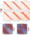
Extended Data Fig. 9. PARIS activation results in DNA compactization akin to inhibition of translation with chloramphenicol.
a, Hi-C contact maps (bin: 4kb). From left to right: WT (pFR66 + pFD245 control vectors), PARIS (pFR85) + PPhlF-ocr (pFD250), and chloramphenicol treated cells. Top and bottom rows correspond to 15 and 30 min after induction with DAPG or treatment with chloramphenicol. b, Ratio between contact maps of WT at t = 30 min and contact maps 30 min after either PARIS activation (left) or chloramphenicol treatment (right).
Extended Data Fig. 10
Extended Data Fig. 10. Identification of the T5 genomic region required for infection of PARIS+ cells.
a, Growth of PARIS- or PARIS+ cells infected with PARIS-sensitive phage T5Mos at low or high MOI. The phage was added at t = 0. b, Phage T5 variants with non-essential deletions in the region encoding tRNAs are sensitive to PARIS defense. Boundaries of the deletions are shown on the right, tRNA genes are highlighted in red. c, Overexpression of Fragment 1 (31885–32870) derived from the deletion in the phage T5123 partially rescues T5123 infection of the PARIS+ culture. d, Comparison of E. coli and T5 tRNALys highlights substitution mutations near the anticodon stem loop that we hypothesize prevents targeting by AriB. e, Swapping U-A for A-U (Mut1) on the E. coli tRNALys confers resistance to PARIS.
Extended Data Fig. 11
Extended Data Fig. 11. tRNALys cleavage product and phylogenetics of PARIS.
a, Alignment of the B803 and B806 Northern Blot probes to the E. coli and T5 tRNALys. b, Northern Blot with probe B806 in the presence or absence of the T5 tRNALys expressed from a pBAD. c, Mapping of the tRNALys cleavage site by reverse-transcription, adapter ligation, PCR and Sanger sequencing. The cleavage site, identified as the junction between the tRNA sequence and the ligated adapter, is marked with a vertical red line. d, Alignment of homologs of AriA representative of the different PARIS clades. Different domains of AriA are indicated. Grey scale represents % of identity between homologs. e, Phylogenetic tree of DUF4435 domain of AriB using M5 Ribonuclease (TOPRIM) as an outgroup.

References

    1. Chevallereau, A. & Westra, E. R. Bacterial immunity: mobile genetic elements are hotspots for defence systems. Curr. Biol.32, R923–R926 (2022). - PubMed
    1. Rousset, F. et al. Phages and their satellites encode hotspots of antiviral systems. Cell Host Microbe30, 740–753 (2022). - PMC - PubMed
    1. Zhang, T. et al. Direct activation of a bacterial innate immune system by a viral capsid protein. Nature612, 132–140 (2022). - PMC - PubMed
    1. Wein, T. & Sorek, R. Bacterial origins of human cell-autonomous innate immune mechanisms. Nat. Rev. Immunol.22, 629–638 (2022). - PubMed
    1. Makarova, K. S., Wolf, Y. I., Snir, S. & Koonin, E. V. Defense islands in bacterial and archaeal genomes and prediction of novel defense systems. J. Bacteriol.193, 6039–6056 (2011). - PMC - PubMed

MeSH terms