. 2024 Aug 7;12(8):e009403.
doi: 10.1136/jitc-2024-009403.

EphA3 CAR T cells are effective against glioblastoma in preclinical models

Affiliations

EphA3 CAR T cells are effective against glioblastoma in preclinical models

Paulo Martins et al. J Immunother Cancer. .

Abstract

Background: Adoptive T-cell therapy targeting antigens expressed in glioblastoma has emerged as a potential therapeutic strategy to prevent or delay recurrence and prolong overall survival in this aggressive disease setting. Ephrin receptor A3 (EphA3), which is highly expressed in glioblastoma; in particular, on the tumor vasculature and brain cancer stem cells, is an ideal target for immune-based therapies.

Methods: We have designed an EphA3-targeted chimeric antigen receptor (CAR) using the single chain variable fragment of a novel monoclonal antibody, and assessed its therapeutic potential against EphA3-expressing patient-derived glioblastoma neurospheres, organoids and xenografted glioblastoma tumors in immunodeficient mice.

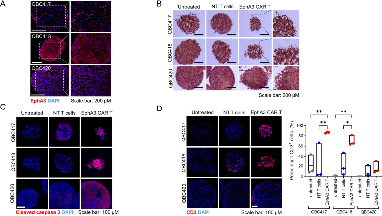
Results: In vitro expanded EphA3 CAR T cells from healthy individuals efficiently recognize and kill EphA3-positive glioblastoma cells in vitro. Furthermore, these effector cells demonstrated curative efficacy in an orthotopic xenograft model of glioblastoma. EphA3 CAR T cells were equally effective in targeting patient-derived neurospheres and infiltrate, disaggregate, and induce apoptosis in glioblastoma-derived organoids.

Conclusions: This study provides compelling evidence supporting the therapeutic potential of EphA3 CAR T-cell therapy against glioblastoma by targeting EphA3 associated with brain cancer stem cells and the tumor vasculature. The ability to target patient-derived glioblastoma underscores the translational significance of this EphA3 CAR T-cell therapy in the pursuit of effective and targeted glioblastoma treatment strategies.

Keywords: Adoptive cell therapy - ACT; Chimeric antigen receptor - CAR; T cell.


Conflict of interest statement

Competing interests: RK and PM are listed as inventors on the patent application describing EphA3 CAR T-cell therapy.

Figures

Figure 1
Figure 1. Novel 3C3-1 anti-EphA3 monoclonal antibody has a high affinity for human EphA3 which is expressed on glioblastoma (A) ELISA titration curve of 10 EphA3-specific monoclonal antibody clones. Monoclonal antibody 3C3-1 was selected for further analysis. (B) Surface plasmon resonance sensorgram of clone 3C3-1 binding to EphA3 protein. Kinetic data of antibody binding to EphA3: association (Ka) 2.22×10⁸±1.59×10⁷ (1\Ms), dissociation (Kd) 3.85×10⁻⁵±2.79×10⁻⁵ (1/s) and equilibrium (KD) 1.14×10⁻¹³±7.54×10⁻¹⁴ (M). The reported values are the mean±SD of at least two independent experiments (C) immunohistochemistry staining of EphA3 in glioblastoma tissue. Sections were stained with anti-EphA3 3C3-1 antibody and brown areas are positive reactions stained by DAB. Images capture the spatial distribution of EphA3 within the tissue, highlighting tumor heterogeneity. (D) EphA3 protein quantification. The percentage of EphA3-positive cells per section on patient sample was calculated from tissue sections from QCell (n=1 section per patient), clinical trial P1228-QGBM01 and TMA GL805-L51 (n=1–3 section(s) per patient). (E) Two GBO cryosections generated from patient QBC420. Organoids were co-stained with anti-EphA3 (green) and the endothelial cell marker CD31 (red). DAPI (blue) was used for nuclear staining. Images with an enlarged area from each tumor are shown. EphA3, erythropoietin-producing hepatoma type-A receptor 3; GBO, glioblastoma organoid; TMA, tumor micro array; GBM, glioblastoma; DAPI, 4′,6-diamidino-2-phenylindole; DAB, 3,3′-Diaminobenzidine.
Figure 2
Figure 2. In vitro therapeutic assessment of EphA3 CAR T cells (A) schematic of EphA3 CAR construct. The EF1 promotor drives transcription of the CAR containing the 3C3-1 scFv and CD28 derived co-stimulatory domain followed by the CD3ζ activation sequence. (B) Peripheral blood mononuclear cells from four healthy participants (GR036, GR066, GR103, and GR170) were stimulated with anti-CD3/CD28, transduced with EphA3-CAR encoding lentivirus and cultured for 14 days. Cells were stained with anti-CD3, anti-CD4 anti-CD8, and anti-mouse IgG antibodies, to detect surface CAR protein. Representative FACS plots of CAR T-cell fraction, CAR-CD4 and CAR-CD8 subsets, and memory populations as determined using CCR7 and CD45RA subgating. (C) Proportion of CD4 and CD8 EphA3-CARs and (D) T-cell memory subsets defined as naïve (N: CCR7+CD45RA+), central memory (CM: CCR7CD45RA+), effector memory (EM: CCR7CD45RA), and effector memory RA+ (EMRA: CCR7CD45RA+). EphA3-CAR T cells demonstrated cytotoxic activity against glioblastoma cell line U251 (EphA3-expressing glioblastoma cell line). (E) In a real-time killing assay, EphA3 CAR T cells were added to monolayers of U251 or U87 cells at a 5:1 effector-to-target ratio, and the percentage cytolysis was recorded over time. The average cytolysis of three triplicates±SD is shown. (F) Cytolytic potential of EphA3-CAR T cells from four donors was tested against U251 cells. Targets were incubated at a 1:5 effector-to-target ratio. Show are mean percentage cytolysis±SD (n=2–3 replicates) at 24, 48 and 72 hours post addition of effectors. CAR, chimeric antigen receptor; EphA3, erythropoietin-producing hepatoma type-A receptor 3; VH, variable heavy; VL, variable light; ScFv, single chain variable fragment.
Figure 3
Figure 3. EphA3 CAR T cells demonstrate selective EphA3-targetted killing in vivo. (A) Schematic representation of in vivo experiment. NRG mice were transplanted with luciferase-expressing U251 (EphA3+) or U87 (EphA3) cells subcutaneously in the flank. Mice were treated once tumors reached approximately 60 mm3. NT (non-transduced) T cells, CD19-CAR (non-glioblastoma specific CAR T cells), or EphA3-CAR T cells were administered intravenously on days 11 and 17 (dotted lines) post-engraftment with two doses of 20×106 T cells (CAR-positive cells ranged between 22% and 36% of products). (A&C) Tumor size was measured over the course of the experiment. Data points represent the mean±SD of one experiment (n=5 mice per group). CAR, chimeric antigen receptor; EphA3, erythropoietin-producing hepatoma type-A receptor 3; IV, intravenous; SC, subcutaneous.
Figure 4
Figure 4. EphA3-CAR T cells prevent the growth of glioblastoma tumors in an orthotopic xenograft mode. (A) Luciferase-expressing cells U251 were orthotopically transplanted in the right cerebral hemisphere of NRG mice. Once tumor had established mice were regrouped so as to normalize the average starting tumor burden per group. Mice received two IV infusions of EphA3-CAR T cells as treatment, and CD19-CAR T cells were administered to control groups. (B) Tumor formation was monitored by bioluminescence imaging of mice up to 100 days post tumor engraftment. Representative image of two independent experiments. (C) Kaplan-Meier survival curve of the mice of two independent experiments (n=17 mice per group). Mice were sacrificed in accordance with institutional ethical guidelines, and not limited to symptoms related to tumor growth. Log-rank (Mantel-Cox) test. (D) Flow cytometry analysis of peripheral blood of mice identifying absolute cell number of circulating human CD45+CD3+ cells. Shown is mean±SD, each symbol is representative of one mouse (n=7 from one experiment). Statistical analysis by the Wilcoxon matched-pairs signed rank test. ***p<0.001, **p<0.01, *p<0.05. CAR, chimeric antigen receptor; EphA3, erythropoietin-producing hepatoma type-A receptor 3; IV, intravenous.
Figure 5
Figure 5. EphA3-CAR T cells target and kill primary glioblastoma cells. (A) The 3C3-1 monoclonal antibody was used to evaluate, by flow cytometry, EphA3-expression of patient-derived glioblastoma primary cell cultures from the QCell bank. Histograms depict the percentage of viable EphA3-positive cells stained with clone 3C3-1 compared with secondary anti-IgG antibody alone (dotted line), from two independent experiments. (B) EphA3-CAR T-cell cultures were co-incubated overnight with the patient-derived glioblastoma samples at a 1:1 ratio (T cell: glioblastoma target). Frequency of total CD4+TNF+ and polyfunctional CD4+TNF+interferon-γ+ cells in response to glioblastoma targets. Shown is the mean±SD of at least two independent experiments with triplicates. (C) Glioblastoma cells were cultured as monolayers on laminin-coated E-plates for xCELLignece. Glioblastoma cells were co-cultured with EphA3-CAR T cells, or control CD19-CAR T cells. Data represent the mean percentage cytolysis±SD (n=2 replicates). (D) A sigmoidal dose-response curve (variable slope) was extrapolated from each killing data curve, and the half-maximal killing time (KT50) was determined. Data represent KT50 average±SE (n=2 replicates). (E) Glioblastoma primary cultures were plated at low density and propagated as floating aggregates. Glioblastoma spheroids were established from all cultures except FPW1 which grew as a monolayer. Glioblastoma cells were co-cultured with EphA3-CAR T cells or CD19 CAR T. Cytotoxicity was measured kinetically using the IncuCyte platform over 4 days, and cell death indicated by uptake of FITC-conjugated annexin V. Shown are representative merged bright field and fluorescent microscopy images of cultures at day 4. Green represents annexin V uptake by the neurospheres. CAR, chimeric antigen receptor; EphA3, erythropoietin-producing hepatoma type-A receptor 3; TNF, Tumor necrosis factor; FITC, Fluorescein isothiocyanate.
Figure 6
Figure 6. EphA3-CAR T cells infiltrate glioblastoma. (A) Immunofluorescence staining of EphA3 (red) in GBOs was established from three patients undergoing glioblastoma resection surgery. (B) H&E images of untreated, non-transduced (NT) and EphA3-CAR T cell treated GBOs of all three samples, after 48 hours incubation. (C) Immunofluorescence staining of cleaved caspase 3 (red) in untreated, NT control and EphA3-CAR T cell treated GBOs of all three samples, after 48 hours incubation. (D) Immunofluorescence staining and quantification of CD3 (red) in untreated, NT control and EphA3-CAR T cell treated GBOs of all three samples, after 48 hours incubation. Statistical analysis by multiple comparisons of an ordinary one-way analysis of variance, **p<0.01, *p<0.05. CAR, chimeric antigen receptor; EphA3, erythropoietin-producing hepatoma type-A receptor 3; GBO, glioblastoma organoid; DAPI, 4′,6-diamidino-2-phenylindole.

References

    1. Stupp R, Mason WP, van den Bent MJ, et al. Radiotherapy plus concomitant and adjuvant temozolomide for glioblastoma. N Engl J Med. 2005;352:987–96. doi: 10.1056/NEJMoa043330. - DOI - PubMed
    1. Cantrell JN, Waddle MR, Rotman M, et al. Progress toward long-term survivors of glioblastoma. Mayo Clin Proc. 2019;94:1278–86. doi: 10.1016/j.mayocp.2018.11.031. - DOI - PubMed
    1. Kochenderfer JN, Wilson WH, Janik JE, et al. Eradication of B-lineage cells and regression of lymphoma in a patient treated with autologous T cells genetically engineered to recognize CD19. Blood. 2010;116:4099–102. doi: 10.1182/blood-2010-04-281931. - DOI - PMC - PubMed
    1. Grupp SA, Kalos M, Barrett D, et al. Chimeric antigen receptor–modified T cells for acute lymphoid leukemia. N Engl J Med. 2013;368:1509–18. doi: 10.1056/NEJMoa1215134. - DOI - PMC - PubMed
    1. Pasquale EB. Eph-ephrin bidirectional signaling in physiology and disease. Cell. 2008;133:38–52. doi: 10.1016/j.cell.2008.03.011. - DOI - PubMed

LinkOut - more resources