. 2024 Aug;632(8026):885-892.
doi: 10.1038/s41586-024-07746-w. Epub 2024 Aug 7.

ILC2-derived LIF licences progress from tissue to systemic immunity

Affiliations

ILC2-derived LIF licences progress from tissue to systemic immunity

Mayuri Gogoi et al. Nature. 2024 Aug.

Abstract

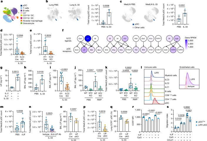
Migration and homing of immune cells are critical for immune surveillance. Trafficking is mediated by combinations of adhesion and chemokine receptors that guide immune cells, in response to chemokine signals, to specific locations within tissues and the lymphatic system to support tissue-localized immune reactions and systemic immunity1,2. Here we show that disruption of leukaemia inhibitory factor (LIF) production from group 2 innate lymphoid cells (ILC2s) prevents immune cells leaving the lungs to migrate to the lymph nodes (LNs). In the absence of LIF, viral infection leads to plasmacytoid dendritic cells (pDCs) becoming retained in the lungs where they improve tissue-localized, antiviral immunity, whereas chronic pulmonary allergen challenge leads to marked immune cell accumulation and the formation of tertiary lymphoid structures in the lung. In both cases immune cells fail to migrate to the lymphatics, leading to highly compromised LN reactions. Mechanistically, ILC2-derived LIF induces the production of the chemokine CCL21 from lymphatic endothelial cells lining the pulmonary lymphatic vessels, thus licensing the homing of CCR7+ immune cells (including dendritic cells) to LNs. Consequently, ILC2-derived LIF dictates the egress of immune cells from the lungs to regulate tissue-localized versus systemic immunity and the balance between allergen and viral responsiveness in the lungs.


Conflict of interest statement

A.N.J.M. is on the scientific advisory board of SinoMab. The other authors declare no competing interests.

Figures

Fig. 1
Fig. 1. ILC2s are required for normal homing of pDCs.
ac, Uniform manifold approximation and projection of lung and MedLN immune cell clusters (a) and quantification of pDCs (b,c) from flow cytometry analysis following PBS and IL-33 intranasal treatment of lung (b) and MedLN (c) in WT mice (n = 4). d,e, Flow cytometry analysis of Il7rCre and Il7rCreRoraflox/flox pDCs in MedLN (d, n = 10) and lung (e, n = 9). f, Heatmap of ligand and receptor expression derived from RNA-seq data. gj, ELISA of LIF from purified ILC2s cultured with IL-2 + IL-7, with (n = 4) or without (n = 3) IL-33 (g); BAL of mice treated with IL-33 (h, WT, n = 4; i, Il7rCre, n = 12; ILC2KO, n = 15); and with either ragweed protein (RWP, j, WT, n = 5; ST2KO, n = 6) or PBS (WT, n = 5; ST2KO, n = 5). k, Flow cytometry analysis of pDCs (WT: PBS, n = 10; RWP, n = 9; ST2KO: PBS, n = 8; RWP, n = 11). l, Flow cytometry analysis of LIF receptor (LIFR) on indicated cell types. m,n, Flow cytometry analysis of pDCs in WT mice following rLIF treatment (m, n = 10) and anti-LIF neutralizing antibody (Ab) and IL-33 treatment (n, n = 10). o, ELISA of BAL LIF following IL-33 challenge (n = 5). p,q, Flow cytometry analysis of pDCs in Il7rCre (n = 7) and LIF-cKO (n = 10) (p), and lung CCR7+ pDC (q) percentage (n = 5), following IL-33 challenge. r, Flow cytometry analysis of CCR7 expression by CpG-activated pDCCre and LIFR-cKO pDCs with or without rLIF for 16 h (pDCCre, n = 4; pDCCre + CpG, n = 4; pDCCre + CpG + rLIF, n = 5; LIFR-cKO, n = 5; LIFR-cKO + CpG, n = 5; LIFR-cKO + CpG + rLIF, n = 5). s, Chemotaxis of CpG-activated pDCCre or LIFR-cKO pDCs to CCL21 with or without rLIF for 16 h (pDCCre + CpG, n = 3; pDCCre + CpG + CCL21, n = 3; pDCCre + CpG + rmLIF + CCL21, n = 7; LIFR-cKO + CpG, n = 3; LIFR-cKO + CpG + CCL21, n = 3; LIFR-cKO + CpG + rmLIF + CCL21, n = 7). be,gi,ms, Unpaired two-sided t-test; j,k, one-way analysis of variance (ANOVA) with Tukey’s multiple-comparisons test; data (mean ± s.e.m.) are either representative (ac,g,h,j,l,o,qs) or pooled from two independent experiments (d,e,i,k,m,n,p). Lineage (Lin) contains CD3, CD4, CD8a, CD19, CD11b, CD11c and FcER1 antibodies; RPKM, reads per kilobase per million mapped reads. Source Data
Fig. 2
Fig. 2. ILC2-derived LIF is required for all CCR7+ immune cell egress to LNs.
a, Lung histology from Il7rCre and LIF-cKO mice following PVM challenge at day 8. b, Lung viral load following PVM challenge in Il7rCre (n = 5) and LIF-cKO (n = 8) mice. c, BAL type I IFN (IFN-I):IL-5 ratio from ELISA data following PVM challenge by Il7rCre (n = 10) and LIF-cKO (n = 9) mice. d, Histology of Il7rCre and LIF-cKO mouse MedLN following PVM challenge. B cells were stained with B220 antibody (green), T cells with CD3e antibody (red). e,f, Flow cytometry analysis of CD45+ cell numbers following PVM challenge in Il7rCre (PBS, n = 5; PVM, n = 5) and LIF-cKO (PBS, n = 5; PVM, n = 4) mice (e) and in BIC (n = 5) and BIC × Lifflox/flox (ILC2LIFKO, n = 6) mice (f). g, Flow cytometry analysis of CCR7+CD45+ cell numbers following PVM challenge in Il7rCre and LIF-cKO mice (n = 5). h, CD45+ cell numbers in Il7rCre and LIF-cKO mice treated with rLIF and infected with PVM (Il7rCre + PVM, n = 8; LIF-cKO + PVM, n = 8; LIF-cKO + PVM + rLIF, n = 8). b,c,f,g Unpaired two-sided t-test; e,h, one-way ANOVA with Tukey’s multiple-comparisons test. Data presented as mean ± s.e.m. a,dg, Data are representative of two independent experiments with similar results. b,c,h, Experiments are pooled data from two independent experiments. Scale bars, 1,000 µm (a), 200 µm (d). Source Data
Fig. 3
Fig. 3. ILC2-derived LIF induces CCL21 production by LECs.
a,b, Flow cytometry analysis of LIFR expression in lung LECs and BECs in naive mice (a) and CCL21 expression in lung LECs and BECs following PVM challenge (b). c,d, Flow cytometry analysis of phosphorylated STAT3+ in cultured LECs following rLIF treatment (Il7rCre, n = 8; LIF-cKO, n = 8) (c) and CCL21+ LEC percentage and CCL21 median fluorescence intensity (MFI) following rLIF treatment (Il7rCre, n = 6; LIF-cKO, n = 6) (d). e, ELISA of CCL21 concentration in in vitro cultured LEC-conditioned media following rLIF treatment (control, n = 5; rLIF, n = 6). f,g, Flow cytometry analysis of CCL21 MFI of lung LECs in WT mice following rLIF challenge at the indicated time points (n = 4) (f) and CCL21+ lung LEC numbers in Il7rCre and LIF-cKO mice following PVM challenge (Il7rCre, n = 4; LIF-cKO, n = 5) (g). h, qPCR analysis of Ccl21 relative expression in purified lung LECs from Il7rCre and LIF-cKO mice following PVM challenge (Il7rCre, n = 6; LIF-cKO, n = 5). i, Flow cytometry analysis of MedLN and lung CCR7+CD45+ cell numbers in BIC and ILC2LIFKO mice following PVM challenge (n = 5). c,d,fi, Unpaired two-sided t-test; e, one-way ANOVA with Tukey’s multiple-comparisons test. Data presented as mean ± s.e.m. ad,fi, Data are representative of two independent experiments with similar results; e, experiments are pooled data from two independent experiments. Source Data
Fig. 4
Fig. 4. Defective immunity and MedLN colonization persist even following viral reinfection.
af, Flow cytometry analysis of CD45+ (a), CD4Teff cell (b), CD8Teff cell and TCM cell (c), cDC (d), pDC (e) and B cell numbers (f) in Il7rCre and LIF-cKO mice following PVM rechallenge (n = 5). g, ELISA of serum IgE in Il7rCre and LIF-cKO mice following PVM primary (Il7rCr, n = 5; LIF-cKO, n = 5) and secondary (Il7rCre, n = 4; LIF-cKO, n = 5) challenge. h, Lung viral load in Il7rCre and LIF-cKO mice following PVM rechallenge (Il7rCre, n = 8; LIF-cKO, n = 9). i, In vitro viral neutralization assay with Il7rCre and LIF-cKO serum following PVM rechallenge. dCT was calculated as CT(PVM)-CT (hamster GAPDH) (control, n = 4; PVM rechallenged Il7rCre serum, n = 8; PVM rechallenged LIF-cKO serum, n  =8). af,h,i, Unpaired two-sided t-test; g, one-way ANOVA with Tukey’s multiple-comparisons test. Data presented as mean ± s.e.m. ag,i, Data are representative of two independent experiments with similar results; h, experiments are pooled data from two independent experiments. PFU, plaque-forming units. Source Data
Fig. 5
Fig. 5. Chronic allergen challenge leads to marked iBALT accumulation in the absence of ILC2-derived LIF.
a,b, Lung histology (a) and immunofluorescence (b) of Il7rCre and LIF-cKO mice following chronic RWP challenge. B cells were stained with B220 antibody (green), T cells with CD3e antibody (red) and nuclei with DAPI (blue). c, Immunofluorescence as in b, with B cells stained with B220 antibody (green), T cells with CD3e antibody (red) and lymphatic vessels with VEGFR3 (grey). d, Representative image of MedLN from Il7rCre and LIF-cKO mice following PBS or chronic RWP challenge. ei, Flow cytometry analysis of numbers of CD45+ cells (e), CD4Teff cells (f), CD4+ TCM cells (g), cDCs (h) and B cells (i) from Il7rCre and LIF-cKO mice following chronic RWP challenge (Il7rCre, n = 5; LIF-cKO, n = 4). j, Immunofluorescence of MedLN from Il7rCre and LIF-cKO mice following chronic RWP challenge. B cells were stained with B220 antibody (green) and T cells with CD3e antibody (red). k, ELISA of serum IgE from Il7rCre and LIF-cKO mice following chronic RWP challenge (n = 10). lp, Flow cytometry analysis of CD45+ cells (l), CD4Teff cells (m), CD4+ TCM cells (n), cDCs (o) and B cells (p) from BIC and ILC2LIFKO mice following chronic RWP challenge (n = 4). q, Immunofluorescence of MedLN from BIC and ILC2LIFKO mice following chronic RWP challenge. B cells were stained with B220 antibody (green) and T cells with CD3e antibody (red). r, ELISA of serum IgE from BIC and ILC2LIFKO mice following chronic RWP challenge (n = 4). ei,kp,r, Unpaired two-sided t-test. Data presented as mean ± s.e.m. aj,lr, Data are representative of two independent experiments with similar results; k, experiments are pooled data from two independent experiments. Scale bars, 10 µm (a), 70 µm (b), 45 µm (c), 10 mm (d), 50 µm (j), 70 µm (q). Source Data
Extended Data Fig. 1
Extended Data Fig. 1. ILC2 production of LIF during type-2 immune response.
a, Gene expression of indicated genes in pDCs from bulk RNAseq data with five biological replicates per group. b. Representative flow cytometry plots of lung pDCs stained for ST2. Flow cytometry analysis of pDC numbers in c, MedLN, Rag2–/– (PBS = 8, IL-33 = 11) and Rag2–/–Il2rgc–/– (Rag2–/–gc–/–) (PBS = 3, IL-33 = 3), and d, lung Rag2–/– (PBS = 10, IL-33 = 10) and Rag2–/–gc–/– (PBS = 8, IL-33 = 10) following IL-33 intranasal challenge. e, Lif expression in ILC2 purified from lymph node (LN) (n = 3), lung (Lung) (n = 3), small intestine lamina propria (LP) (n = 3), adipose tissue (FAT) (n = 3), bone marrow ILC2 progenitor (ILC2P) (n = 2) from naïve mice. ELISA of LIF in the BAL f, from Rag2–/– (PBS = 10, IL-33 = 8) mice following IL-33 intranasal challenge, and g, from wildtype mice (WT) following ragweed protein extract (RWP) challenge (PBS = 5, RWP = 5), and from h, Il7rCre (PBS = 5, RWP = 5) and Il7rCre x Roraflox/flox (ILC2KO) (PBS = 5, RWP = 5) mice treated with RWP. i, Flow cytometry analysis of lung pDCs in WT (PBS = 10, RWP = 10 IL-33 receptor-deficient (ST2KO) (PBS = 9, RWP = 11) mice following RWP intranasal challenge. j, Flow cytometry analysis of LIF receptor (LIFR) staining on lung myeloid cells. Flow cytometry analysis of k, phospho-STAT3 positive cells following activation as indicated, ILC2 conditioned media (ILC2 CM), IL-33 activated ILC2 conditioned media (IL-33 activated ILC2 CM), anti-LIF neutralizing antibody (n = 4), and l, lung pDCs following rLIF treatment in wildtype mice (n = 10), and m, lung pDCs in wild type mice following treatment with anti-LIF neutralizing antibody or isotype and IL-33 intranasal challenge (n = 10), and n, pDCs in the MedLN (PBS = 10, 6 h = 9, 24 h = 9) and lung (PBS = 9, 6 h = 9, 24 h = 10) at the indicated timepoints following one dose of rLIF challenge, and o, MedLN pDC Ki67 expression in wild type mice following IL-33 challenge (n = 6). p, Trans-well chemotaxis of pDCs to rLIF. (control = 5, LIF (500 ng/ml) = 6, LIF (1000 ng/ml) = 6). q, Il7rCre x Lifflox/flox (LIF-cKO) generation schematic. r, Schematic of Lifflox/flox mouse generation. s, Southern analysis of Lifflox/flox ES cells. t, qPCR analysis of Lif expression in ILC2s purified from Il7rCre and Il7rCre x Lifflox/flox (LIF-cKO) mice. u, Flow cytometry analysis of lung pDC numbers in Il7rCre (n = 9) and LIF-cKO (n = 10) following IL-33 challenge. v, ELISA of BAL LIF from RWP challenged Il7rCre and LIF-cKO mice (Il7rCre = 5 and LIF-cKO =5), and flow cytometry analysis of MedLN pDCs (Il7rCre = 5 and LIF-cKO =7) and lung pDCs (Il7rCre = 5 and LIF-cKO =7). Flow cytometry analysis of w, MedLN, and x, lung pDCs in recipient ILC2 KO mice transplanted with Il7rCre or LIF-cKO ILC2s and subsequently challenged with RWP. (Il7rCre = 4 and LIF-cKO =5). y, Schematic of mouse FITC-dextran and IL-33 intranasal challenge. z, Flow cytometry gating strategy and MedLN FITC-dextran+ pDCs numbers in Il7rCre and LIF-cKO mice following PVM and FITC-dextran intranasal challenge (Il7rCre = 8 and LIF-cKO =8). f-h,m-p,u-x,z, unpaired two-sided t-test, c,d,i,k,l one-way ANOVA with Tukey’s multiple comparisons test. Data mean ± SEM. Data are representative of two (b, g, h, j, k, o, p, v, w, x) independent experiments with similar results. Experiments in (c, d, f, i, l, m, n, u, z) are pooled data from two independent experiments. RNA-seq data in a are based on five biological replicates per group. Source Data
Extended Data Fig. 2
Extended Data Fig. 2. pDC expression of LIFR and Pneumovirus infection of conditional LIF-deficient mice.
a, Schematic of pDC-specific LIF receptor (LIFR) deficient pDCCre x Lifr –/flox (LIFR cKO) mice generation. b, LIF receptor (LIFR) expression in lung pDCs in SiglechCre/Cre x Lifr –/+ (pDCCre) and SiglechCre/Cre x Lifr –/flox (LIFR cKO) mice. Flow cytometry analysis of pDCCre and LIFR cKO c, MedLN, and d, lung pDCs following RWP challenge (pDCCre =4 and LIFR cKO = 5). e, Flow cytometry analysis of phospho-STAT3 positive cells following activation as indicated, ILC2 conditioned media (ILC2 CM), IL-33 activated ILC2 conditioned media (IL-33 + ILC2 CM), anti-LIF neutralizing antibody (control pDCCre = 4, control LIFR cKO =3, rLIF pDCCre = 4, rLIF LIFR cKO =3, IL-33 = 4, ILC2 CM = 4, IL-33 + ILC2 CM = 4, IL-33 + ILC2 CM+Anti-LIF Ab =4). f, Volcano plot of all differentially expressed genes in bulk RNAseq of lung pDCs from PBS or IL-33 challenged wildtype mice. g, KEGG pathway analysis of significant differentially expressed genes from (f). h, Heatmap of significant differentially expressed chemokine receptor genes from lung pDC bulk RNAseq. Flow cytometry analysis of lung i, CCR7+pDCs in LIF-cKO mice after RWP challenge (Il7rCre = 5 and LIF-cKO =7), and j, CCR7+pDCs in naïve LIF-cKO mice (Il7rCre = 5 and LIF-cKO =4). k, schematic of the experimental protocol for pneumovirus of mouse (PVM) infection. l, ELISA of LIF in the BAL from Il7rCre and LIF-cKO mice following PVM challenge (n = 10). m, qPCR analysis of Lif gene expression in ILC2s, CD4+ and CD8+ T cells purified from indicated mice following PVM intranasal challenge. Il7rCre, ILC2KO and LIF-cKO mice (Il7rrer ILC2 = 2, Il7rCre CD4+T = 5, Il7rCre CD8+ T = 4, ILC2KO CD4+T =4, ILC2KO CD8+ T = 5, LIF-cKO CD4+T =4, LIF-cKO CD8+ T = 5). n, Lung histology infiltration score of Il7rCre and LIF-cKO mice following PVM challenge (Il7rCre = 5 and LIF-cKO =5). o, Flow cytometry analysis of lung pDCs in Il7rCre and LIF-cKO mice following PVM challenge (Il7rCre = 9 and LIF-cKO =10). ELISA of: p, IFN-I in BAL (Il7rCre = 10 and LIF-cKO =10);and q, IL-5 in BAL (Il7rCre = 9 and LIF-cKO =9) from Il7rCre and LIF-cKO mice following PVM challenge. r, Flow cytometry analysis of lung IL-5+ILC2s in lung in Il7rCre and LIF-cKO mice after PVM challenge (Il7rCre = 10 and LIF-cKO =10). s, Flow cytometry analysis of MedLN and lung eosinophil, neutrophil, alveolar macrophage (AM) and monocyte numbers following PVM challenge (Il7rCre = 5 and LIF-cKO =4). Flow cytometry analysis of t, lung CD45+.cell numbers Il7rCre (PBS = 5, PVM = 5) and LIF-cKO (PBS = 5, PVM = 4); u, T cell numbers; v, B cells; w, cDC; x, pDCs from MedLN and lung from Il7rCre and LIF-cKO mice following PVM challenge (Il7rCre = 5 and LIF-cKO =4). y, Flow cytometry analysis of lung pDCs for CCR7 expression in Il7rCre and LIF-cKO mice following PVM challenge (Il7rCre = 10 and LIF-cKO =11). z, Flow cytometry analysis of lung pDCs for CCR9 (Il7rCre = 5 and LIF-cKO =5), CXCR3 (Il7rCre = 4 and LIF-cKO =4), CXCR4 (Il7rCre = 4 and LIF-cKO =4), and CCR5 (Il7rCre = 4 and LIF-cKO =4) in Il7rCre and LIF-cKO mice following PVM challenge. c,d,i,j,n-s,u-z unpaired two-sided t-test, e,l,t one-way ANOVA with Tukey’s multiple comparisons test. Data mean ± SEM. Data are representative of two (b, c, d, e, l, i,j,m,n,s-x,z) independent experiments with similar results. Experiments in (l,o-r,y) are pooled data from two independent experiments. RNA-seq data in a are based on five biological replicates per group. Source Data
Extended Data Fig. 3
Extended Data Fig. 3. Assessment of circulating and tissue-resident immune cells in conditional LIF-deficient mice during PVM infection.
a, Schematic of FITC-dextran and PVM intranasal challenge. b, Flow cytometry gating strategy and FITC-dextran+ CD45+ MedLN cell numbers in Il7rCre and LIF-cKO mice following PVM and FITC-dextran intranasal challenge. Flow cytometry analysis of: c, MedLN FITC-dextran+ cell numbers in Il7rCre and LIF-cKO mice following PVM and FITC-dextran intranasal challenge (Il7rCre = 7 and LIF-cKO =7); d, MedLN FITC-dextran+ DCs, B cells, and other immune cell numbers (Il7rCre = 7 and LIF-cKO =7) and percentages in Il7rCre and LIF-cKO mice following PVM and FITC-dextran intranasal challenge (Il7rCre = 8 and LIF-cKO =8). e, Schematic of intravenous anti-CD45-APC antibody labelling after PVM infection. Flow cytometry analysis of lung and MedLN intravenous anti-CD45-APC antibody labelled: f, CD45+ immune cells, g, T cells, h, B cells, i, pDCs, and j, cDCs after PVM infection (Il7rCre = 7 and LIF-cKO =10). k, ILC2LIFKO generation schematic. l, qPCR analysis of Lif expression in lung ILC2, T cells and B cells purified from Boolean Cre (BIC) and BIC x Lifflox/flox (ILC2LIFKO) mice after PVM challenge (BIC = 3, ILC2LIFKO = 3). m, Lung viral load BIC and ILC2LIFKO mice following PVM challenge (BIC = 5, ILC2LIFKO = 5). n, Flow cytometry analysis of lung eosinophil (BIC = 5, ILC2LIFKO = 5), neutrophils (BIC = 5, ILC2LIFKO = 6), alveolar macrophages (AM) (BIC = 5, ILC2LIFKO = 6) and monocytes (BIC = 5, ILC2LIFKO = 6) in BIC and ILC2LIFKO mice following PVM intranasal challenge. Flow cytometry analysis of: o, lung CD45+ cells (BIC = 5, ILC2LIFKO = 6); p, T cells from MedLN (BIC = 6, ILC2LIFKO = 6) and lung (BIC = 6, ILC2LIFKO = 6); q, B cells from MedLN (BIC = 5, ILC2LIFKO = 6) and lung (BIC = 6, ILC2LIFKO = 6); r, cDCs from MedLN (BIC = 5, ILC2LIFKO = 6) and lung (BIC = 6, ILC2LIFKO = 6); s, pDCs from MedLN (BIC = 5, ILC2LIFKO = 6) and lung (BIC = 6, ILC2LIFKO = 6). c,d,f-j,l-s unpaired two-sided t-test. Data mean ± SEM. Data are representative of two (b, l,m-s) independent experiments with similar results. c,d,f-j unpaired two-sided t-test. Data mean ± SEM. Data are pooled from two independent experiments. Source Data
Extended Data Fig. 4
Extended Data Fig. 4. CCR7-positive immune cell analysis in conditional LIF-deficient mice.
a, Flow cytometry gating strategy for CCR7+ CD45+ cells in Il7rCre and LIF-cKO mice following PVM challenge. b-d, Il7rCre and LIF-cKO mice following PVM challenge (Il7rCre = 5 and LIF-cKO =5). b, Flow cytometry analysis of CCR7+ cell numbers in MedLN; c, Flow cytometry analysis of CCR7+ cell numbers in lung. d, Flow cytometry analysis of CCR7+ cell percentages. e, Schematic of PVM infection combined with PBS or rLIF intranasal treatment. f, Flow cytometry analysis of MedLN T cells, B cells, pDCs and cDCs in Il7rCre and LIF-cKO mice treated with rLIF and infected with PVM (Il7rCre = 8 and LIF-cKO =8). b-d, unpaired two-sided t-test, f, one-way ANOVA with Tukey’s multiple comparisons test. Data mean ± SEM. Data are representative of two (a, b, c, d) independent experiments with similar results. Experiment (f) are pooled data from two independent experiments. Source Data
Extended Data Fig. 5
Extended Data Fig. 5. FTY720 treatment of conditional LIF-deficient mice.
a, Schematic of PVM intranasal infection with PBS or FTY720 treatment. Flow cytometry analysis of b, MedLN CD45+ cells; c, lung CD45+ cells; d, MedLN and lung pDCs; e, MedLN and lung T cells; f, MedLN and lung B cells; g, MedLN and lung cDCs; h, spleen CD45+ cells; and i, thymus CD45+ cells in Il7rCre and LIF-cKO mice treated with PBS or FTY720 following PVM challenge. (n = 3). b-i, one-way ANOVA with Tukey’s multiple comparisons test. Data mean ± SEM. Data are representative of two (b-i) independent experiments with similar results. Source Data
Extended Data Fig. 6
Extended Data Fig. 6. Adhesion molecule and chemokine receptor expression by lung LECs.
Flow cytometry analysis of: a, lung LEC for adhesion molecules VCAM1, ICAM1, CD73, CD206, CD34, CD31, Podoplanin (PDPN) from Il7rCre and LIF-cKO mice following PVM challenge (n = 4), b, CCL21+ lung LEC numbers in wildtype mice following PVM challenge at indicated timepoints (n = 2). c, ELISA of CCL19, CCL25, CXCL9, CXCL10 concentration in in-vitro cultured LEC conditioned media following rLIF treatment for 6 h or 24 h or 48 h (n = 6). d, Flow cytometry analysis of CCL21+ lung LEC numbers in wildtype mice following rLIF challenge at indicated timepoint (PBS = 3, rLIF =4). e, Flow cytometry analysis of MedLN and lung CCR7+ T cells, CCR7+ B cells, and CCR7+ cDCs from BIC and ILC2LIFKO mice following PVM challenge (n = 5). a,d,e, unpaired two-sided t-test, c, one-way ANOVA with Tukey’s multiple comparisons test. Data mean ± SEM. Data are representative of two (a, b, d, e) independent experiments with similar results. Experiments in (c) are pooled data from two independent experiments. Source Data
Extended Data Fig. 7
Extended Data Fig. 7. Pneumovirus reinfection in conditional LIF-deficient mice.
a, Schematic of PVM intranasal primary and secondary infection. Flow cytometry analysis of lung: b, CD45+; c, eosinophil, neutrophil, alveolar macrophage (AM) and monocyte; d, CD4+TEff cells, CD8+TEff cells and TCM cells, DCs, pDCs and B cell numbers from Il7rCre and LIF-cKO mice following PVM rechallenge (n = 5). Flow cytometry analysis of e, MedLN CD45+ cells; f, lung CD45+ cells; g, lung eosinophils, neutrophils, alveolar macrophages (AM) and monocytes; h, MedLN and lung CD4+ TEff cells; i, MedLN and lung CD8+ TEff and CD8+ TCM cells; j, MedLN and lung cDCs; k, MedLN and lung pDCs, and l, MedLN and lung B cells in BIC and ILC2LIFKO mice following PVM rechallenge (BIC = 4 and ILC2LIFKO = 5). m, ELISA of serum IgE from BIC and ILC2LIFKO mice following PVM primary and secondary challenge (PVM challenged BIC = 3, PVM challenged ILC2LIFKO = 5, PVM rechallenged BIC = 5, PVM rechallenged ILC2LIFKO = 5;). n, Lung viral load in BIC and ILC2LIFKO mice following PVM rechallenge (BIC = 7 and ILC2LIFKO = 9). o, In vitro viral neutralization assay with BIC and ILC2LIFKO mice serum following PVM rechallenge. dCT was calculated as CT(PVM)-CT(hamster GAPDH) (control = 4, PVM rechallenged BIC serum =10 and PVM rechallenged ILC2LIFKO serum =10). b-o, unpaired two-sided t-test. Data mean ± SEM. Data are representative of two (b-m) independent experiments with similar results. Experiments in (n,o) are pooled data from two independent experiments. Source Data
Extended Data Fig. 8
Extended Data Fig. 8. Chronic RWP challenge of conditional LIF-deficient mice.
a, Schematic of chronic RWP intranasal challenge protocol. b, ELISA of LIF in BAL from Il7rCre and LIF-cKO mice following chronic RWP challenge (Il7rCre = 5, LIF-cKO =4). c, Lung histology scores from Il7rCre and LIF-cKO mice following chronic RWP challenge (Il7rCre = 8, LIF-cKO =10). d, Lung iBALT scores from Il7rCre and LIF-cKO mice following chronic RWP challenge. (Il7rCre = 6, LIF-cKO =8). e, Flow cytometry analysis of lung eosinophils, neutrophils, alveolar macrophages and monocytes in Il7rCre and LIF-cKO mice following chronic RWP challenge (Il7rCre = 5, LIF-cKO =4). f, Immunofluorescence histology of lung showing tertiary lymphoid tissue formation in BIC and ILC2LIFKO mice following chronic RWP challenge, B cells stained with B220 antibody (green), T cells with CD3e antibody (red) and nuclei with DAPI (blue). g, Immunofluorescence histology of lung from LIF-cKO mouse after chronic RWP intranasal challenge. KLRG1 (green), CD3e (red) and VEGFR3 (grey). Flow cytometry analysis of h, MedLN T cells and pDCs (Il7rCre = 5, LIF-cKO =4); i, lung CD45+ cells (Il7rCre = 5, LIF-cKO =5), j, lung CD4+ TEff cells (Il7rCre = 5, LIF-cKO =4), k, lung B cells (Il7rCre = 5, LIF-cKO =5), I, lung cDCs (Il7rCre = 5, LIF-cKO =5) and pDCs (Il7rCre = 5, LIF-cKO =4) from Il7rCre and LIF-cKO mice following chronic RWP challenge. m, Flow cytometry analysis of lung eosinophils, neutrophils, alveolar macrophages (AM) and monocytes in Boolean Cre (BIC) and BIC x Lifflox/flox (ILC2LIFKO) mice following chronic RWP challenge (n = 4). n, ELISA of LIF in BAL from BIC and ILC2LIFKO mice following chronic RWP challenge (BIC = 9, ILC2LIFKO = 10). Flow cytometry analysis of o, MedLN pDCs; p, lung CD45+ cells; q, lung CD4+ TEff cells; r, lung B cells; s, lung cDCs and pDCs, and t, inguinal lymph node (pLN) CD45+ cells in BIC and ILC2LIFKO mice following chronic RWP challenge (n = 4). u, qPCR analysis of Lif expression in lung ILC2, CD4+ and CD8+ T cell purified from Il7rCre and LIF-cKO mice following chronic RWP challenge (n = 2-4). b-e, h-t unpaired two-sided t-test. Data mean ± SEM. Data are representative of two (b, e, f, g, h, i, j, k, l, m, o, p, q, r, s, t, u) independent experiments with similar results. Experiments in (c, d, n) are pooled data from two independent experiments. Source Data
Extended Data Fig. 9
Schematic of the roles of ILC2-derived LIF regulation of immune cell migration during pulmonary responses to viral infection and allergen challenge. Source Data

References

    1. Arasa, J., Collado-Diaz, V. & Halin, C. Structure and immune function of afferent lymphatics and their mechanistic contribution to dendritic cell and T cell trafficking. Cells10.3390/cells10051269 (2021). - PMC - PubMed
    1. Griffith, J. W., Sokol, C. L. & Luster, A. D. Chemokines and chemokine receptors: positioning cells for host defense and immunity. Annu. Rev. Immunol.32, 659–702 (2014). 10.1146/annurev-immunol-032713-120145 - DOI - PubMed
    1. Rodriguez-Rodriguez, N., Gogoi, M. & McKenzie, A. N. J. Group 2 innate lymphoid cells: team players in regulating asthma. Annu. Rev. Immunol.39, 167–198 (2021). 10.1146/annurev-immunol-110119-091711 - DOI - PMC - PubMed
    1. Clementi, N. et al. Viral respiratory pathogens and lung injury. Clin. Microbiol. Rev.10.1128/CMR.00103-20 (2021). - PMC - PubMed
    1. Molofsky, A. B. & Locksley, R. M. The ins and outs of innate and adaptive type 2 immunity. Immunity56, 704–722 (2023). 10.1016/j.immuni.2023.03.014 - DOI - PMC - PubMed

MeSH terms