Review
. 2024 Aug 7;24(1):278.
doi: 10.1186/s12935-024-03464-5.

Harnessing exosomes as cancer biomarkers in clinical oncology

Affiliations
Review

Harnessing exosomes as cancer biomarkers in clinical oncology

Subhrojyoti Ghosh et al. Cancer Cell Int. .

Abstract

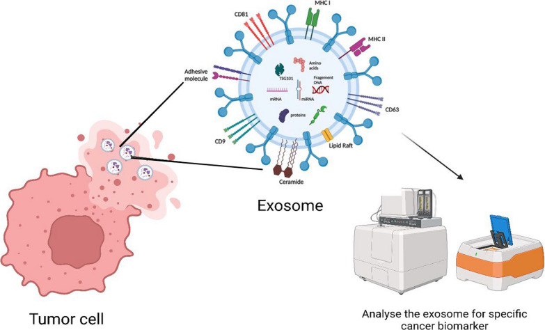
Exosomes are extracellular vesicles well known for facilitating cell-to-cell communication by distributing essential macromolecules like proteins, DNA, mRNA, lipids, and miRNA. These vesicles are abundant in fluids distributed throughout the body, including urine, blood, saliva, and even bile. They are important diagnostic tools for breast, lung, gastrointestinal cancers, etc. However, their application as cancer biomarkers has not yet been implemented in most parts of the world. In this review, we discuss how OMICs profiling of exosomes can be practiced by substituting traditional imaging or biopsy methods for cancer detection. Previous methods like extensive imaging and biopsy used for screening were expensive, mostly invasive, and could not easily provide early detection for various types of cancer. Exosomal biomarkers can be utilized for routine screening by simply collecting body fluids from the individual. We anticipate that the use of exosomes will be brought to light by the success of clinical trials investigating their potential to enhance cancer detection and treatment in the upcoming years.

Keywords: Cancer biomarkers; Cancer diagnosis; Clinical signature; Diagnostic tool; Exosomes; Molecular profiling; Prognostic indicator; Tumor-derived exosomes.


Conflict of interest statement

The authors declare that the research was conducted without any commercial or financial relationships that could be construed as a potential conflict of interest.

Figures

Fig.1
Fig.1
Biogenesis, Secretion, and Uptake of Exosomes. Exosomes form through the process of inward budding during endocytosis. Specific cargos are sorted into these exosomes within multivesicular bodies (MVBs), where early and late sorting endosomes are assembled. Commonly, Exosomes consist of proteins, lipids, RNAs, and genetic material. The protein content of EVs includes various types, such as transmembrane or lipid-bound proteins found on the cell surface (CD63, CD9, CD81, etc.). Additionally, Exosomes contain lipids like ceramide, different types of RNAs such as messenger RNA (mRNA) and microRNA (miRNA), and DNA fragments. Exosomes are taken up by cells using several mechanisms, including direct fusion of exosomes with the cell membrane of the recipients, receptor-ligand interactions, and endocytosis. EVs transport their contents within the cells comprising proteins, RNAs, and DNAs, releasing them into the cytoplasm or endoplasmic reticulum. MVB: multivesicular body; CD: cluster of differentiation, Created with BioRender.com
Fig. 2
Fig. 2
Exosomes and Cancer. The role played by exosomes in cancer metastasis and progression. Tumor cells release pathogenic exosomes that inhibit immune cell functions (recognition of cancer cells, cytolytic effects, etc.) and pre-mesenchymal niche formation, Created with BioRender.com
Fig. 3
Fig. 3
Exosome as Cancer Biomarkers. Each tumor cell releases specific exosomes that contain microRNA, DNA, proteins, and metabolites that can be used as biomarkers of specific cancer, Created with BioRender.com
Fig. 4
Fig. 4
Different Isolation Strategies for Exosomes. Traditional methods for isolating exosomes include size exclusion chromatography (SEC) and differential ultracentrifugation (DUC). SEC involves using biofluids as a mobile phase against a porous stationary phase to elute molecules based on their size, with larger particles eluting first, followed by smaller exosomes, resulting in a longer elution time due to increased path length. In addition to these conventional methods, more innovative techniques are available for exosome isolation. In addition to these conventional methods, more innovative techniques are available for exosome isolation. One such technique is PEG-based precipitation, which facilitates the aggregation of exosomes in large numbers using a polymer solution. Another approach is immunoaffinity (IA) capture, where antibodies targeted against exosomal surface proteins are used to isolate specific exosome populations. Microfluidics (MF) technology, utilizing chips with specific antibody-mediated binding, enables efficient capture of exosomes, Created with BioRender.com
Fig. 5
Fig. 5
Representation of single exosome profiling methods. Created with BioRender.com
Fig. 6
Fig. 6
OMICS Profiling of Exosomes. This profiling 3 ways: Transcriptomics and genomics using the messenger RNA (mRNA), microRNA (miRNA), and DNA present in the cancer-specific exosome; Proteomics using the proteins, histones, transporters present in the exosome; Metabolomics using the metabolites such as glucose, pyruvate, nucleotides, amino acids (AAs) specific for cancer. Created with BioRender.com
Fig. 7
Fig. 7
Application of Single-Exosome Profiling in Cancer. Step 1: A sample containing exosomes (small vesicles) is collected and processed. Step 2: Exosomes are isolated using ultracentrifugation or microfluidics, then captured and trapped using nanoscale traps or microfluidic chambers. Step 3: The surface proteins and biomarkers on the trapped exosome are identified and analyzed using mass spectrometry or fluorescence microscopy techniques. Step 4: The cargo contents (e.g., RNA, DNA, proteins) of the exosome are extracted and analyzed using techniques like qRT-PCR. Step 5: The data from the analysis is integrated to create a comprehensive profile of the individual exosome, including its size, shape, surface markers, and cargo contents. Created with Biorender.com

References

    1. Brennan K, et al. A comparison of methods for the isolation and separation of extracellular vesicles from protein and lipid particles in human serum. Sci Rep. 2020;10(1):1039. 10.1038/s41598-020-57497-7. 10.1038/s41598-020-57497-7 - DOI - PMC - PubMed
    1. Colombo M, Raposo G, Théry C. Biogenesis, secretion, and intercellular interactions of exosomes and other extracellular vesicles. Annu Rev Cell Dev Biol. 2014;30(1):255–89. 10.1146/annurev-cellbio-101512-122326. 10.1146/annurev-cellbio-101512-122326 - DOI - PubMed
    1. Gruenberg J, Stenmark H. The biogenesis of multivesicular endosomes. Nat Rev Mol Cell Biol. 2004;5(4):317–23. 10.1038/nrm1360. 10.1038/nrm1360 - DOI - PubMed
    1. Piper RC, Katzmann DJ. Biogenesis and function of multivesicular bodies. Annu Rev Cell Dev Biol. 2007;23(1):519–47. 10.1146/annurev.cellbio.23.090506.123319. 10.1146/annurev.cellbio.23.090506.123319 - DOI - PMC - PubMed
    1. Weidle UH, Birzele F, Kollmorgen G, Rüger R. The multiple roles of exosomes in metastasis. Cancer Genom Proteom. 2017;14(1):1–16. 10.21873/cgp.20015.10.21873/cgp.20015 - DOI - PMC - PubMed

LinkOut - more resources