. 2024 Dec;20(12):2752-2768.
doi: 10.1080/15548627.2024.2389607. Epub 2024 Aug 15.

Pharmacological inhibition of USP14 delays proteostasis-associated aging in a proteasome-dependent but foxo-independent manner

Affiliations

Pharmacological inhibition of USP14 delays proteostasis-associated aging in a proteasome-dependent but foxo-independent manner

Jin Ju Lim et al. Autophagy. 2024 Dec.

Abstract

Aging is often accompanied by a decline in proteostasis, manifested as an increased propensity for misfolded protein aggregates, which are prevented by protein quality control systems, such as the ubiquitin-proteasome system (UPS) and macroautophagy/autophagy. Although the role of the UPS and autophagy in slowing age-induced proteostasis decline has been elucidated, limited information is available on how these pathways can be activated in a collaborative manner to delay proteostasis-associated aging. Here, we show that activation of the UPS via the pharmacological inhibition of USP14 (ubiquitin specific peptidase 14) using IU1 improves proteostasis and autophagy decline caused by aging or proteostatic stress in Drosophila and human cells. Treatment with IU1 not only alleviated the aggregation of polyubiquitinated proteins in aging Drosophila flight muscles but also extended the fly lifespan with enhanced locomotive activity via simultaneous activation of the UPS and autophagy. Interestingly, the effect of this drug disappeared when proteasomal activity was inhibited, but was evident upon proteostasis disruption by foxo mutation. Overall, our findings shed light on potential strategies to efficiently ameliorate age-associated pathologies associated with perturbed proteostasis.Abbreviations: AAAs: amino acid analogs; foxo: forkhead box, sub-group O; IFMs: indirect flight muscles; UPS: ubiquitin-proteasome system; USP14: ubiquitin specific peptidase 14.

Keywords: Autophagy; IU1; foxo; proteostasis; ubiquitin-proteasome system; ubiquitin-specific peptidase 14.


Conflict of interest statement

No potential conflict of interest was reported by the author(s).

Figures

Figure 1.
Figure 1.
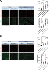
IU1 alleviates age-induced accumulation of poly-ubiquitin protein aggregates and the decline in autophagy in fly muscles. Immunostaining of the IFMs was performed in wild-type flies of three different ages (20-day, 30-day, 34-day) with or without IU1 treatment. (A,B) increased ref(2)P (green in A) with poly-ubiquitinated protein aggregates (red in A and B) is observed as flies age. IU1 decreased the number of ref(2)P and poly-ubiquitinated protein aggregates, especially in old flies. The colocalization of ref(2)P and poly-ubiquitinated protein aggregates is indicated by white arrows in (A). IU1 increased the number of immune signals of LC3 (green in B; representative LC3 puncta are indicated by yellow arrows) in flies. The results of immune signal quantification are shown in A and B. Data are presented as mean ± SEM. n > 10 male flies under each condition. The p values compared to the respective controls are denoted in the graphs. Scale bar: 20 µm (magnification, 400 ×).
Figure 2.
Figure 2.
IU1 alleviates aaa-induced accumulation of poly-ubiquitin protein aggregates and the decline in autophagy in fly muscles. Immunostaining was performed on the IFMs of wildtype flies administered three treatments (control, AAA, AAA+IU1). (A, B) AAA increased the number of ref(2)P (green in A), decreased LC3 (green in B; representative LC3 puncta are indicated by yellow arrows), and increased poly-ubiquitinated protein aggregates (red in A and B). Co-administration of IU1 and AAA decreased the number of ref(2)P and poly-ubiquitinated protein aggregates (A) and increased the number of immune signals of LC3 (B) in flies. The results of immune signal quantification are shown in A and B. Data are presented as mean ± SEM. n > 8 male flies under each condition. The p values compared to the respective controls are denoted in the graphs. Scale bar: 20 µm (magnification, 400 ×).
Figure 3.
Figure 3.
IU1 rescues the aaa-induced decline in UPS activity in fly muscles. (A) UPS activity gradually declined as flies aged as revealed by an increase in the CL1-GFP reporter (green) that is specifically degraded by UPS activity. (B) AAA decreased UPS activity, which was rescued by treatment with IU1. The results of immune signal quantification are shown in a and b. Data are presented as mean ± SEM. n > 15 male flies under each condition. The p values compared to the respective controls are denoted in the graphs. Scale bar: 20 µm (magnification, 400 ×).
Figure 4.
Figure 4.
ProteoDN expression in IFMs aggravates the accumulation of poly-ubiquitin protein aggregates and decline in autophagy during aging. Immunostaining was performed on the IFMs in flies of two ages (10-day and 20-day) with or without ProteoDN expression in IFMs. (A, B) both ref(2)P (green in A) and poly-ubiquitinated protein aggregates (red in A and B) were increased by the expression of ProteoDN as flies aged. LC3 (green in B; representative LC3 puncta are indicated by yellow arrows) was decreased by the expression of ProteoDN as flies aged. The flies were tested during the indicated days at the restricted temperature (29°C). The results of immune signal quantification are shown in a and b. Data are presented as mean ± SEM. n > 18 male flies under each condition. The p values compared to the respective controls are denoted in the graphs. Scale bar: 20 µm (magnification, 400 ×).
Figure 5.
Figure 5.
ProteoDN expression in IFMs abolishes the improvement effect of IU1 on proteostasis and autophagy during aging. Immunostaining was performed on the IFMs of flies of two ages (10-day and 20-day) under various conditions, including the presence or absence of ProteoDN expression and IU1 treatment. (A, B) treatment with IU1 failed to decrease both ref(2)P (green in A) and poly-ubiquitinated protein aggregates (red in A and B) upon expression of ProteoDN as flies aged. Treatment with IU1 failed to increase LC3 (green in B; representative LC3 puncta are indicated by yellow arrows) upon expression of ProteoDN as flies aged. The flies were tested during the indicated days at the restricted temperature (29°C). The results of immune signal quantification are shown in A and B. Data are presented as mean ± SEM. n > 13 male flies under each condition. The p values compared to the respective controls are denoted in the graphs. Scale bar: 20 µm (magnification, 400 ×).
Figure 6.
Figure 6.
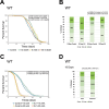
IU1 extends fly lifespan with increased locomotive activity. (A) IU1 was administered to flies in three different life phases [adult eclosion until death (0 d – death); adult eclosion until middle age (0 d − 30 d); and middle age until death (30 d – death)]. Treatment with IU1 during 0 d – death or 0 d − 30 d significantly prolonged the lifespan of flies as depicted by the survival curve. n = 190 (control), n = 282 (0 d – death), n = 282 (0–30 d), n = 190 (30 d – death). (B) IU1 increased the locomotive activity decreased by aging as revealed by the ability to climb from the bottom of the vial. (C) IU1 significantly increased fly lifespan which was decreased by AAA. AAA rapidly and dose-dependently shortened fly lifespan, while co-treatment of IU1 alleviated the aaa-induced shortening of the lifespan. 1×AAA (5 mM canavanine +0.5 mM L-azetidine-2-carboxylic acid) or 2×AAA (10 mM canavanine +1 mM L-azetidine-2-carboxylic acid) was used. n = 583 (control +1×AAA), n = 565 (IU1 + 1×AAA), n = 439 (control +2×AAA), n = 407 (IU1 + 2×AAA). (D) Decreased locomotive activity induced by AAA was improved by subsequent treatment with IU1 as revealed by the ability of flies to climb from the bottom of the vial. For A and C, the p values for the lifespan curves were calculated using the log-rank test and all lifespan experiments were repeated with similar results; representative experiments are shown. For B and D, the p value for the locomotive bar graph compared to the respective control was calculated using Student’s t-test.
Figure 7.
Figure 7.
Foxo mutation does not abolish the ability of IU1 to delay proteostasis-associated aging. (A, B) immunostaining of the IFMs was performed for foxo mutant flies of two different ages (10-day and 20-day) with or without IU1 treatment. IU1 significantly decreased both ref(2)P (green in A) and poly-ubiquitinated protein aggregates (red in A and B) when foxo was mutated as flies aged. The co-localization of ref(2)P and poly-ubiquitinated protein aggregates is indicated by white arrows in A. IU1 significantly increased LC3 (green in B; representative LC3 puncta are indicated by yellow arrows) when foxo was mutated as flies aged. The results of immune signal quantification are shown in A and B. n > 16 male flies under each condition. The p values compared to the respective controls are denoted in the graphs. Scale bar: 20 µm (magnification, 400 ×). (C) IU1 significantly prolonged the lifespan of the foxo mutant flies as revealed by the survival curve. n = 534 (control), n = 529 (IU1 0.1 mM). The p values for the lifespan curves were calculated using the log-rank test. All lifespan experiments were repeated, yielding similar results; representative experiments are presented. (D) IU1 increased the locomotive activity of foxo mutant flies decreased by aging based on the ability of flies to climb from the bottom of the vial. The p value for the locomotive bar graph compared to the respective control was calculated using Student’s t-test.
Figure 8.
Figure 8.
Knockdown of atg gene abolishes the ability of IU1 to delay proteostasis-associated aging. (A, B) immunostaining was performed on the IFMs of flies of three ages (10-day, 20-day and 30-day) under various conditions, including the presence or absence of Atg2 knockdown in IFMs and IU1 treatment. Treatment with IU1 failed to decrease both ref(2)P (green in A) and poly-ubiquitinated protein aggregates (red in A and B) upon Atg2 knockdown as flies aged. Treatment with IU1 failed to increase LC3 (green in B; representative LC3 puncta are indicated by yellow arrows) upon Atg2 knockdown as flies aged. The results of immune signal quantification are shown in A and B. Data are presented as mean ± SEM. n > 10 male flies under each condition. The p values compared to the respective controls are denoted in the graphs. Scale bar: 20 µm (magnification, 400 ×). (C) IU1 failed to increase the locomotive activity of Atg2 knockdown flies decreased by aging based on the ability of flies to climb from the bottom of the vial. The p value for the locomotive bar graph compared to the respective control was calculated using Student’s t-test.
Figure 9.
Figure 9.
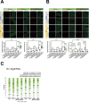
Usp14 mutation abolishes the ability of IU1 to delay proteostasis-associated aging. (A, B) immunostaining of the IFMs was performed for Usp14 mutant flies of three different ages (10-day, 20-day and 30-day) with or without IU1 treatment. IU1 failed to significantly decrease both ref(2)P (green in A) and poly-ubiquitinated protein aggregates (red in A and B) when Usp14 was mutated as flies aged. The co-localization of ref(2)P and poly-ubiquitinated protein aggregates is indicated by white arrows in A. IU1 failed to significantly increase LC3 (green in B; representative LC3 puncta are indicated by yellow arrows) when Usp14 was mutated as flies aged. The results of immune signal quantification are shown in A and B. n > 12 male flies under each condition. The p values compared to the respective controls are denoted in the graphs. Scale bar: 20 µm (magnification, 400 ×). (C) IU1 failed to significantly prolong the lifespan of the Usp14 mutant flies in the presence or absence of AAA as revealed by the survival curve. 1×AAA (5 mM canavanine +0.5 mM L-azetidine-2-carboxylic acid). n = 150 (control), n = 149 (IU1 0.1 mM), n = 150 (control +1×AAA), n = 150 (IU1 + 1×AAA). The p values for the lifespan curves were calculated using the log-rank test. All lifespan experiments were repeated, yielding similar results; representative experiments are presented. (D) IU1 failed to increase the locomotive activity of Usp14 mutant flies decreased by aging based on the ability of flies to climb from the bottom of the vial. The p value for the locomotive bar graph compared to the respective control was calculated using Student’s t-test.
Figure 10.
Figure 10.
IU1 treatment in human cells rescues the decreased activity of proteostasis and autophagy induced by AAA in a proteasome-dependent manner. (A) Human bj-hTERT cells were exposed to AAA in the absence or presence of IU1, and then analyzed using immunoblotting with anti-ub. (B) Scheme of compound treatment is shown. IMR90-hTERT cells were incubated with IU1, AAA, or baf at the indicated time points and the lysates were analyzed to determine the autophagic flux by LC3B immunoblotting. IU1 clearly rescues the autophagic activity impaired by AAA. As shown in graph, the LC3B-II levels were quantified based on three independent results, including the representative immunoblotting presented in (B). Data are expressed as mean ± SD. The p value was calculated using Student’s t-test. *p < 0.1 and **p < 0.05. (C) Upper panel indicates the treatment scheme of compounds. IMR90-hTERT cells were incubated with IU1, and then treated with the proteasome inhibitor, MG-132, prior to treatment with baf at the indicated time intervals. Immunoblotting of LC3B was performed using the lysates. The data indicate that proteasome inhibition hinders the autophagic activation induced by IU1. AAA (2 mM canavanine plus 0.2 mM L-azetidine-2-carboxylic acid), 50 μM IU1, 100 nM baf or 20 μM MG-132 was used throughout the experiments. All panels show SDS-PAGE/immunoblotting and quantification data. % LC3B-II protein was normalized based on the GAPDH level.

References

    1. Augustin H, Partridge L.. Invertebrate models of age-related muscle degeneration. BBA-Gen Subj. 2009;1790(10):1084–1094. doi: 10.1016/j.bbagen.2009.06.011 - DOI - PubMed
    1. Nair KS. Aging muscle. Am J Clin Nutr. 2005;81(5):953–963. doi: 10.1093/ajcn/81.5.953 - DOI - PubMed
    1. Zheng A, Edelman SW, Tharmarajah G, et al. Differential patterns of apoptosis in response to aging in Drosophila. Proc Natl Acad Sci USA. 2005;102(34):12083–12088. doi: 10.1073/pnas.0503374102 - DOI - PMC - PubMed
    1. Herndon LA, Schmeissner PJ, Dudaronek JM, et al. Stochastic and genetic factors influence tissue-specific decline in ageing C-elegans. Nature. 2002;419(6909):808–814. doi: 10.1038/nature01135 - DOI - PubMed
    1. Boyle PA, Buchman AS, Wilson RS, et al. Association of muscle strength with the risk of Alzheimer disease and the rate of cognitive decline in community-dwelling older persons. Arch Neurol. 2009;66(11):1339–1344. doi: 10.1001/archneurol.2009.240 - DOI - PMC - PubMed

Publication types

MeSH terms

LinkOut - more resources