. 2024 Aug;33(8):e15150.
doi: 10.1111/exd.15150.

Pro-inflammatory activity of Cutibacterium acnes phylotype IA1 and extracellular vesicles: An in vitro study

Affiliations

Pro-inflammatory activity of Cutibacterium acnes phylotype IA1 and extracellular vesicles: An in vitro study

Caroline T Cheung et al. Exp Dermatol. 2024 Aug.

Abstract

Acne is a chronic inflammatory skin condition that involves Cutibacterium acnes (C. acnes), which is classified into six main phylotypes (IA1, IA2, IB, IC, II and III). Acne development is associated with loss of C. acnes phylotype diversity, characterised by overgrowth of phylotype IA1 relative to other phylotypes. It was also shown that purified extracellular vesicles (EVs) secreted by C. acnes can induce an acne-like inflammatory response in skin models. We aimed to determine if the inflammatory profile of EVs secreted by C. acnes phylotype IA1 from an inflammatory acne lesion was different from C. acnes phylotype IA1 from normal skin, thus playing a direct role in the severity of inflammation. EVs were produced in vitro after culture of two clinical strains of C. acnes phylotype IA1, T5 from normal human skin and A47 from an inflammatory acne lesion, and then incubated with either human immortalised keratinocytes, HaCaT cells, or skin explants obtained from abdominoplasty. Subsequently, quantitative PCR (qPCR) was performed for human β-defensin 2 (hBD2), cathelicidin (LL-37), interleukin (IL)-1β, IL-6, IL-8, IL-17α and IL-36γ, and ELISA for IL-6, IL-8 and IL-17α. We found that EVs produced in vitro by C. acnes derived from inflammatory acne lesions significantly increased the pro-inflammatory cytokines and anti-microbial peptides at both transcriptional and protein levels compared with EVs derived from normal human skin. We show for the first time that C. acnes EVs from inflammatory acne play a crucial role in acne-associated inflammation in vitro and that C. acnes phylotype IA1 collected from inflammatory acne lesion and normal skin produce different EVs and inflammatory profiles in vitro.

Keywords: Cutibacterium acnes; acne; extracellular vesicles; inflammation; skin.


Conflict of interest statement

The authors declare that the research was conducted with funding from industrial sources.

Figures

FIGURE 1
FIGURE 1
Growth and morphological differences between the two different clinical isolates of Cutibacterium acnes (C. acnes): T5 (phylotype IA1, normal skin) and A47 (phylotype IA1, lesional acne). (A) Growth analysis of C. acnes strains T5 (phylotype IA1, normal skin) and A47 (phylotype IA1, acne lesion). Bacterial cultures of T5 and A47 were inoculated into fresh medium at an optical density (OD) of 0.1, and then OD measurements of the bacterial cultures were taken at 10 days post‐inoculation. (B) EVs derived from C. acnes T5 and A47 were subjected to scanning transmission electron microscopy (STEM), and EV size was measured from at least five images from each sample. (C) Images of EVs derived from C. acnes T5 and A47 by SEM with STEM detector. Experiments were performed at least three times in triplicate. Significance was considered when p < 0.05 as determined by Student's t‐test. ***p < 0.001, ****p < 0.0001.
FIGURE 2
FIGURE 2
Transcriptional analysis by qPCR for hBD2, IL‐1β, IL‐6, IL‐8 and IL‐36ɣ in extracellular vesicles (EVs)‐treated immortalised human keratinocytes (HaCaT cells). qPCR for innate immune markers (A) hBD2, (B) IL‐1β, (C) IL‐6, (D) IL‐8 and (E) IL‐36ɣ in HaCaT cells treated for 24 h with EVs isolated from Cutibacterium acnes strains T5 (phylotype IA1, normal skin) and A47 (phylotype IA1, acne skin). Vehicle control was the incubation of cells with the same volume of phosphate‐buffered saline (PBS). Experiments were performed at least three times in triplicate. Significance was considered when p < 0.05 as determined by multiple comparisons using ANOVA analysis. *p < 0.05, **p < 0.01, ***p < 0.001, ****p < 0.0001.
FIGURE 3
FIGURE 3
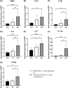
Transcriptional analysis by qPCR for hBD2, LL‐37, IL‐1β, IL‐6, IL‐8, IL‐17α and IL‐36ɣ in extracellular vesicles (EVs)‐treated human skin explants. qPCR for innate immune markers (A) hBD2, (B) LL‐37, (C) IL‐1β, (D) IL‐6, (E) IL‐8, (F) IL‐17α and (G) IL‐36ɣ in ex vivo skin explants treated for 24 h with EVs derived from Cutibacterium acnes (C. acnes) strains T5 (phylotype IA1, normal skin) and A47 (phylotype IA1, acne skin). Experiments were performed at least three times in triplicate. Significance was considered when p < 0.05 as determined by multiple comparisons using ANOVA analysis. *p < 0.05, **p < 0.01, ****p < 0.0001.
FIGURE 4
FIGURE 4
Protein analysis by ELISA for innate immune markers IL‐6, IL‐8 and IL‐17α in extracellular vesicles (EVs)‐treated immortalized human keratinocytes (HaCaT cells) and human skin explants. ELISA for innate immune markers (A) IL‐6 and (B) IL‐8 in HaCaT cells, and (C) IL‐6, (D) IL‐8, and (E) IL‐17α in human skin explants treated for 24 h with EVs derived from Cutibacterium acnes (C. acnes) strains T5 (phylotype IA1, normal skin) and A47 (phylotype IA1, acne skin). Experiments were performed at least three times in triplicate. Significance was considered when p < 0.05 as determined by multiple comparisons using ANOVA analysis. *p < 0.05, **p < 0.01, ***p < 0.001, ****p < 0.0001.

References

    1. Dréno B, Dagnelie MA, Khammari A, Corvec S. The skin microbiome: a new actor in inflammatory acne. Am J Clin Dermatol. 2020;21(1):18‐24. doi:10.1007/S40257-020-00531-1/FIGURES/2 - DOI - PMC - PubMed
    1. Dréno B, Bettoli V, Araviiskaia E, Sanchez Viera M, Bouloc A. The influence of exposome on acne. J Eur Acad Dermatol Venereol. 2018;32(5):812‐819. doi:10.1111/jdv.14820 - DOI - PMC - PubMed
    1. Barnard E, Nagy I, Hunyadkürti J, Patrick S, McDowell A. Multiplex touchdown PCR for rapid typing of the opportunistic pathogen Propionibacterium acnes. J Clin Microbiol. 2015;53(4):1149‐1155. doi:10.1128/JCM.02460-14 - DOI - PMC - PubMed
    1. Corvec S, Dagnelie MA, Khammari A, Dréno B. Taxonomy and phylogeny of Cutibacterium (formerly Propionibacterium) acnes in inflammatory skin diseases. Ann Dermatol Venereol. 2019;146(1):26‐30. doi:10.1016/J.ANNDER.2018.11.002 - DOI - PubMed
    1. Kilian M, Scholz CFP, Lomholt HB. Multilocus sequence typing and phylogenetic analysis of Propionibacterium acnes. J Clin Microbiol. 2012;50(4):1158‐1165. doi:10.1128/JCM.r06129-11 - DOI - PMC - PubMed

Supplementary concepts