. 2024 Jul 15;14(11):4331-4351.
doi: 10.7150/thno.98587. eCollection 2024.

Unlocking potential of oxytocin: improving intracranial lymphatic drainage for Alzheimer's disease treatment

Affiliations

Unlocking potential of oxytocin: improving intracranial lymphatic drainage for Alzheimer's disease treatment

Caihua Ye et al. Theranostics. .

Abstract

Background: The impediment to β-amyloid (Aβ) clearance caused by the invalid intracranial lymphatic drainage in Alzheimer's disease is pivotal to its pathogenesis, and finding reliable clinical available solutions to address this challenge remains elusive. Methods: The potential role and underlying mechanisms of intranasal oxytocin administration, an approved clinical intervention, in improving intracranial lymphatic drainage in middle-old-aged APP/PS1 mice were investigated by live mouse imaging, ASL/CEST-MRI scanning, in vivo two-photon imaging, immunofluorescence staining, ELISA, RT-qPCR, Western blotting, RNA-seq analysis, and cognitive behavioral tests. Results: Benefiting from multifaceted modulation of cerebral hemodynamics, aquaporin-4 polarization, meningeal lymphangiogenesis and transcriptional profiles, oxytocin administration normalized the structure and function of both the glymphatic and meningeal lymphatic systems severely impaired in middle-old-aged APP/PS1 mice. Consequently, this intervention facilitated the efficient drainage of Aβ from the brain parenchyma to the cerebrospinal fluid and then to the deep cervical lymph nodes for efficient clearance, as well as improvements in cognitive deficits. Conclusion: This work broadens the underlying neuroprotective mechanisms and clinical applications of oxytocin medication, showcasing its promising therapeutic prospects in central nervous system diseases with intracranial lymphatic dysfunction.

Keywords: Alzheimer's disease; Oxytocin; cerebral hemodynamics; intracranial lymphatic system; meningeal lymphangiogenesis; β-amyloid clearance.


Conflict of interest statement

Competing Interests: The authors have declared that no competing interest exists.

Figures

Scheme 1
Scheme 1
Schematic representation of intracranial lymphatic drainage in AD and after OT administration. (A) AD pathological status. In the glymphatic system, AQP4 depolarization results in reduced Aβ exchange between the ISF and CSF, while cerebral hemodynamic disorders exacerbate drainage inefficiency. In the meningeal lymphatic system, the damage to the MLVs causes occlusion of drainage to the dCLNs, leading to the inability to remove Aβ deposits from the brain. (B) Therapeutic effect of intranasal OT administration. Exogenous OT can enter the brain through the olfactory nerve bundle to simultaneously exert multiple regulatory functions, including enhancing cerebral hemodynamics, inhibiting AQP4 depolarization, and promoting lymphangiogenesis, ultimately restoring the structural integrity and Aβ clearance efficiency of intracranial lymphatic drainage system.
Figure 1
Figure 1
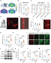
Dysfunctional intracranial lymphatic drainage in middle-old-aged but not young APP/PS1 mice. (A) In vivo brain ventral images of 3-month-old and 11-month-old APP/PS1 and WT mice at 2 h after i.c.m. injection of OVA-647. (B) Fluorescence quantification of OVA-647 in the cervical region of mice (n = 5). (C), (E) Representative fluorescence images of LYVE-1 staining in (C) the dCLNs and (E) the meninges of mice at 2 h after i.c.m. injection of OVA-647. Scale bars, (C) 200 μm; (E) 100 μm. (D), (F) Area fraction analysis of LYVE-1 and OVA-647 in (D) the dCLNs and (F) the meninges (n = 5). Data are presented as mean ± SD and analyzed by unpaired Student's t tests and one-way ANOVA followed by Bonferroni's post hoc test.
Figure 2
Figure 2
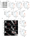
Improvement of cerebral hemodynamics and glymphatic drainage by OT administration in middle-old-aged APP/PS1 mice. (A) Coronal atlas of mouse brain anatomical template annotated with ROIs and representative ASL pseudo-color images of different groups. (B-C) CBF analysis in (B) whole brain and (C) specific brain regions (n = 5). (D) Visualization of penetrating arterioles 100 μm below the cortical surface by in vivo two-photon fluorescence angiography. Green lines, vertical line of the vessel axis drawn based on X-T line scanning. Scar bar, 20 μm. (E) Left, vessel diameter measurement. Scale bar, 10 μm. Right, vessel diameter quantification (n = 9). (F) Measurement of the distance (Δx) and time (Δt) of RBC movement along the intravascular scan line. (G) RBC velocity and (H) volume flux quantification (n = 9). (I) Time-lapsein in vivo two-photon imaging of CSF tracer influx into the cortex after i.c.m. injection in AD mice. Scale bar, 50 μm. (J-M) Representative immunoblots and quantitative analysis for (K) M1/M23-AQP4, (L) GFAP, and (M) MMP-9 (n = 4). Data are presented as mean ± SD and analyzed by one-way ANOVA followed by Bonferroni's post hoc test.
Figure 3
Figure 3
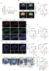
Improvement of meningeal lymphatic structure by OT administration in middle-old-aged APP/PS1 mice. (A-D) Representative immunoblots and quantitative analysis for (B) VEGF-C, (C) LYVE-1, and (D) Prox1 in the meninges (n = 4). (E-G) Quantitative analysis of the mRNA levels of (E) VEGF-C, (F) LYVE-1, and (G) Prox1 in the meninges by RT-qPCR (n = 3). (H) Representative confocal LYVE-1-stained images depicting MLV sprouts along the transverse sinuses (left) and MLV loops near lymphatic hotspots (right). Red arrow: the location of sprouts or loops. Scar bar, 1 mm (left) and 100 μm (right). (I-J) Quantification of the number of sprouts (I) and loops (J) in whole meningeal mounts (n = 5). Data are presented as mean ± SD and analyzed by one-way ANOVA followed by Bonferroni's post hoc test.
Figure 4
Figure 4
Improvement of meningeal lymphatic drainage by OT administration in middle-old-aged APP/PS1 mice. (A) Z-spectrum asymmetry curve of the whole mouse brain. Black arrows, 1 ppm location. (B) Representative T2-weighted (T2WI) and CEST images (MTRasym maps at 1.0 ppm) of different groups. (C) MTR% analysis at 1.0 ppm in the whole brains (n = 5). (D-G) Representative fluorescence images of LYVE-1 staining in (D) the meninges and (G) the dCLNs of mice at 30 min after i.c.m. injection of OVA-647. Scale bars, (D) 100 μm; (G) 200 μm. (E-F), (H, I) Area fraction analysis of LYVE-1 and OVA-647 in (E, F) the meninges and (H, I) the dCLNs (n = 5). (J) In vivo brain ventral images of AD mice with dCLN ligation at 30 min after i.c.m. injection of OVA-647. (K) Fluorescence quantification of OVA-647 in the cervical region of mice (n = 5). Data are presented as mean ± SD and analyzed by one-way ANOVA followed by Bonferroni's post hoc test.
Figure 5
Figure 5
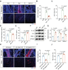
Enhancement of intracranial lymphatic clearance of Aβ by OT administration in middle-old-aged APP/PS1 mice. (A) Representative fluorescence images of Aβ staining in the meninges and brain sections. Scale bar, 100 μm. (B) Area fraction analysis of Aβ in the meninges (n = 5). (C) Area fraction analysis of Aβ in the cortex and hippocampus. (n = 3). (D-E) Levels of Aβ in the hippocampus and the CSF measured by ELISA (n = 3). (F) Representative immunoblots and quantitative analysis for (G) IL-1β, IL-6, and TNF-α (n = 4). (H) Representative fluorescence images of LYVE-1 and Aβ staining in the meninges and brain sections of mice with ligation. Scale bar, 100 μm. (I) Area fraction analysis of Aβ in the meninges (n = 4). (J) Area fraction analysis of Aβ in the cortex and hippocampus (n = 3). Data are presented as mean ± SD and analyzed by one-way ANOVA followed by Bonferroni's post hoc test.
Figure 6
Figure 6
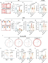
Cognitive impairment in AD mice ameliorated by OT administration. (A) Representative trajectories, (B) time spent in the center zone, and (C) total traveled distance in the OFT (n = 15). (D) Spontaneous alternation ratio in Y-maze test (n = 15). (E) Representative traces, (F) percentage of time spent with novel objects, (G) recognition index, and (H) discrimination index in the NOR test (n = 15). (I) Representative swimming paths in the hidden-platform probe phase of two-day WM test. (J) Escape latency for each trial in day 1 (visible trials) and day 2 (hidden trials), (K) escape latency analysis on day 1 trial 4 (D1T4) and day 2 trial 1 (D2T1), and (L) latency ratio by D2T1/D1T4 (n = 15). Data are presented as mean ± SD. *p < 0.05, **p < 0.01, ***p < 0.001, one-way ANOVA followed by Bonferroni's post hoc test.
Figure 7
Figure 7
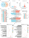
Dysregulated meningeal transcriptional profiles restored by OT administration. (A) Venn diagram showing intersection DEGs between the different comparison groups. (B-C) Volcano plots of significantly changed genes (two-fold change, p < 0.05). (D) Heatmap showing the top 30 DEGs in AD mice and altered after OT administration (n = 3). (E) The top five biological processes of significantly up- and down-regulated genes (p < 0.05). Red, GO terms for up-regulated genes; Blue, GO terms for down-regulated genes. (F) Expression of genes related to adaptive immune response among the top 30 DEGs (n = 3). (G) Scatter plots of the top 20 KEGG enrichment pathways in AD mice vs. OT-treated mice. (H) Scatter plots of the top 20 Reactome enrichment pathways in AD mice vs. OT-treated mice.

References

    1. Sun BL, Wang LH, Yang T, Sun JY, Mao LL, Yang MF. et al. Lymphatic drainage system of the brain: A novel target for intervention of neurological diseases. Prog Neurobiol. 2018;163-164:118–43. - PubMed
    1. Chen J, Wang L, Xu H, Wang Y, Liang Q. The lymphatic drainage system of the CNS plays a role in lymphatic drainage, immunity, and neuroinflammation in stroke. J Leukoc Biol. 2021;110:283–91. - PubMed
    1. Louveau A, Plog BA, Antila S, Alitalo K, Nedergaard M, Kipnis J. Understanding the functions and relationships of the glymphatic system and meningeal lymphatics. J Clin Invest. 2017;127:3210–9. - PMC - PubMed
    1. Bower NI, Hogan BM. Brain drains: new insights into brain clearance pathways from lymphatic biology. Journal of Molecular Medicine. 2018;96:383–90. - PubMed
    1. Xu JQ, Liu QQ, Huang SY, Duan CY, Lu HB, Cao Y. et al. The lymphatic system: a therapeutic target for central nervous system disorders. Neural Regen Res. 2023;18:1249–56. - PMC - PubMed

Publication types

MeSH terms