

Unenhanced computed tomography as a diagnostic tool in suspected pulmonary hypertension: a retrospective cross-sectional pilot study
- PMID: 39113847
- PMCID: PMC11303945
- DOI: 10.12688/wellcomeopenres.16853.2
Unenhanced computed tomography as a diagnostic tool in suspected pulmonary hypertension: a retrospective cross-sectional pilot study
Abstract
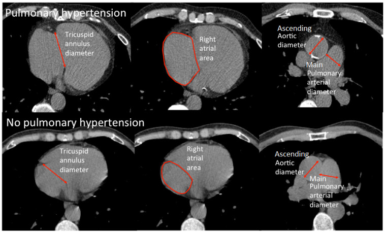
Background: Computed tomography pulmonary angiography (CTPA) has been proposed to be diagnostic for pulmonary hypertension (PH) in multiple studies. However, the utility of the unenhanced CT measurements diagnosing PH has not been fully assessed. This study aimed to assess the diagnostic utility and reproducibility of cardiac and great vessel parameters on unenhanced computed tomography (CT) in suspected pulmonary hypertension (PH).
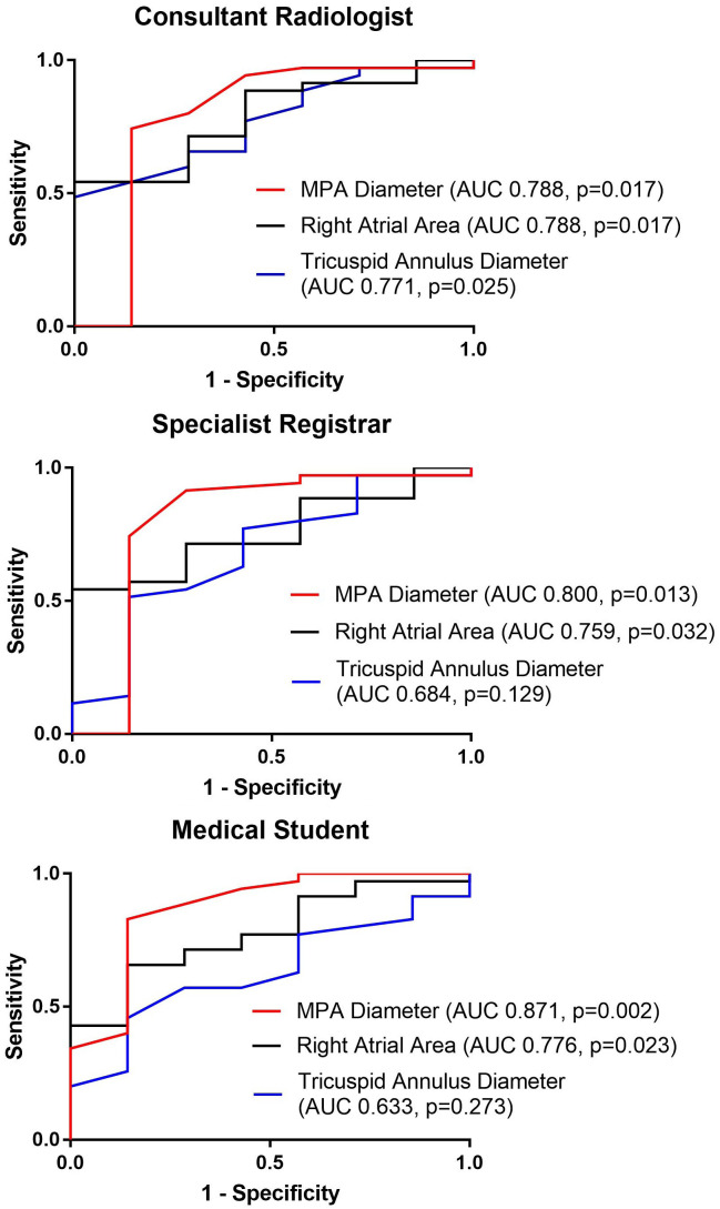
Methods: In total, 42 patients with suspected PH who underwent unenhanced CT thorax and right heart catheterization (RHC) were included in the study. Three observers (a consultant radiologist, a specialist registrar in radiology, and a medical student) measured the parameters by using unenhanced CT. Diagnostic accuracy of the parameters was assessed by area under the receiver operating characteristic curve (AUC). Inter-observer variability between the consultant radiologist (primary observer) and the two secondary observers was determined by intra-class correlation analysis (ICC).
Results: Overall, 35 patients were diagnosed with PH by RHC while 7 patients were not. Main pulmonary arterial (MPA) diameter was the strongest (AUC 0.79 to 0.87) and the most reproducible great vessel parameter. ICC comparing the MPA diameter measurement of the consultant radiologist to the specialist registrar's and the medical student's were 0.96 and 0.92, respectively. Right atrial area was the cardiac measurement with highest accuracy and reproducibility (AUC 0.76 to 0.79; ICC 0.980, 0.950) followed by tricuspid annulus diameter (AUC 0.76 to 0.79; ICC 0.790, 0.800).
Conclusions: MPA diameter and right atrial areas showed high reproducibility. Diagnostic accuracies of these were within the range of acceptable to excellent, and might have clinical value. Tricuspid annular diameter was less reliable and less diagnostic and was therefore not a recommended diagnostic measurement.
Keywords: Computed tomography; Diagnosis; Right Ventricle; Pulmonary hypertension.
Plain language summary
Pulmonary hypertension (PH) is a condition characterized by elevated pressure in the pulmonary artery and may lead to right heart failure. Several studies have demonstrated the diagnostic value of non-invasive techniques computed tomography (CT) with contrast in identifying PH. Therefore, we aim to investigate the diagnostic accuracy of non-contrast CT, which is commonly performed in patients with suspected lung diseases who are at risk of PH.
Copyright: © 2024 Goh ZM et al.
Conflict of interest statement
No competing interests were disclosed.
Figures
References
-
- Galiè N, Humbert M, Vachiery JL, et al. : 2015 ESC/ERS guidelines for the diagnosis and treatment of pulmonary hypertension: The Joint Task Force for the Diagnosis and Treatment of Pulmonary Hypertension of the European Society of Cardiology (ESC) and the European Respiratory Society (ERS): Endorsed by: Association for European Paediatric and Congenital Cardiology (AEPC), International Society for Heart and Lung Transplantation (ISHLT). Eur Respir J. European Respiratory Society;2015;46(4):903–75. 10.1183/13993003.01032-2015 - DOI - PubMed
Associated data
Grants and funding
LinkOut - more resources
Full Text Sources
