. 2024 Jul 15;14(7):3483-3496.
doi: 10.62347/PASE2970. eCollection 2024.

Functional analysis of circSNYJ1/miR-142-5p/CCND1 regulatory axis in non-small cell lung cancer

Affiliations

Functional analysis of circSNYJ1/miR-142-5p/CCND1 regulatory axis in non-small cell lung cancer

Jie Yan et al. Am J Cancer Res. .

Abstract

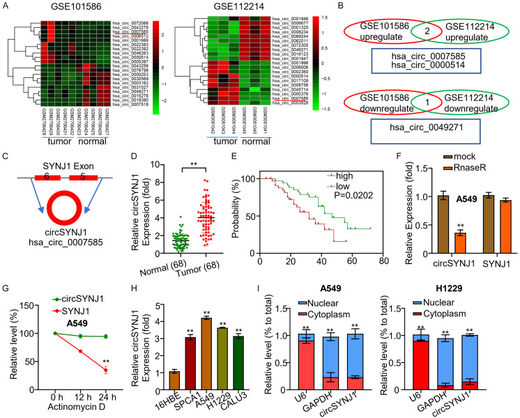
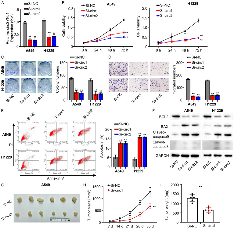
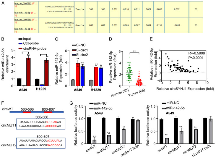
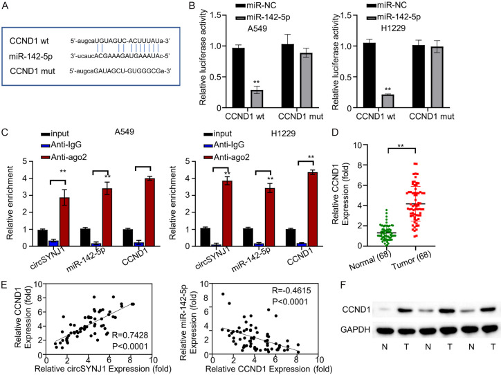
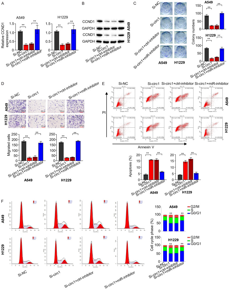
Non-small cell lung cancer (NSCLC), the most prevalent form of lung cancer, accounts for approximately 85% of all lung cancer diagnoses. Circular RNAs (circRNAs) are non-coding RNAs that play an active role in gene expression regulation, influencing cell growth, migration, and apoptosis. Here, we aimed to investigate the function of circSNYJ1 in NSCLC. In the present study, we found that circSNYJ1 expression level was increased in NSCLC tissues and cell lines. Knockdown of circSNYJ1 suppressed NSCLC cell proliferation, colony formation and migration while promoting apoptosis. Mechanistically, we demonstrated that circSNYJ1 sponged miR-142-5p, thereby regulating the expression of CCND1, a well-known cell cycle regulator. In conclusion, this study uncovered a novel circSNYJ1/miR-142-5p/CCND1 axis involved in NSCLC progression, providing potential diagnostic and prognostic biomarkers for treating NSCLC.

Keywords: CCND1; CircSNYJ1; NSCLC; biomarkers; miR-142-5p.


Conflict of interest statement

None.

Figures

Figure 1
Figure 1
CircSYNJ1 expression in NSCLC tissues and cells. A. Heatmaps illustrate the top upregulated and downregulated circRNAs in NSCLC and adjacent tissues from two databases (GSE112214 and GSE101586). B. A comparison of differentially expressed circRNAs between NSCLC and normal tissues. C. The diagram illustrates the circular architecture and origin of circSYNJ1. D. qRT-PCR analysis of circSYNJ1 expression in NSCLC and normal tissues from 68 patients. E. A Kaplan-Meier plot assessing the correlation between circSYNJ1 expression and the survival rates in NSCLC patients. F. qRT-PCR results showing RNase R digestion of SYNJ1 mRNA in A549 cells. G. The resistance of circSYNJ1 and SYNJ1 mRNA to RNase R treatment. H. Quantitative real-time PCR evaluation of circSYNJ1 expression in NSCLC cell lines and normal bronchial epithelial cells. I. Subcellular localization of circSYNJ1 in A549 and H1229 cells.
Figure 2
Figure 2
CircSNJY1 expression and impact on proliferation and metastasis in NSCLC. A. qRT-PCR analysis of circSNJY1 expression in H1229 and A549 cells transfected with si-circ1 and si-circ2. B. The CCK-8 assay results indicating that suppression of circSNJY1 expression reduced cell viability in H1229 and A549 cells. C. Colony formation assay results demonstrating circSNJY1 knockdown impaired colony formation ability in H1229 and A549 cells. D. The transwell assay indicating that circSNJY1 knockdown impaired cell migration in H1229 and A549 cells. E. Flow cytometry analysis examining cell apoptosis in H1229 and A549 cells post circSNJY1 knockdown. F. Western blot analysis of apoptosis-related cytokines in H1229 and A549 cells following circSNJY1 knockdown. G-I. Tumor formation assay illustrating the reduced tumorigenic potential of A549 cells with circSNJY1 knockdown.
Figure 3
Figure 3
The interaction between circSNJY1 and miR-142-5p in NSCLC. A. Circinteractome analysis predicting miR-142-5p binding sites within circSNJY1. B. RNA Pulldown assay demonstrating the direct interaction between circSNJY1 and miR-142-5p. C. qRT-PCR analysis showing that dramatically increased miR-142-5p expression following circSNJY1 knockdown in H1229 and A549 cells. D. qRT-PCR results indicating lower expression levels of miR-142-5p in NSCLC tissues compared to adjacent normal tissues. E. Pearson correlation analysis revealing a significantly negative correlation between circSNJY1 and miR-142-5p expression in NSCLC patients. F. The fluorescent reporter construct containing wild-type and mutated miR-142-5p binding sites in circSNJY1. G. Transfection of H1229 and A549 cells with the reporter construct carrying wild-type, mutated, or partially mutated miR-143-5p binding sites, along with miR-142-5p or a control miRNA (miR-NC).
Figure 4
Figure 4
CCND1 is the target gene of miR-142-5p. A. Prediction by Starbase identifying miR-142-5p as a regulator of the CCND1 gene. B. Effects of miR-142-5p on the expression of wild-type and mutant fluorescence reporter genes, which include miR-142-5p and CCND1 binding site sequences, in H1229 and A549 cells. C. RIP-QRT-PCR analysis revealed an enrichment of AGO2 protein on complex involving circSNYJ1, miR-142-5p, and CCND1 mRNA in H1229 and A549 cells. D. Comparative qRT-PCR analysis of CCND1 expression in malignant and adjacent normal tissues from lung cancer patients. E. Pearson correlation analysis examining the relationship between CCND1, circSNYJ1, and miR-142-5p expression across 68 lung cancer patients. F. WB study of EEF2 protein expression in in tumor and adjacent normal tissues from lung cancer patients.
Figure 5
Figure 5
CircSNYJ1-mediated modulation of the miR-142-5p/CCND1 axis. A, B. qRT-PCR and WB analyses assessing CCND1 expression in H1229 and A549 cells across different experimental groups. C. Clonogenic assay evaluating the clonogenic ability of H1229 and A549 cells in various groups. D. The Transwell assay (without matrix gel) examining the migratory capabilities of H1229 and A549 cells in different groups. E. Flow cytometry analysis determining the apoptotic rates of H1229 and A549 cells across the experimental groups. F. Cell cycle analysis assessing the cell cycle phases of H1229 and A549 cells from different groups.

Similar articles

References

    1. Molina JR, Yang P, Cassivi SD, Schild SE, Adjei AA. Non-small cell lung cancer: epidemiology, risk factors, treatment, and survivorship. Mayo Clin Proc. 2008;83:584–594. - PMC - PubMed
    1. Chen Z, Fillmore CM, Hammerman PS, Kim CF, Wong KK. Non-small-cell lung cancers: a heterogeneous set of diseases. Nat Rev Cancer. 2014;14:535–546. - PMC - PubMed
    1. Larsen JE, Minna JD. Molecular biology of lung cancer: clinical implications. Clin Chest Med. 2011;32:703–740. - PMC - PubMed
    1. Worley S. Lung cancer research is taking on new challenges: knowledge of tumors’ molecular diversity is opening new pathways to treatment. P T. 2014;39:698–714. - PMC - PubMed
    1. Wang M, Yu F, Wu W, Zhang Y, Chang W, Ponnusamy M, Wang K, Li P. Circular RNAs: a novel type of non-coding RNA and their potential implications in antiviral immunity. Int J Biol Sci. 2017;13:1497–1506. - PMC - PubMed

LinkOut - more resources