. 2024 Aug 1;21(10):1990-1999.
doi: 10.7150/ijms.98343. eCollection 2024.

Aspirin Inhibits Colorectal Cancer via the TIGIT-BCL2-BAX pathway in T Cells

Affiliations

Aspirin Inhibits Colorectal Cancer via the TIGIT-BCL2-BAX pathway in T Cells

Jiayu Liu et al. Int J Med Sci. .

Abstract

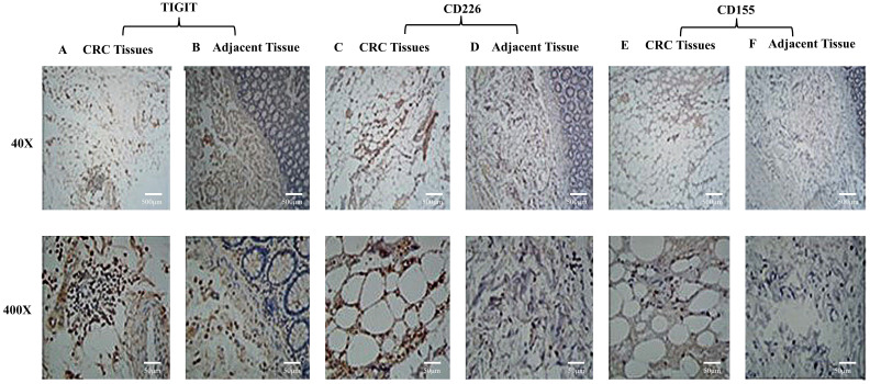
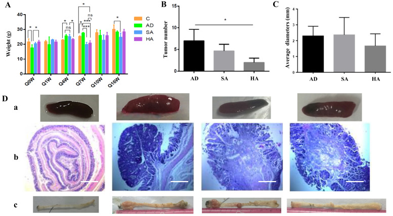
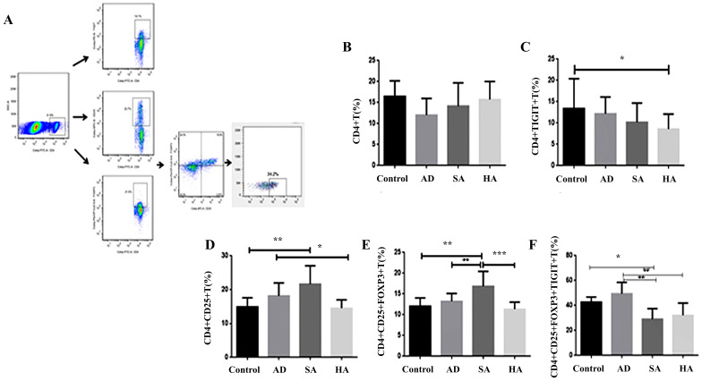
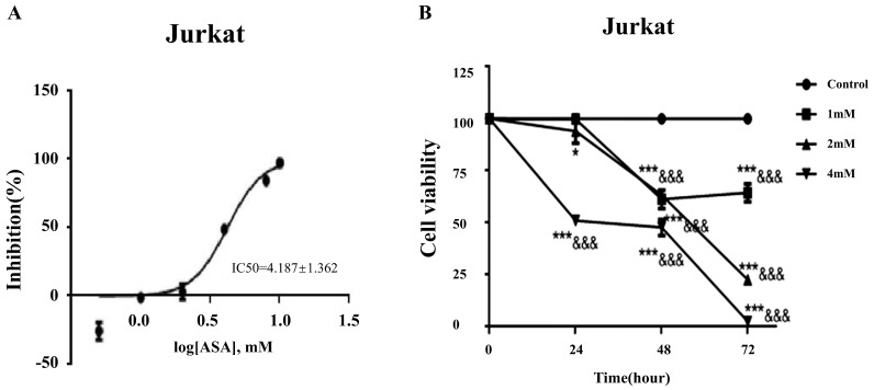
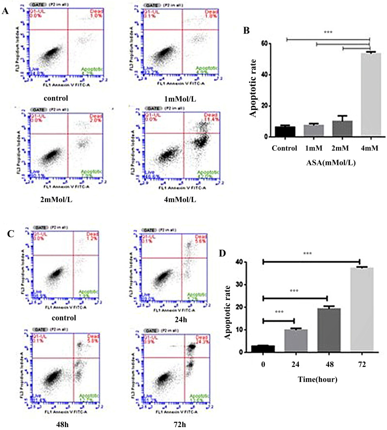
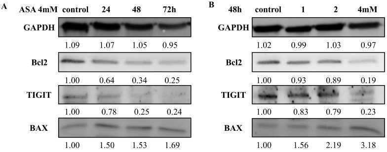
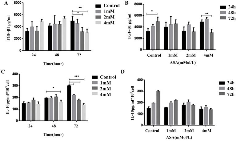
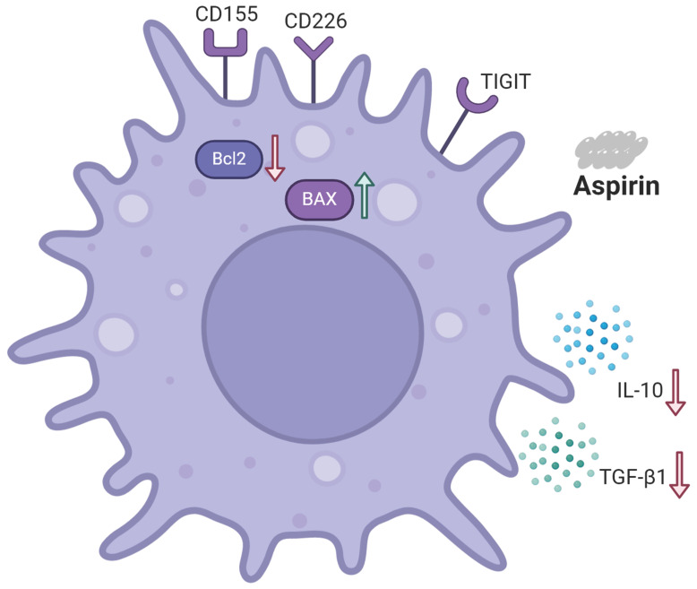
The T cell immunoglobulin and ITAM domain (TIGIT) is a recently discovered synergistic co-suppressor molecule that plays an important role in immune response and tumor immune escape in the context of cancer. Importantly, CD155 acts as a receptor for TIGIT, and CD155 signaling to immune cells is mediated through interactions with the co-stimulatory immune receptor CD226 (DNAM-1) and the inhibitory checkpoint receptors TIGIT and CD96. Aspirin (ASA) has been shown to reduce the growth and survival of colorectal cancer (CRC) cells, but the immunological mechanisms involved have not been sufficiently elucidated. In the present study the effects of aspirin on CRC in mice and on Jurkat cells were investigated. Aspirin may suppress the expression of TIGIT on T cells and Regulatory T cells (Tregs) and inhibit T cell viability, and therefore induce tumor cell apoptosis. TIGIT is expressed at higher levels on infiltrating lymphocytes within CRC tumor tissue than adjacent. Further, aspirin could inhibit Jurkat cell proliferation and induce apoptosis via downregulation of TIGIT expression and the anti-apoptosis B cell lymphoma 2 (BCL2) protein and upregulation of BCL2-associated X protein (BAX) expression. The present study suggests that aspirin can inhibit specific aspects of T cell function by reducing interleukin-10 and transforming growth factor-β1 secretion via the TIGIT-BCL2-BAX signaling pathway, resulting in improved effector T cell function that inhibits tumor progression.

Keywords: CD155; CD226; TIGIT.; aspirin; colorectal cancer.


Conflict of interest statement

Competing Interests: The authors have declared that no competing interest exists.

Figures

Figure 1
Figure 1
TIGIT, CD226, and CD155 expression by lymphocytes in CRC tissue and adjacent tissue. TIGIT and CD226 were expressed by lymphocytes in colorectal carcinomas. CD155 was not expressed by lymphocytes. (A) Colon adenocarcinoma tissue stained positively with polyclonal anti-TIGIT. (B) A section of adjacent tissue stained with an irrelevant polyclonal anti-TIGIT antibody (x40, x400). (C) Colon adenocarcinoma tissue stained positively with polyclonal anti-CD226 (x40, x400). (D) A section of adjacent tissue stained with an irrelevant polyclonal anti-CD226 antibody (x40, x400). (E) Colon adenocarcinoma tissue stained positively with polyclonal anti-CD155 (x40, x400). (F) A section of adjacent tissue stained with an irrelevant polyclonal anti-CD155 antibody (x40, x400). Scale bar, 500 μm and 50 μm.
Figure 2
Figure 2
Effects of aspirin on pathological changes in a murine CRC model. (A) Bodyweights in CRC mice treated with aspirin, measured every 3 weeks. (B, C) Tumor numbers and tumor diameters after treatment with aspirin. (Da) Splenic changes. (Db) Hematoxylin-eosin staining depicting colorectal changes after treatment with aspirin (original magnification x200). Scale bar, 1000 μm. (Dc) Tumor number and tumor position. The results represent means ± the standard deviation. Control--normal mice, AD--CRC mice without aspirin, SA--low-dose aspirin added to feed, HA-- high-dose aspirin added to feed. *p < 0.05, **p < 0.01 in comparison with the control.
Figure 3
Figure 3
Low TIGIT expression in splenic lymphocytes from CRC mice. (A) Representative flow cytometry plots of Tregs as determined by TIGIT and chemokine receptor expression gated on CD4+ T cells in healthy controls (n = 5). (B) CD4+ T cells. (C) TIGIT expression on CD4+ T cells. (D) CD4+CD25+ T cells. (E) CD4+CD25+FOXP3+ T cells. (F) TIGIT expression on CD4+CD25+FOXP3+ T cells.
Figure 4
Figure 4
Aspirin inhibited the viability of Jurkat cells. (A) The IC50 curve of aspirin intervention Jurkat. (B) Viability of Jurkat cells treated with 1, 2, or 4mM concentrations of aspirin for 24, 48, and 72 h. *p < 0.05, **p < 0.01, ***p < 0.001 compared to the control at that same timepoint. &p < 0.05, &&p < 0.001, &&&p < 0.0001 compared to the control at 0 h.
Figure 5
Figure 5
Aspirin induced Jurkat cell apoptosis. (A, B) Jurkat cells were stained with annexin-V-FITC and propidium iodide after exposure to 1, 2, and 4mM concentrations of aspirin for 48 h. (C, D) Jurkat cells were stained with annexin-V-FITC and propidium iodide after exposure to a 4 mM concentration of aspirin for 0, 24, 48, or 72 h. Data are indicative of three separate experiments. The statistical significance of the results was assessed using one-way analysis of variance followed by Fisher's least significant difference test. *p < 0.05, **p < 0.01, ***p < 0.001 compared to the control.
Figure 6
Figure 6
Western blotting of TIGIT, BAX, and BCL2 expression by Jurkat cells after exposure to aspirin. (A) Effects of exposure to 1, 2, and 4mM concentrations of aspirin for 48 h. (B) Effects of exposure to a 4 mM concentration of aspirin for 24, 48, and 72 h. An anti-GAPDH antibody was used as a loading control. *p < 0.05, **p < 0.01, ***p < 0.001 compared to the control.
Figure 7
Figure 7
Dynamic changes in cytokine levels in Jurkat cell culture supernatants. Enzyme-linked immunosorbent assays were used to determine (A, B) TGF-β1 and (C, D) IL-10 levels in the supernatants of Jurkat cell cultures after exposure to 1, 2, or 4mM concentrations of aspirin for 24, 48, and 72 h. All data are expressed as means ± the standard deviation of three separate experiments. *p < 0.05, **p < 0.01, ***p < 0.001 compared to the control.
Figure 8
Figure 8
Schematic representation of the effects of aspirin on Tregs and Jurkat cells. Aspirin induced Jurkat cell apoptosis by upregulating the expression of pro-apoptotic protein BAX and downregulating the expression of anti-apoptotic protein BCL2. Aspirin can induce apoptosis of Tregs through the TIGIT pathway and inhibit the secretion of TGF-β1 and IL-10 by Jurkat cells to improve effector T cell function and ultimately inhibit tumors.

Similar articles

Cited by

References

    1. Sung H, Ferlay J, Siegel RL. et al. Global cancer statistics 2020: globocan estimates of incidence and mortality worldwide for 36 cancers in 185 countries. Ca Cancer J Clin. 2021;71:209–49. - PubMed
    1. Morgan E, Arnold M, Gini A. et al. Global burden of colorectal cancer in 2020 and 2040: incidence and mortality estimates from globocan. Gut. 2023;72:338–44. - PubMed
    1. Drew DA, Chan AT. Aspirin in the prevention of colorectal neoplasia. Annu Rev Med. 2021;72:415–30. - PMC - PubMed
    1. Menter DG, Bresalier RS. An aspirin a day: new pharmacological developments and cancer chemoprevention. Annu Rev Pharmacol Toxicol. 2023;63:165–86. - PubMed
    1. Ren G, Ma Y, Wang X. et al. Aspirin blocks ampk/sirt3-mediated glycolysis to inhibit nsclc cell proliferation. Eur J Pharmacol. 2022;932:175208. - PubMed

MeSH terms