. 2024 Jul 16;21(10):1890-1902.
doi: 10.7150/ijms.95952. eCollection 2024.

ICOS-ICOSL pathway enhances NKT-like cell antiviral function in pregnant women with COVID-19

Affiliations

ICOS-ICOSL pathway enhances NKT-like cell antiviral function in pregnant women with COVID-19

Lu Zong et al. Int J Med Sci. .

Abstract

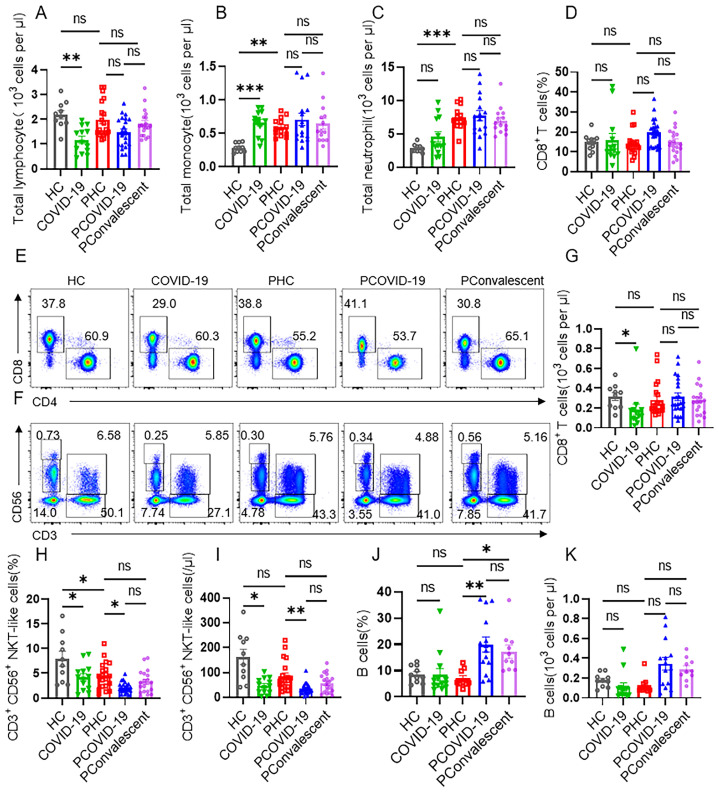
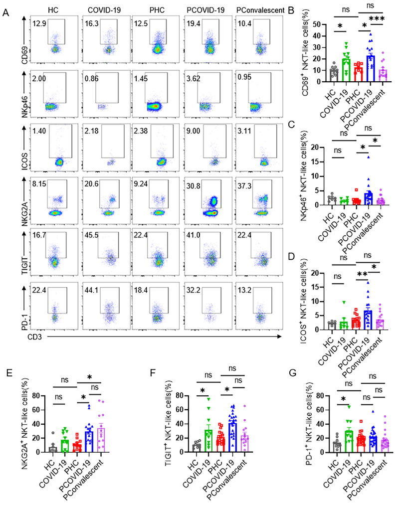
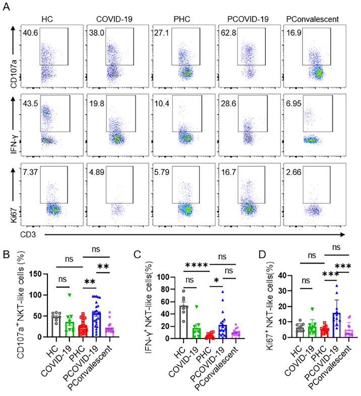
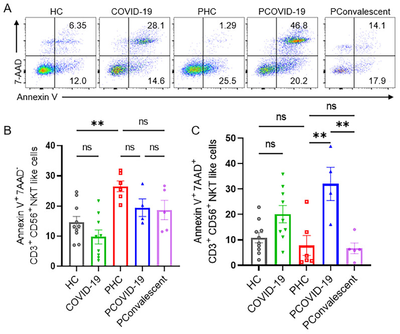
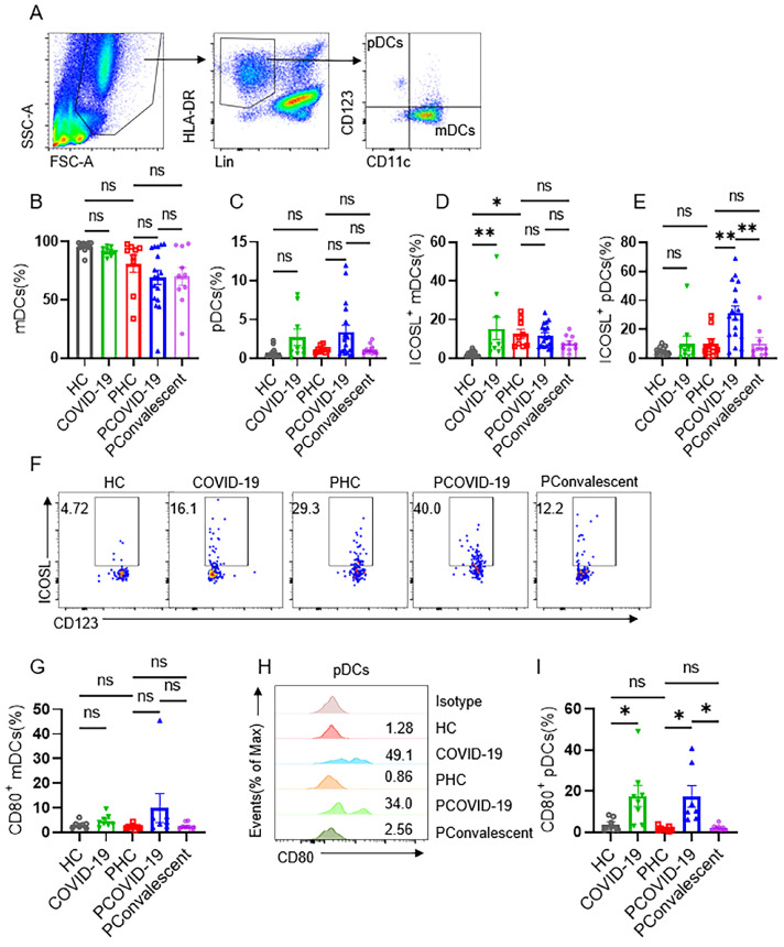
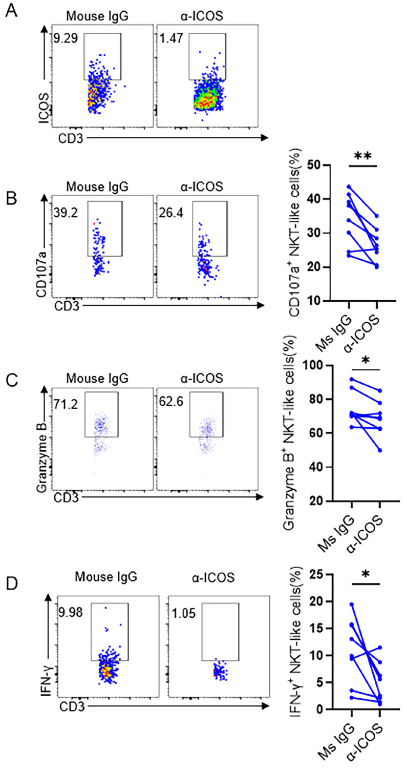
Objective: The immune response initiated by SARS-CoV-2 infection in pregnancy is poorly elucidated. We aimed to access and compare the antiviral cellular responses and lymphocytes activation between healthy pregnancies and pregnant women infected with SARS-CoV-2. Methods: We detected the immunological changes of lymphocytes in peripheral blood of healthy non-pregnant women, non-pregnant women with COVID-19, healthy pregnant women, pregnant women with COVID-19 and convalescent group by flow cytometry. In vitro blockade was used to identify NKT-like cell activation through ICOS-ICOSL pathway. Results: We found that CD3+CD56+ NKT-like cells decreased significantly in COVID-19 positive pregnant women compared to healthy pregnant women. NKT-like cells of pregnant women expressed higher level of activating receptors CD69 and NKp46 after SARS-CoV-2 infection. Particularly, they also increased the expression of the co-stimulatory molecule ICOS. NKT-like cells of pregnant women with COVID-19 up-regulated the expression of IFN-γ, CD107a and Ki67. Meanwhile, we found that ICOSL expression was significantly increased on pDCs in pregnant women with COVID-19. Blocking ICOS in vitro significantly decreased the antiviral activity of NKT-like cells in COVID-19 positive pregnant women, suggesting that ICOS-ICOSL may play an important role in the virus clearance by NKT-like cells. Conclusions: During SARS-CoV-2 infection, NKT-like cells of pregnant women activated through ICOS-ICOSL pathway and played an important role in the antiviral response.

Keywords: CD3+CD56+ NKT-like cells; COVID-19; ICOS; Pregnancy.


Conflict of interest statement

Competing Interests: The authors have declared that no competing interest exists.

Figures

Figure 1
Figure 1
CD3+CD56+ NKT-like cells decrease dramatically in pregnant women with COVID-19 compared with healthy pregnant women. (A-C) The cellularity of lymphocytes, monocytes and neutrophils in peripheral blood of healthy control (HC group), non-pregnant women with COVID-19 (COVID-19 group), healthy pregnant women control (PHC group), pregnant women with COVID-19 (PCOVID-19 group) and convalescent patients (PConvalescent group). (E-F) Representative flow cytometry graphs of CD4+ T cells, CD8+ T cells, CD56bri NK cells, CD56dim NK cells, CD3+CD56+ NKT-like cells and T cells. (D, G-K) Flow cytometry detected the percentages and absolute number of (D and G) CD8+ T cells, (H-I) CD3+CD56+ NKT-like T cells and (J-K) CD19+ B cells among HC group, COVID-19 group, PHC group, PCOVID-19 group, and PConvalescent group. Data are shown as mean ± SEM. One-way ANOVA was used to determine statistical significance. *P< 0.05; **P <0.01; ***P <0.001; ns, not significant.
Figure 2
Figure 2
The phenotype of CD3+CD56+ NKT-like cells in COVID-19 positive pregnant women. (A) The representative flow cytometry graphs of CD69+, NKp46+, ICOS+, NKG2A+, TIGIT+, PD-1+ NKT-like cells among the HC group, COVID-19 group, PHC group, PCOVID-19 group and PConvalescent group. (B-G) The expression of (B) CD69, (C) NKp46, (D) ICOS, (E) NKG2A, (F) TIGIT and (G) PD-1 on NKT-like cells among the HC group, COVID-19 group, PHC group, PCOVID-19 group and PConvalescent group. Results are shown as mean ± SEM. One-way ANOVA was used to determine statistical significance. *P< 0.05; **P <0.01; ***P <0.001; ns, not significant.
Figure 3
Figure 3
NKT-like cells of pregnant women enhanced their antiviral ability and proliferation rate after SARS-CoV-2 infection. (A) The representative flow cytometry graphs of CD107a+ NKT-like cells, IFN-γ+ NKT-like cells and Ki67+ NKT-like cells among the HC group, COVID-19 group, PHC group, PCOVID-19 group, PConvalescent group. (B-D) The expression of (B) CD107a, (C) IFN-γ and (D) Ki67 on NKT-like cells among the HC group, COVID-19 group, PHC group, PCOVID-19 group, PConvalescent group. Results are shown as mean ± SEM. One-way ANOVA was used to determine statistical significance. *P< 0.05; **P <0.01; ***P <0.001; ****P <0.001; ns, not significant.
Figure 4
Figure 4
The apoptotic levels of NKT-like cells among the HC group, COVID-19 group, PHC group, PCOVID-19 group and PConvalescent group. (A) Representative flow graphs of Annexin V and 7-AAD staining of NKT-like cells. (B-C) The statistical diagram of (B) Annexin V+7-AAD- NKT-like cells and (C) Annexin V+7-AAD+ NKT-like cells among the HC group, COVID-19 group, PHC group, PCOVID-19 group and PConvalescent group. Results are shown as mean ± SEM. One-way ANOVA was used to determine statistical significance **P <0.01; ns, not significant.
Figure 5
Figure 5
Expression of ICOSL on plasmacytoid DCs and myeloid DCs in COVID-19 positive pregnant women. (A) Gating strategy for myeloid DCs (mDCs): Lin- (CD3, CD19, CD20, CD56, CD66b) HLA-DR+CD123-CD11c+ and plasmacytoid DCs (pDCs): Lin- HLA-DR+CD123+CD11c-. (B-C) The proportion of (B) mDCs and (C) pDCs among the HC group, COVID-19 group, PHC group, PCOVID-19 group, PConvalescent group. (D-E) The expression of ICOSL on (D) mDCs and (E) pDCs among the HC group, COVID-19 group, PHC group, PCOVID-19 group, PConvalescent group. (F)The representative flow cytometry graphs of ICOSL on pDCs among the HC group, COVID-19 group, PHC group, PCOVID-19 group and PConvalescent group. Expression of CD80 on (G) mDCs and (I) pDCs among the HC group, COVID-19 group, PHC group, PCOVID-19 group and PConvalescent group. (H) Representative histogram graphs for the expression of CD80 on pDCs. Results are shown as mean ± SEM. One-way ANOVA was used to determine statistical significance. *P <0.05; **P <0.01; ns, not significant.
Figure 6
Figure 6
ICOS blockade reduced the cytokine secretion level of NKT-like cells in COVID-19 positive pregnant women. (A) In vitro blocking effect of anti-ICOS on NKT-like cells. (B-D) The frequency of (B) CD107a+ NKT-like cells, (C) Granzyme B+ NKT-like cells and (D) IFN-γ+ NKT-like cells of COVID-19 positive pregnant women in the presence of anti-ICOS mAb or isotype mAb. Data are shown as mean ± SEM. Paired Student's t-tests were performed. *P< 0.05; **P <0.01; ns, not significant.

Similar articles

Cited by

  • Single-cell RNA-sequencing highlights a curtailed NK cell function in convalescent COVID-19 pregnant women.
    Salker MS, Abd Aziz NH, Prodan NC, Yang Z, Lankapalli AK, Lazar K, Shafiee MN, Kocak E, Ganesan H, Pal SS, Khalid O, Kasim NM, Kalok A, Fuad NA, Bissinger AL, Ganzenmueller T, Iftner T, Kagan KO, Ossowski S, Casadei N, Brucker SY, Riess O, Singh Y; Deutsche COVID-19 OMICS Initiative (DeCOI). Salker MS, et al. Front Immunol. 2025 Jun 30;16:1560391. doi: 10.3389/fimmu.2025.1560391. eCollection 2025. Front Immunol. 2025. PMID: 40661959 Free PMC article.

References

    1. Liang WZ, Wang SQ, Wang H, Li XS, Meng QX, Zhao Y. et al. When 3D genome technology meets viral infection, including SARS-CoV-2. Journal of Medical Virology. 2022;94:5627–39. - PMC - PubMed
    1. Li S, Li XB, Liang HW, Yu KK, Zhai JB, Xue MZ. et al. SARS-CoV-2 ORF7a blocked autophagy flux by intervening in the fusion between autophagosome and lysosome to promote viral infection and pathogenesis. Journal of Medical Virology. 2023;95:e29200. - PubMed
    1. Li S, Zhang H, Li WC, Zhai JB, Li XB, Zheng CF. The role of SARS-CoV-2 ORF7a in autophagy flux disruption: implications for viral infection and pathogenesis. Autophagy. 2024;20:1449–1451. - PMC - PubMed
    1. Ander SE, Diamond MS, Coyne CB. Immune responses at the maternal-fetal interface. Sci Immunol. 2019;4:eaat6114. - PMC - PubMed
    1. Xi CX, Yan ZH, Bai DD, Zhang YL, Wang BY, Han XX. et al. Immune rebalancing at the maternal-fetal interface of maternal SARS-CoV-2 infection during early pregnancy. Protein Cell. 2024;15:460–73. - PMC - PubMed

MeSH terms