. 2024 Jun 5;4(5):100287.
doi: 10.1016/j.xjidi.2024.100287. eCollection 2024 Sep.

Guselkumab Reduces Disease- and Mechanism-Related Biomarkers More Than Adalimumab in Patients with Psoriasis: A VOYAGE 1 Substudy

Affiliations

Guselkumab Reduces Disease- and Mechanism-Related Biomarkers More Than Adalimumab in Patients with Psoriasis: A VOYAGE 1 Substudy

Andrew Blauvelt et al. JID Innov. .

Abstract

Background: Psoriasis is an immune-mediated inflammatory disease characterized by activation of IL-23-driven IL-17-producing T cell and other IL-23 receptor-positive IL-17-producing cell responses. Selective blockade of IL-23p19 with guselkumab was superior to blockade of TNF-α with adalimumab (ADA) in treating moderate-to-severe psoriasis. Objective: Pharmacodynamic responses of guselkumab versus ADA were compared in patients with psoriasis in VOYAGE 1.

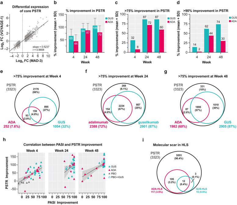
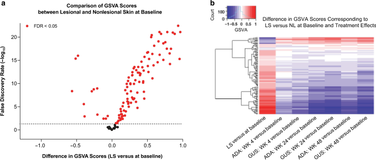
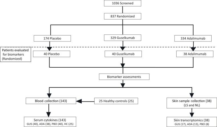
Design: Inflammatory cytokine serum levels were assessed (n = 118), and lesional and nonlesional skin biopsies were collected (n = 38) in patient subsets at baseline and 4, 24, and 48 weeks after treatment to evaluate pharmacodynamic responses of guselkumab versus those of ADA.

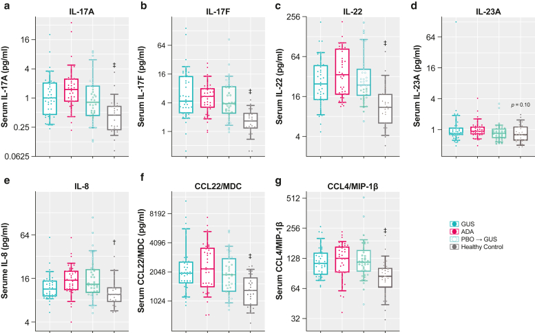
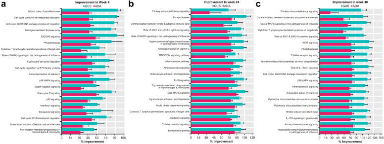
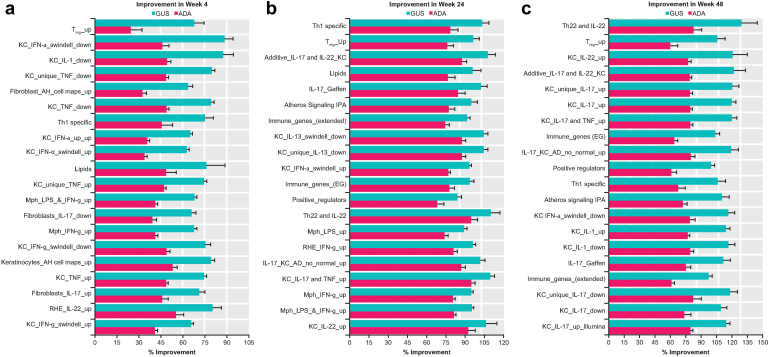
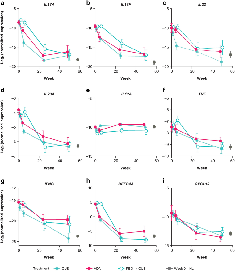
Results: Guselkumab provided rapid reductions in serum IL-17A, IL-17F, and IL-22 levels by week 4 versus at baseline, which were maintained through weeks 24 and 48 (P < .001). The magnitude of reduction of IL-17A and IL-22 at week 48 and IL-17F at weeks 4, 24, and 48 were greater with guselkumab than with ADA (all P < .05). In the skin, guselkumab reduced the expression of IL-23/IL-17 pathway-associated and psoriasis-associated genes.

Conclusion: These data provide extensive characterization of pharmacodynamic anti-inflammatory responses to IL-23p19 and TNF-α inhibition in human blood and tissue over time with FDA-approved doses of guselkumab and ADA. Trial registration:ClinicalTrials.govClinicalTrials.gov (NCT02207231).

Keywords: Adalimumab; Biomarkers; Guselkumab; IL-23/IL-17 pathway; Psoriasis.


Figures

Figure 1
Figure 1
Serum cytokine levels in psoriasis at baseline versus normal healthy serum control. (a) IL-17A, (b) IL-17F, (c) IL-22, (d) IL-23A, (e) IL-8, (f) CCL22/MDC, and (g) CCL4/MIP-1β. Data are presented as concentration (pg/ml, y-axis in log scale) of a subset of the VOYAGE 1 study population (n = 118) compared with that of serum samples obtained from an independent demographically matched healthy control cohort (n = 25). Boxplots show median (25th–75th percentile) and 10th–90th percentiles (whiskers). P-values were derived from Welch’s t-test comparing all biomarker substudy patients with psoriasis with healthy controls; adjusted P-values were calculated using the Benjamini–Hochberg procedure for correction of multiple comparisons (P < .01 and P < .001). ADA, adalimumab; GUS, guselkumab; MDC, macrophage-derived chemokine; PBO, placebo.
Figure 2
Figure 2
Selective blockade of IL-23 attenuates IL-17 and IL-22 effector cytokines. Data are presented as mean log2 concentration (pg/ml) over time (plotted means are linear model derived expected LS mean [emmean] plus SE). (a) IL-17A, (b) IL-17F, (c) IL-22, (d) IL-23, (e) MDC, and (f) MIP-1β. PD effects by treatment group at weeks 4, 24, and 48 were evaluated using a paired t-test against baseline (§P < .05, P < .01, and P < .001). Differences in PD effects by GUS (n = 40) versus ADA (n = 38) at weeks 4, 24, and 48 were further evaluated by ANCOVA (∗P < .05), with baseline concentration as a covariate. The PBO group (n = 40) crossed over to GUS at week 16. Healthy control sera (n = 25) were included as reference. Error bars represent SEs. ADA, adalimumab; ANCOVA, analysis of covariance; GUS, guselkumab; LS, lesional skin; MDC, macrophage-derived chemokine; PBO, placebo; PD, pharmacodynamic; SE, standard error.
Figure 3
Figure 3
Psoriatic skin transcriptomic profiling. (a) Association of differential expression of core PSTR genes in this study (VOYAGE 1) versus a previous meta-analysis (MAD-3) (Tian et al, 2012). (b–d) Percentage improvement in PSTR and percentage of PSTR with >75% or >90% improvement over time. Venn diagrams of PSTR with >75% improvement at weeks (e) 4, (f) 24, and (g) 48 are shown. (h) Correlations between PASI improvement and improvement in PSTR using Pearson’s correlation coefficient (r = 0.68). (i) Venn diagram of PSTR at baseline and molecular scars in HLS after treatment with GUS (n = 17) and ADA (n = 13). ADA, adalimumab; FC, fold change; GUS, guselkumab; HLS, healed lesional skin; MAD-3, meta-analysis derived-3; PBO, placebo; PSTR, psoriatic skin transcriptomic signature; SD, standard deviation.
Figure 4
Figure 4
Normalization of disease-associated gene expression by guselkumab versus adalimumab in PSTR-enriched pathways. Fifty-six canonical pathways in QIAGEN IPA were selected as enriched with PSTR genes. Percentage improvement (mean + SE) in the PSTR among the top 20 pathways ranked by the difference in improvement between GUS and ADA at weeks (a) 4, (b) 24, and (c) 48. ADA, adalimumab; BTG, B-cell translocation gene; GADD, growth arrest and DNA damage; GUS, guselkumab; iNOS, inducible nitric oxide synthase; IPA, Ingenuity Pathway Analysis; MSP, macrophage-stimulating protein; PSTR, psoriatic skin transcriptomic signature; RON, recepteur d'origine Nantais; SE, standard error.
Figure 5
Figure 5
Treatment effect on immune/skin inflammation gene sets by GSVA. (a) Volcano plot comparing GSVA scores of 124 gene sets between LS and NL at baseline. The 79 gene sets enriched with psoriasis DEG1 between LS and NL at baseline are highlighted in red. Dashed horizontal line indicates an FDR = 0.05. Gray dots represent the remaining gene sets not enriched by psoriasis DEG. (b) Heatmap of difference in GSVA scores in 108 gene sets (rows), corresponding to comparisons (columns). 1Gene set with ≥6% and >20% of genes differentially expressed (FC > 1.5) between LS and NL at baseline. ADA, adalimumab; DEG, differentially expressed gene; FC, fold change; FDR, false discovery rate; GSVA, gene set variation analysis; GUS, guselkumab; LS, lesional skin; NL, nonlesional skin; WK, week.
Figure 6
Figure 6
Normalization of disease-associated gene expression by GUS versus ADA in immune cell and skin inflammation gene sets. GSVA was used to evaluate differential expression of 124 gene sets between baseline LS and NL. Percentage improvement (mean + SE) in the PSTR among the top 20 sets ranked by the difference in improvement between GUS and ADA at weeks (a) 4, (b) 24, and (c) 48 is shown. AH gene sets are from (Asifa) (Haider et al, 2008). AD, atopic dermatitis; ADA, adalimumab; Atheros, atherosclerosis; GSVA, gene set variation analysis; GUS, guselkumab; IPA, Ingenuity Pathway Analysis; KC, keratinocyte; LPS, lipopolysaccharide; LS, lesional skin; Mph, macrophage; NL, nonlesional skin; PSTR, psoriatic skin transcriptomic signature; RHE, epithelial cell; SE, standard error; Th, T helper; Treg, regulatory T cell.
Figure 7
Figure 7
GUS-treated patients showed faster and greater reductions of IL-17a and IL-22 expression than ADA-treated patients. Results from 3 of the rerun genes, including (a) IL17A, (b) IL17F, and (c) IL22 plus 6 other genes that included (d) IL23A, (e) IL12A, (f) TNF, (g) IFNG, (h) DEFB4A, and (i) CXCL10, profiled using Fluidigm RT-PCR panel (mean + SE). The y-axis (log2 normalized expression) was calculated as negative delta ct to the ct values of the reference probe in the qPCR panel. GUS treated, n = 17; ADA treated, n = 13; PBO to GUS, n = 8. ADA, adalimumab; GUS, guselkumab; NL, nonlesional skin; PBO, placebo; SE, standard error.
Figure 8
Figure 8
Consort flowchart. patient stratification for the VOYAGE 1 study. ADA, adalimumab; GUS, guselkumab; LS, lesional skin; NL, nonlesional skin; PBO, placebo.

Similar articles

Cited by

References

    1. Basu R., O’Quinn D.B., Silberger D.J., Schoeb T.R., Fouser L., Ouyang W., et al. Th22 cells are an important source of IL-22 for host protection against enteropathogenic bacteria. Immunity. 2012;37:1061–1075. - PMC - PubMed
    1. Benham H., Norris P., Goodall J., Wechalekar M.D., FitzGerald O., Szentpetery A., et al. Th17 and Th22 cells in psoriatic arthritis and psoriasis. Arthritis Res Ther. 2013;15:R136. - PMC - PubMed
    1. Blauvelt A., Chiricozzi A. The immunologic role of IL-17 in psoriasis and psoriatic arthritis pathogenesis. Clin Rev Allergy Immunol. 2018;55:379–390. - PMC - PubMed
    1. Blauvelt A., Lebwohl M.G., Bissonnette R. IL-23/IL-17A dysfunction phenotypes inform possible clinical effects from anti-IL-17A therapies. J Invest Dermatol. 2015;135:1946–1953. - PMC - PubMed
    1. Blauvelt A., Leonardi C.L., Gooderham M., Papp K.A., Philipp S., Wu J.J., et al. Efficacy and safety of continuous risankizumab therapy vs treatment withdrawal in patients with moderate to severe plaque psoriasis: a phase 3 randomized clinical trial. JAMA Dermatol. 2020;156:649–658. - PMC - PubMed

Associated data