. 2024 Aug 8;9(15):e178050.
doi: 10.1172/jci.insight.178050.

GSK3 inhibition reduces ECM production and prevents age-related macular degeneration-like pathology

Affiliations

GSK3 inhibition reduces ECM production and prevents age-related macular degeneration-like pathology

Sophia M DiCesare et al. JCI Insight. .

Abstract

Malattia Leventinese/Doyne honeycomb retinal dystrophy (ML/DHRD) is an age-related macular degeneration-like (AMD-like) retinal dystrophy caused by an autosomal dominant R345W mutation in the secreted glycoprotein, fibulin-3 (F3). To identify new small molecules that reduce F3 production in retinal pigmented epithelium (RPE) cells, we knocked-in a luminescent peptide tag (HiBiT) into the endogenous F3 locus that enabled simple, sensitive, and high-throughput detection of the protein. The GSK3 inhibitor, CHIR99021 (CHIR), significantly reduced F3 burden (expression, secretion, and intracellular levels) in immortalized RPE and non-RPE cells. Low-level, long-term CHIR treatment promoted remodeling of the RPE extracellular matrix, reducing sub-RPE deposit-associated proteins (e.g., amelotin, complement component 3, collagen IV, and fibronectin), while increasing RPE differentiation factors (e.g., tyrosinase, and pigment epithelium-derived factor). In vivo, treatment of 8-month-old R345W+/+ knockin mice with CHIR (25 mg/kg i.p., 1 mo) was well tolerated and significantly reduced R345W F3-associated AMD-like basal laminar deposit number and size, thereby preventing the main pathological feature in these mice. This is an important demonstration of small molecule-based prevention of AMD-like pathology in ML/DHRD mice and may herald a rejuvenation of interest in GSK3 inhibition for the treatment of retinal degenerative diseases, including potentially AMD itself.

Keywords: Drug screens; Extracellular matrix; Ophthalmology; Retinopathy.


Conflict of interest statement

Conflict of interest: The authors have declared that no conflict of interest exists.

Figures

Figure 1
Figure 1. Design and validation of fibulin-3 HiBiT tagging in ARPE-19 cells using CRISPR.
(A) Schematic of CRISPR editing of exon 2 of the fibulin-3 (F3) gene to knockin a 2xFLAG-VS-HiBiT sequence immediately proceeding the signal sequence cleavage site (upward arrow). (B) Successful editing was verified by gDNA amplification of exon 2. An additional band corresponding to insertion of the 87 bp 2x FLAG VS HiBiT tag was identified with a calculated editing efficiency of approximately 15%. (C) F3 HiBiT tagging results in a single extracellular protein species of correct molecular weight (~55 kDa), as identified by immunoprecipitation (FLAG beads) followed by elution and HiBiT blotting. (D) siRNA verifies that more than 95% of the HiBiT signal can be attributed to F3 gene translation. n = 3 independent experiments performed in biological triplicates. ****P ≤ 0.0001, 1-way ANOVA with Dunnett’s multiple comparison test vs. nontargeting siRNA.
Figure 2
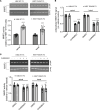
Figure 2. Structurally diverse glycogen synthase kinase 3 inhibitors reduce F3 transcripts and extracellular/intracellular levels without causing toxicity.
(A and B) A series of chemically unrelated glycogen synthase kinase 3 (GSK3) inhibitors significantly reduced (A) extracellular and (B) intracellular HiBiT F3 levels after 72 hours of treatment. n = 3 independent experiments, mean ± SD. *P ≤ 0.05, **P ≤ 0.01, ***P ≤ 0.001, 1-sample t test vs. hypothetical unchanged value of 1 (vehicle treated). (C) CHIR99021-dependent HiBiT assay results in A were confirmed at the protein level by HiBiT blotting. Representative image from n ≥ 3 independent experiments. (D) Seventy-two-hour CHIR99021 treatment reduced F3 mRNA expression. Representative data from n = 3 independent experiments; mean ± SD of technical triplicates. (E) Treatment with CHIR did not elevate release of cytosolic lactate dehydrogenase (LDH). n = 3 independent experiments performed in biological triplicate. *P ≤ 0.05, ****P ≤ 0.0001, 1-way ANOVA with Dunnett’s multiple comparison test vs. vehicle-treated (DMSO) levels. BIO, 6-bromoindirubin-3-oxime; LiCl, lithium chloride.
Figure 3
Figure 3. Genetic knockdown of GSK3 isoforms also reduces HiBiT F3 levels, paralleling pharmacologic GSK3 inhibitor effects.
(A) Knockdown (96 h) of either GSK3α or GSK3β significantly lowered F3 production in ARPE-19 cells, and the effects of knocking down both α and β isoforms were additive. n = 3 independent experiments performed in biological triplicate. **P ≤ 0.01, ***P ≤ 0.001, ****P ≤ 0.0001, 1-way ANOVA with Dunnett’s T3 multiple comparison test. (B) Protein-level knockdown effects were verified by Western and HiBiT blotting, (C) followed by quantification of intracellular HiBiT F3. Representative images from n = 3 independent experiments. **P ≤ 0.01, ****P ≤ 0.0001, 1-way ANOVA with Dunnett’s multiple comparison test vs. siNT (nontargeting).
Figure 4
Figure 4. Low-level, prolonged CHIR99021 treatment reduces F3 production while avoiding triggering TCF4-dependent Wnt activation.
(A) Seventy-two-hour CHIR99021 treatment dose-dependently reduced HiBiT F3 extracellular levels (B) without necessarily triggering TCF4-dependent green fluorescent protein (GFP) expression. n = 3 independent experiments performed in at least biological triplicate, with representative data presented. Scale bar: 200 μm. (C) One-week treatment with CHIR99021 (1 μM) significantly lowered HiBiT F3 extracellular levels (D) without activating TCF4-dependent firefly luciferase (FLuc). n = 3 independent experiments performed in biological replicates. (E) Prolonged low-level CHIR99021 did not increase cell death as indicated by LDH release. n = 3 independent experiments performed in biological replicates. *P ≤ 0.05, **P ≤ 0.01, ***P ≤ 0.001, ****P ≤ 0.0001, 1-way ANOVA with Dunnett’s multiple comparison test vs. vehicle (DMSO) treatment.
Figure 5
Figure 5. Low-level, 1-week CHIR99021 treatment reduces extracellular matrix and collagen-associated transcripts while upregulating RPE differentiation-associated genes.
(A) Bulk RNA-Seq analysis of ARPE-19 cells treated with CHIR99021 indicated significant transcript reduction (≥2-fold) of genes implicated in extracellular matrix (ECM) formation and sub-RPE deposit formation while upregulating (≥2-fold) RPE differentiation genes. n = 3 independent experiments combined to produce these data. P adjusted < 0.05 using a negative binomial distribution model, green and red dots are significant. (B) Pathway enrichment of significantly altered genes from RNA-Seq data set using Gene ontology enrichment analysis.
Figure 6
Figure 6. CHIR99021 reduces R345W F3-associated matrix metalloproteinase 2 alterations in culture.
(A) ARPE-19 cells expressing HiBiT R345W F3 demonstrate significantly elevated apical and basal matrix metalloproteinase 2 (MMP2) activity levels compared with HiBiT WT F3 cells after 1 week on Transwells. n = 3 independent experiments, performed in sextuplet biological replicates. ****P ≤ 0.0001, t test vs. HiBiT WT F3 cells. (B) Treatment (1 week, 1 μM) with CHIR99021 significantly decreased HiBiT WT and R345W F3 both apically and basally in Transwell format (C) while simultaneously significantly decreasing MMP2 activity. n = 3 independent experiments, performed in triplicate biological replicates. **P ≤ 0.01, ***P ≤ 0.001, ****P ≤ 0.0001, t test vs. each respective vehicle-treated control.
Figure 7
Figure 7. One-month CHIR99021 treatment does not affect retinal function or gross structure.
(A and B) Eight-month-old R345W+/+ C57BL/6 mice were injected i.p. with vehicle (PBS) or CHIR99021 for 1 month (every weekday). Scotopic electroretinogram (ERG) readings demonstrated no difference between groups in either a-wave (outer retina, A) or b-wave (inner retina, B). Values were not significant by an ANOVA test. Box-and-whisker plots show average and minimum and maximum values. (C) An example H&E histology image of 8-month-old untreated WT C57BL/6 mice. (D and E) After completion of ERG evaluation, R345W+/+ mice were sacrificed and 1 eye was prepared for H&E histology, which demonstrated no observable differences between the 2 groups (vehicle, D; CHIR, E). n = 4 mice/treatment group, 2 male, 2 female. Scale bar: 100 μm.
Figure 8
Figure 8. CHIR99021 significantly reduces the formation of basal laminar deposits in vivo.
(A) Representative transmission electron microscopy (TEM) image of an 8-month-old WT C57BL/6 mice. (B and C) R345W+/+ mice after 1 month of vehicle (B) or CHIR99021 (C) treatment. Mice in B and C were 9 months old when sacrificed for basal laminar deposit (BLamD) (arrows) evaluation by TEM. Representative fields of view (FOV) of 22 fields are presented for vehicle or CHIR99021 treatment. Scale bar: 2 μm. (D and E) A masked observer systematically quantified (D) the number of FOV containing any BLamD (****P ≤ 0.0001, χ2 test) as well as (E) the average size of the BLamD, if at all present (**P ≤ 0.01, t test vs. vehicle-treated samples).

Update of

References

    1. Daniel S, et al. Fibulin-3 knockout mice demonstrate corneal dysfunction but maintain normal retinal integrity. J Mol Med (Berl) 2020;98(11):1639–1656. doi: 10.1007/s00109-020-01974-z. - DOI - PMC - PubMed
    1. Daniel S, Hulleman JD. Exploring ocular fibulin-3 (EFEMP1): anatomical, age-related, and species perspectives. Biochim Biophys Acta Mol Basis Dis. 2024;1870(6):167239. doi: 10.1016/j.bbadis.2024.167239. - DOI - PMC - PubMed
    1. Sura AA, et al. Measuring the contributions of basal laminar deposit and Bruch’s membrane in age-related macular degeneration. Invest Ophthalmol Vis Sci. 2020;61(13):19. doi: 10.1167/iovs.61.13.19. - DOI - PMC - PubMed
    1. Fernandez-Godino R, et al. Extracellular matrix alterations and deposit formation in AMD. Adv Exp Med Biol. 2016;854:53–58. doi: 10.1007/978-3-319-17121-0_8. - DOI - PubMed
    1. Fernandez-Godino R, et al. Changes in extracellular matrix cause RPE cells to make basal deposits and activate the alternative complement pathway. Hum Mol Genet. 2018;27(1):147–159. doi: 10.1093/hmg/ddx392. - DOI - PMC - PubMed

MeSH terms

Supplementary concepts