. 2024 Sep 29;45(36):3751-3763.
doi: 10.1093/eurheartj/ehae476.

KCNQ1 suppression-replacement gene therapy in transgenic rabbits with type 1 long QT syndrome

Affiliations

KCNQ1 suppression-replacement gene therapy in transgenic rabbits with type 1 long QT syndrome

Sahej Bains et al. Eur Heart J. .

Abstract

Background and aims: Type 1 long QT syndrome (LQT1) is caused by pathogenic variants in the KCNQ1-encoded Kv7.1 potassium channels, which pathologically prolong ventricular action potential duration (APD). Herein, the pathologic phenotype in transgenic LQT1 rabbits is rescued using a novel KCNQ1 suppression-replacement (SupRep) gene therapy.

Methods: KCNQ1-SupRep gene therapy was developed by combining into a single construct a KCNQ1 shRNA (suppression) and an shRNA-immune KCNQ1 cDNA (replacement), packaged into adeno-associated virus serotype 9, and delivered in vivo via an intra-aortic root injection (1E10 vg/kg). To ascertain the efficacy of SupRep, 12-lead electrocardiograms were assessed in adult LQT1 and wild-type (WT) rabbits and patch-clamp experiments were performed on isolated ventricular cardiomyocytes.

Results: KCNQ1-SupRep treatment of LQT1 rabbits resulted in significant shortening of the pathologically prolonged QT index (QTi) towards WT levels. Ventricular cardiomyocytes isolated from treated LQT1 rabbits demonstrated pronounced shortening of APD compared to LQT1 controls, leading to levels similar to WT (LQT1-UT vs. LQT1-SupRep, P < .0001, LQT1-SupRep vs. WT, P = ns). Under β-adrenergic stimulation with isoproterenol, SupRep-treated rabbits demonstrated a WT-like physiological QTi and APD90 behaviour.

Conclusions: This study provides the first animal-model, proof-of-concept gene therapy for correction of LQT1. In LQT1 rabbits, treatment with KCNQ1-SupRep gene therapy normalized the clinical QTi and cellular APD90 to near WT levels both at baseline and after isoproterenol. If similar QT/APD correction can be achieved with intravenous administration of KCNQ1-SupRep gene therapy in LQT1 rabbits, these encouraging data should compel continued development of this gene therapy for patients with LQT1.

Keywords: AAV9; Gene therapy; KCNQ1; Long QT syndrome; Transgenic LQT1 rabbits.


Figures

Structured Graphical Abstract
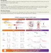
KCNQ1 suppression-replacement (SupRep) gene therapy was developed by combining into a single construct a KCNQ1 shRNA (suppression) and an shRNA-immune KCNQ1 cDNA (replacement). This hybrid SupRep gene therapy was packaged into adeno-associated virus serotype 9 (AAV9) and delivered in vivo via intra-aortic root injections (1E10 vg/kg). To ascertain the efficacy of SupRep, 12-lead electrocardiograms (ECGs) were assessed in adult type 1 long QT syndrome (LQT1) and wild-type (WT) rabbits. Patch-clamp and calcium transient experiments were performed on isolated ventricular cardiomyocytes (VCMs), both at baseline and under β-adrenergic stimulation with isoproterenol. In LQT1 transgenic rabbits, treatment with KCNQ1-SupRep resulted in significant shortening of the pathologically prolonged QT interval, QT index, and cellular action potential duration (APD) and Ca2+T towards WT levels. Furthermore, unlike sham-treated LQT1 rabbits, SupRep-treated LQT1 rabbits demonstrated a physiological behaviour under β-adrenergic stimulation.
Figure 1
Figure 1
KCNQ1 suppression-and-replacement gene therapy in LQT1 rabbits. KCNQ1-SupRep gene therapy consists of two therapeutic components expressed from a single viral vector. The suppression component (depicted in yellow in the left panel) utilizes a U6 promoter element driven KCNQ1 shRNA (shKCNQ1) to silence the expression of KCNQ1 transcripts. This approach targets the gene itself rather than specific variants. Simultaneously, the replacement component (in red in the right panel) of KCNQ1-SupRep restores the lost transcripts by expressing a ‘shRNA-immune’ (shIMM) version of the KCNQ1 cDNA, featuring synonymous variants at each wobble base within the shRNA target site. Importantly, this does not alter the WT amino acid sequence but prevents shRNA binding, making KCNQ1-shIMM resistant to knockdown. When expressed in tandem, suppression-replacement results in the simultaneous suppression of KCNQ1 and the replacement with KCNQ1-shIMM. The outcome is an increase in functional KCNQ1-encoded Kv7.1 potassium channels, rescuing the LQT1 phenotype in transgenic rabbits arising from KCNQ1 loss-of-function. Figure created using Biorender.com
Figure 2
Figure 2
Evaluation of in vitro KCNQ1 SupRep efficacy. (A) Schematic illustration of ventricular cardiomyocyte (VCM) isolation process. Rabbit hearts were extracted, perfused on a Langendorff system, and ventricular CMs were isolated using enzymatic digestion. Following isolation, cells were either untreated or transfected with SupRep or sham (non-targeting scramble RNA) plasmids. Twenty-four hours following transfection, action potential durations at 90% repolarization (APD90) were measured via whole cell patch clamp. Figure created using Biorender.com. (B) Representative whole cell patch clamp action potential recordings in UT-LQT1 (blue), sham-treated LQT1 (light blue), SupRep-treated LQT1 (red), and UT-WT cultured VCMs (black). (C) Whole cell patch-clamp APD90 measurements at 1 Hz stimulation were performed at 37°C in the various treatment groups. Results are expressed as mean ± standard error of the mean (SEM). One-way ANOVA with post hoc Tukey’s multiple comparison was performed for statistical analysis. P < .05 was considered significant
Figure 3
Figure 3
AAV9-KCNQ1-SupRep delivery: route, methodology, and monitoring. AAV9-KCNQ1-SupRep gene therapy was delivered in transgenic LQT1 rabbits via a targeted intra-aortic root injection. Using a Swan–Ganz catheter, the viral construct was perfused during thrice balloon occlusion to obtain a trans-coronary distribution of our gene therapy into the heart. During the surgical procedure, animal vital signs including heart rate (ECG II lead), respiratory rate, oxygen arterial saturation, capnography, invasive blood pressure (in aorta distal to the balloon and in the ear artery), non-invasive Doppler blood pressure, and electroencephalogram (BIS®) were continuously monitored. Chest X-ray imaging was used to guide the catheter and assess the virus perfusion. Figure created using Biorender.com
Figure 4
Figure 4
AAV9-KCNQ1-SupRep shortens the pathologically prolonged QT index in treated transgenic LQT1 rabbits. Panel A depicts the representative 12-lead ECG traces from LQT1 and WT animals. The QT interval of the UT-LQT1 rabbit is shown in blue, SupRep-treated LQT1 rabbit in red, and WT rabbit in black. Compared to the UT-LQT1 rabbit, the QT interval of the SupRep-treated rabbit is shortened and close to the level of the WT rabbit. (B) QTi in LQT1 rabbits before SupRep treatment (baseline, blue), in LQT1 rabbits either 2 (dark red) or 3 weeks (red) post-SupRep treatment, and in WT rabbits at baseline (black) is shown. SupRep treatment significantly shortens the QTi in the treated rabbits close to the level of WT. Results are expressed as mean ± standard error of the mean (SEM). One-way ANOVA (comparing data to LQT1-UT) with post hoc Dunnett’s multiple comparison was performed. P < .05 was considered statistically significant
Figure 5
Figure 5
Treatment with KCNQ1-SupRep shortens significantly the pathologically prolonged APD90 in transgenic LQT1 rabbits. (A) Representative whole cell patch clamp action potential recordings in ventricular cardiomyocytes (VCMs) isolated from UT-LQT1, sham-treated LQT1, 2- or 3-week SupRep-treated LQT1, and UT-WT rabbits. (B) Whole cell patch clamp was used for APD90 measurements at 37°C at 1 Hz stimulation rate. SupRep-treated LQT1 rabbits demonstrate significantly shortened APD90 as compared to the UT or sham-treated LQT1 rabbits. Results are expressed as mean ± standard error of the mean (SEM). One-way ANOVA with post hoc Tukey’s multiple comparison was performed. P < .05 was considered statistically significant
Figure 6
Figure 6
AAV9-KCNQ1-SupRep-treated transgenic LQT1 rabbit behaves like WT rabbit under β-adrenergic stimulation with isoproterenol. Depicted are three pairs of ECGs collected at baseline and after isoproterenol (ISO) perfusion in one sham-treated LQT1 rabbit, one SupRep-treated LQT1 rabbit, and one WT rabbit. The AAV9-KCNQ1-SupRep-treated LQT1 rabbit demonstrates a similar ΔQTi (%) as the WT rabbit after provocation with ISO. A 20% heart rate increase was observed in all three animals after stimulation with ISO. Raw values for RR and QT intervals are also provided both at baseline and under β-adrenergic stimulation with isoproterenol
Figure 7
Figure 7
Ventricular cardiomyocytes isolated from SupRep-treated LQT1 rabbits demonstrate WT-like APD90 and Ca2+T90 under β-adrenergic stimulation. (A) (Top) Representative whole cell patch clamp action potential (AP) traces and (bottom) statistical analysis of the APD90 at baseline (control) and under isoproterenol provocation. Indicated are individual ventricular cardiomyocytes (VCMs) and their changes. Unlike VCMs isolated from sham and UT-LQT1 rabbits, VCMs isolated from SupRep-treated LQT1 rabbits and WT rabbits demonstrated a significant shortening in APD90 at 1 Hz stimulation rate following β-adrenergic stimulation. Paired Student’s t-test was performed. (B) (Top) Representative calcium transient recordings and (bottom) Ca2+T90 statistical analysis at baseline (control) and under isoproterenol provocation. VCMs isolated from SupRep-treated LQT1 and WT rabbits showed a Ca2+T90 shortening after β-adrenergic stimulation. Unpaired Student’s t-test or Mann–Whitney was performed. P < .05 was considered statistically significant

References

    1. Schwartz PJ, Stramba-Badiale M, Crotti L, Pedrazzini M, Besana A, Bosi G, et al. . Prevalence of the congenital long-QT syndrome. Circulation 2009;120:1761–7. 10.1161/CIRCULATIONAHA.109.863209 - DOI - PMC - PubMed
    1. Schwartz PJ, Ackerman MJ. The long QT syndrome: a transatlantic clinical approach to diagnosis and therapy. Eur Heart J 2013;34:3109–16. 10.1093/eurheartj/eht089 - DOI - PubMed
    1. Amin AS, Tan HL, Wilde AA. Cardiac ion channels in health and disease. Heart Rhythm 2010;7:117–26. 10.1016/j.hrthm.2009.08.005 - DOI - PubMed
    1. Huang H, Kuenze G, Smith JA, Taylor KC, Duran AM, Hadziselimovic A, et al. . Mechanisms of KCNQ1 channel dysfunction in long QT syndrome involving voltage sensor domain mutations. Sci Adv 2018;4:eaar2631. 10.1126/sciadv.aar2631 - DOI - PMC - PubMed
    1. Splawski I, Shen J, Timothy KW, Lehmann MH, Priori S, Robinson JL, et al. . Spectrum of mutations in long-QT syndrome genes. KVLQT1, HERG, SCN5A, KCNE1, and KCNE2. Circulation 2000;102:1178–85. 10.1161/01.CIR.102.10.1178 - DOI - PubMed

Substances