. 2024 Aug 13;121(33):e2409167121.
doi: 10.1073/pnas.2409167121. Epub 2024 Aug 8.

A DNA condensation code for linker histones

Affiliations

A DNA condensation code for linker histones

Matthew Watson et al. Proc Natl Acad Sci U S A. .

Abstract

Linker histones play an essential role in chromatin packaging by facilitating compaction of the 11-nm fiber of nucleosomal "beads on a string." The result is a heterogeneous condensed state with local properties that range from dynamic, irregular, and liquid-like to stable and regular structures (the 30-nm fiber), which in turn impact chromatin-dependent activities at a fundamental level. The properties of the condensed state depend on the type of linker histone, particularly on the highly disordered C-terminal tail, which is the most variable region of the protein, both between species, and within the various subtypes and cell-type specific variants of a given organism. We have developed an in vitro model system comprising linker histone tail and linker DNA, which although very minimal, displays surprisingly complex behavior, and is sufficient to model the known states of linker histone-condensed chromatin: disordered "fuzzy" complexes ("open" chromatin), dense liquid-like assemblies (dynamic condensates), and higher-order structures (organized 30-nm fibers). A crucial advantage of such a simple model is that it allows the study of the various condensed states by NMR, circular dichroism, and scattering methods. Moreover, it allows capture of the thermodynamics underpinning the transitions between states through calorimetry. We have leveraged this to rationalize the distinct condensing properties of linker histone subtypes and variants across species that are encoded by the amino acid content of their C-terminal tails. Three properties emerge as key to defining the condensed state: charge density, lysine/arginine ratio, and proline-free regions, and we evaluate each separately using a strategic mutagenesis approach.

Keywords: chromatin; complex coacervation; intrinsically disordered protein; linker histone; phase separation.


Conflict of interest statement

Competing interests statement:The authors declare no competing interest.

Figures

Fig. 1.
Fig. 1.
Chromatin condensation by H1 tails. (A) A minimal model for the study of chromatin condensation by linker histones consists of internucleosomal linker DNA and the C-terminal tail of H1 (CH1) (16). (B) The model system encompasses the possible H1-condensed states of chromatin, forming disordered complexes, phase-separated droplets, and higher-order structures (a liquid crystalline phase), depending on the conditions (see main text). (C) Amino acid residue content of curated set of 94 CH1s by percentage (black bars, Gallus gallus isoform H1.11L and variant H5 as red and green bars, respectively). (D) Fraction of charged residues (Gallus gallus isoform H1.11L and variant H5 are shown as a red circle and a green triangle, respectively). (E) Distributions of κ (charge patterning; blue) and Ω (patterning of charged and proline residues; orange) where 0 = well-mixed, 1 = fully segregated. (F) Sequences of CH1 and CH5 are the wild-type sequences of the C-terminal tails of H1.11L and H5 from Gallus gallus. For the rationale behind the design of CH1R, CH5K, CH1VA, CH1VT, and CH1PA see the main text. Arginine, proline, and valine are highlighted in dark blue, green, and yellow, respectively.
Fig. 2.
Fig. 2.
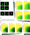
Condensation behavior of CH1, CH5, and mutants. (A) Confocal fluorescence microscopy of droplets formed at 25 µM 1:1 protein:20mer DNA complex, I = 160 mM, 10% of DNA fluorescein amidite (FAM)-labeled. (Scale bar, 5 µm). (B) Csat, measured by DLS (values were consistent across triplicate experiments). Phase diagrams for (C) arginine and charge density series, (D) hydrophobicity series, and (E) proline knockout, vs. concentration and ionic strength. Concentration and turbidity (A340) is of the 1:1 mixture. Turbidity is shown in numbers as well as a yellow-to-green color scale. Errors are <5%.
Fig. 3.
Fig. 3.
Thermodynamics of condensation. ITC of protein (as indicated) into 20 bp DNA, alongside A340 to identify phase separation events, in buffer containing 150 mM NaCl (total I = 160 mM). (A) Raw data, isotherms, and A340 for CH1 and CH5. (B) Isotherms (above) and A340 (below) for the complete arginine and charge density series. (C) Raw data, isotherms, and A340 for the proline knockout. (D) Isotherms (above) and A340 (below) for the hydrophobicity and proline knockouts. (E) Raw data, isotherms, and A340 for protamine titrated into 16 bp DNA. Complete dataset in SI Appendix, Fig. S3.
Fig. 4.
Fig. 4.
CH1PA forms temperature-dependent alpha helix. Far-UV CD of the free proteins for (A) arginine and charge density series, (B) hydrophobicity and proline knockouts, and (C) proline knockout and CH1 vs. temperature. Temperature 25 °C unless indicated, in buffer containing 150 mM NaF (total I = 160 mM).
Fig. 5.
Fig. 5.
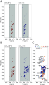
Alpha helix nucleates at several distinct loci in CH1PA. 15N-HSQC NMR of free proteins: (A) CH1 at 25 °C and 0 °C, (B) proline knockout at 25 °C and 0 °C, and (C) proline knockout vs. temperature, with peak assignments. Gray shading present to illustrate chemical shift dispersion in 1HN. Buffer contains 150 mM NaCl (total I = 160 mM).
Fig. 6.
Fig. 6.
Structure and heterogeneity of the dense phase. Far-UV CD of the protein:DNA condensates for (A) arginine and charge density series and (B) CH1 and proline knockouts, with 20 bp DNA, 1:1, at 25 °C in buffer containing 150 mM NaCl (total I = 160 mM). Free DNA (black) is also shown for reference. The position of the ψ-DNA scattering signal is indicated (see the text). (C) FRAP recovery curves and fits. For comparative purposes, these are shown normalized after subtraction of A0 (Materials and Methods). (D) Fitted parameters resulting from mono- or biexponential models, as appropriate. SSR = sum of the squared residuals.
Fig. 7.
Fig. 7.
Proline-free regions across linker histones. (Top) All proline-free regions across the curated set of 94 CH1s are shown as dots corresponding to their length in residues, vs. record number in the database (SI Appendix, Fig. S1). The three known avian CH5s are also shown on the Right. (Bottom) Net charge per residue, and net arginine per residue (fraction arginine). Proteins with long proline-free regions and known gene-repressive functions (see text) are boxed in gray and labeled. CH1 and CH5 are boxed and labeled in red and green, respectively.

References

    1. Gilbert N., Allan J., The many length scales of DNA packaging. Essays Biochem. 63, 13–16 (2019). - PubMed
    1. Kornberg R. D., Lorch Y., Twenty-five years of the nucleosome, fundamental particle of the eukaryote chromosome. Cell 98, 285–294 (1999). - PubMed
    1. Bednar J., Hamiche A., Dimitrov S., H1-nucleosome interactions and their functional implications. Biochim. Biophys. Acta 1859, 436–443 (2016). - PubMed
    1. Thoma F., Koller T. H., Klug A., Involvement of histone H1 in the organization of the nucleosome and of the salt-dependent superstructures of chromatin. J. Cell Biol. 83, 403–427 (1979). - PMC - PubMed
    1. Woodcock C. L., Chromatin fibers observed in situ in frozen hydrated sections. Native fiber diameter is not correlated with nucleosome repeat length. J. Cell Biol. 125, 11–19 (1994). - PMC - PubMed

LinkOut - more resources