. 2024 Aug 27;43(8):114609.
doi: 10.1016/j.celrep.2024.114609. Epub 2024 Aug 7.

FDA-approved disulfiram inhibits the NLRP3 inflammasome by regulating NLRP3 palmitoylation

Affiliations

FDA-approved disulfiram inhibits the NLRP3 inflammasome by regulating NLRP3 palmitoylation

Jie Xu et al. Cell Rep. .

Abstract

The NLRP3 inflammasome is dysregulated in autoinflammatory disorders caused by inherited mutations and contributes to the pathogenesis of several chronic inflammatory diseases. In this study, we discovered that disulfiram, a safe US Food and Drug Administration (FDA)-approved drug, specifically inhibits the NLRP3 inflammasome but not the NLRC4 or AIM2 inflammasomes. Disulfiram suppresses caspase-1 activation, ASC speck formation, and pyroptosis induced by several stimuli that activate NLRP3. Mechanistically, NLRP3 is palmitoylated at cysteine 126, a modification required for its localization to the trans-Golgi network and inflammasome activation, which was inhibited by disulfiram. Administration of disulfiram to animals inhibited the NLRP3, but not NLRC4, inflammasome in vivo. Our study uncovers a mechanism by which disulfiram targets NLRP3 and provides a rationale for using a safe FDA-approved drug for the treatment of NLRP3-associated inflammatory diseases.

Keywords: CP: Immunology; NLRP3; disulfiram; gasdermin D; inflammasome; palmitoylation.


Conflict of interest statement

Declaration of interests The authors declare no competing interests.

Figures
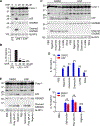
Figure 1.
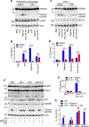
Figure 1.. Disulfiram inhibits caspase-1 activation induced by the activators of the NLRP3 inflammasome in mouse BMDMs
(A and B) LPS-primed BMDMs were treated with DMSO or disulfiram (DSF; the indicated doses, 20 min), followed by stimulation of ATP (5 mM, 40 min). Cell lysates were then analyzed by immunoblotting (A), and IL-1β release was analyzed by ELISA (B). Results are representative of three independent experiments. The bars represent mean ± SD. Results were analyzed by one-way ANOVA. ****p < 0.0001. (C and D) LPS-primed BMDMs were treated with DMSO or DSF (30 μM, 20 min), followed by stimulation of PBS, nigericin (5 μM, 40 min), imiquimod (20 μg/mL, 1 h), gramicidin (0.5 μM, 1 h), L-leucyl-L-leucine methyl ester (LLOMe; 2 μM, 1 h), or nano-silica (200 μg/mL, 1 h). Cell lysates were then analyzed by immunoblotting (C), and IL-1β release was analyzed by ELISA (D). Results are representative of three independent experiments. The bars represent mean ± SD. Results were analyzed by unpaired two-tailed Student’s t test. ****p < 0.0001. (E and F) LPS-primed BMDMs were treated with DMSO or DSF (30 μM, 20 min), followed by stimulation of PBS, Samonella (MOI 30, 1 h), poly(dA:dT) (1 μg/mL, 4 h), or nigericin (5 μM, 40 min). Cell lysates were then analyzed by immunoblotting (E), and IL-1β release was analyzed by ELISA (F). Results are representative of three independent experiments. The bars represent mean ± SD. Results were analyzed by unpaired two-tailed Student’s t test. ****p < 0.0001; ns, not significant. See also Figures S1–S3.
Figure 2.
Figure 2.. Disulfiram inhibits NLRP3 activation in mouse peritoneal macrophages, human THP-1 cells, and Nlrp3D301N/+ BMDMs
(A and B) LPS-primed peritoneal macrophages were treated with DMSO or DSF (30 μM, 20 min), followed by stimulation with PBS, ATP (5 mM, 40 min), nigericin (5 μM, 40 min), or poly(dA:dT) (1 μg/mL, 4 h). Cell lysates were then analyzed by immunoblotting (A), and IL-1β release was analyzed by ELISA (B). ACC, acetyl-CoA carboxylase. Results are representative of three independent experiments. The bars represent mean ± SD. Results were analyzed by unpaired two-tailed Student’s t test. ****p < 0.0001. ns, not significant. (C and D) LPS-primed THP-1 cells were treated with DMSO or DSF (30 μM, 20 min), followed by stimulation with PBS, nigericin (5 μM, 40 min), imiquimod (20 μg/mL, 1 h), or poly(dA:dT) (1 μg/mL, 4 h). Cell lysates were then analyzed by immunoblotting (A), and IL-1β release was analyzed by ELISA (B). ACC, acetyl-CoA carboxylase. Results are representative of three independent experiments. The bars represent mean ± SD. Results were analyzed by unpaired two-tailed Student’s t test. ***p < 0.001 and ****p < 0.0001. (E) Tamoxifen-treated BMDMs from Nlrp3fl(D301N)/+ Esr1-Cre or Nlrp3fl(D301N)/+ mice were incubated with both LPS and DMSO/DSF. IL-1β release was analyzed by ELISA. Results are representative of three independent experiments. The bars represent mean ± SD. Results were analyzed by unpaired two-tailed Student’s t test. ****p < 0.0001. (F and G) LPS-primed BMDMs were treated with PBS, NAC (15 mM, 70 min), GSH (15 mM, 70 min), or vitamin C (15 mM, 70 min) in the presence or absence of DSF (30 μM, 20 min), followed by stimulation of nigericin (5 μM, 40 min). Cell lysates were then analyzed by immunoblotting (G), and IL-1β release was analyzed by ELISA (H). Results are representative of three independent experiments. The bars represent mean ± SD. Results were analyzed by unpaired two-tailed Student’s t test. ****p < 0.0001; ns, not significant. See also Figure S4.
Figure 3.
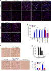
Figure 3.. Disulfiram inhibits ASC speck formation and pyroptosis induced by the activators of NLRP3 inflammasome
(A and B) Representative immunofluorescence images and quantification of endogenous ASC specks (arrowheads) in LPS-primed BMDMs treated with agonists as indicated in the presence of vehicle DMSO or DSF. Cells were stained with anti-ASC (red) and DAPI (blue). Scale bar, 15 μm. Data were analyzed from three combined experiments. The bars represent mean± SD. Results were analyzed by unpaired two-tailed Student’s t test. ***p < 0.001 and ****p < 0.0001. ns, not significant. (C) Representative differential interference contrast images of LPS-primed BMDMs treated with nigericin in the presence or absence of DSF (2 μM, 10 or 50 μM). Arrowheads indicate cells with features of pyroptosis. Scale bar, 15 μm. (D) Quantification of BMDMs that underwent pyroptosis in (C). Data were analyzed from three combined experiments. The bars represent mean ± SD. Results were analyzed by one-way ANOVA. ****p < 0.0001. ns, not significant; DSF, disulfiram. (E) LDH levels in cell culture supernatants of LPS-primed macrophages treated with ATP (5 mM) were analyzed. The bars represent mean ± SD. Results were analyzed by unpaired two-tailed Student’s t test. **p < 0.01.
Figure 4.
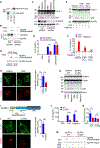
Figure 4.. Palmitoylation at Cys126 promotes NLRP3 activation and is inhibited by disulfiram
(A) Nlrp3−/− immortalized BMDMs (iBMDMs) were reconstituted with FLAG-tagged NLRP3. After LPS priming, NLRP3 was immunoprecipitated, and samples were then analyzed by immunoblotting. (B) zDHHC5 or control shRNA transduced iBMDMs were primed with LPS, followed by acyl-biotin exchange assay. NLRP3 palmitoylation and protein levels were determined by immunoblotting. (C and D) zDHHC5 or control shRNA transfected iBMDMs were primed with LPS and treated with nigericin or Salmonella. Cell lysates were analyzed by immunoblotting (C), and IL-1β release was analyzed by ELISA (D). ACC, acetyl-CoA carboxylase. (E and F) LPS-primed BMDMs were pre-treated with vehicle ethanol, or 2-BP (25 μM, 45 or 90 min) before stimulation with nigericin (5 μM, 40 min). Cell lysates were analyzed by immunoblotting (E), and IL-1β release was analyzed by ELISA (F). (G) LPS-primed iBMDMs were treated with vehicle DMSO or DSF, followed by acyl-biotin exchange assay. NLRP3 palmitoylation and protein levels were determined by immunoblotting. (H) Representative immunofluorescence images and quantification of the percentage of cells with NLRP3 perinuclear puncta. Nlrp3−/− iBMDMs reconstituted with NLRP3-FLAG were treated with vehicle (DMSO, left) or 30 μM DSF (right) and then simultaneously stained with both anti-FLAG (green, top) and anti-TGN38 (red, bottom). The arrowheads indicate the perinuclear puncta where NLRP3 and TGN38 co-localized. Scale bar, 10 μm. Data were analyzed from three combined experiments. The bars represent mean ± SD. Results were analyzed by unpaired two-tailed Student’s t test. *p < 0.05. (I) Domain organization of NLRP3. Amino acid sequences in the polybasic region between the Pyrin and NACHT domains are shown. C126 is highlighted in red. (J) Representative immunofluorescence images and quantification of the percentage of cells with NLRP3 perinuclear puncta. Nlrp3−/− iBMDMs reconstituted with wild-type (WT) NLRP3 (left), or C126S mutant (C126S; right) with FLAG tag were simultaneously stained with both anti-FLAG (green, top) and anti-TGN38 (red, bottom). Arrowheads indicate the perinuclear puncta where NLRP3 and TGN38 co-localized. Scale bar, 10 μm. Data were analyzed from three combined experiments. The bars represent mean ± SD. Results were analyzed by unpaired two-tailed Student’s t test. **p < 0.01. (K and L) LPS-primed Nlrp3−/− iBMDMs reconstituted with NLRP3 WT or C126S mutant (C126S) were stimulated with PBS, ATP, or nigericin. Cell lysates were then analyzed by immunoblotting (K), and IL-1β release was analyzed by ELISA (L). Results are representative of three independent experiments. The bars represent mean ± SD. Results were analyzed by unpaired two-tailed Student’s t test. **p < 0.01 and ***p < 0.001. (M and N) LPS-primed Nlrp3−/− iBMDMs reconstituted with NLRP3 WT or C126S mutant (C126S) were treated with vehicle DMSO or DSF, followed by acyl-biotin exchange assay. NLRP3 palmitoylation and protein levels were determined by immunoblotting. Results are representative of three independent experiments. The bars represent mean ± SD. Results were analyzed by one-way ANOVA. *p < 0.05. ns, not significant. See also Figures S5 and S6.
Figure 5.
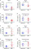
Figure 5.. Disulfiram inhibits NLRP3 inflammasome activation in vivo
(A–D) Mice were pre-treated with DSF (50 mg/kg) or vehicle sesame oil by intraperitoneal injection 24 and 4 h before intraperitoneal LPS challenge (20 mg/kg). IL-1β and TNF-α in serum and peritoneal lavage were measured by ELISA 12 h post-LPS challenge. Results were analyzed by unpaired two-tailed Student’s t test. (E–H) Mice were pre-treated with DSF (50 mg/kg) or vehicle sesame oil by intraperitoneal injection 24 and 4 h before intraperitoneal Salmonella infection (6 × 10⁷ colony-forming units (CFUs). IL-1β and TNF-α in serum and peritoneal lavage were measured by ELISA 6 h after Salmonella infection. Results were analyzed by the Mann-Whitney test. Results were from two experiments. The bars represent mean ± SD. ns, not significant; *p < 0.05 and ***p < 0.001. See also Figures S7.

References

    1. Xu J, and Núñez G (2023). The NLRP3 inflammasome: activation and regulation. Trends Biochem. Sci. 48, 331–344. 10.1016/j.tibs.2022.10.002. - DOI - PMC - PubMed
    1. Mangan MSJ, Olhava EJ, Roush WR, Seidel HM, Glick GD, and Latz E (2018). Targeting the NLRP3 inflammasome in inflammatory diseases. Nat. Rev. Drug Discov. 17, 588–606. 10.1038/nrd.2018.97. - DOI - PubMed
    1. Wang L, Cai J, Zhao X, Ma L, Zeng P, Zhou L, Liu Y, Yang S, Cai Z, Zhang S, et al. (2023). Palmitoylation prevents sustained inflammation by limiting NLRP3 inflammasome activation through chaperone-mediated autophagy. Mol. Cell 83, 281–297.e10. 10.1016/j.molcel.2022.12.002. - DOI - PubMed
    1. Ko PJ, and Dixon SJ (2018). Protein palmitoylation and cancer. EMBO Rep. 19, e46666. 10.15252/embr.201846666. - DOI - PMC - PubMed
    1. Linder ME, and Deschenes RJ (2007). Palmitoylation: policing protein stability and traffic. Nat. Rev. Mol. Cell Biol. 8, 74–84. 10.1038/nrm2084. - DOI - PubMed

Publication types

LinkOut - more resources