. 2024 Oct 8;8(19):5072-5085.
doi: 10.1182/bloodadvances.2024012979.

Activated platelets retain and protect most of their factor XIII-A cargo from proteolytic activation and degradation

Affiliations

Activated platelets retain and protect most of their factor XIII-A cargo from proteolytic activation and degradation

Yaqiu Sang et al. Blood Adv. .

Abstract

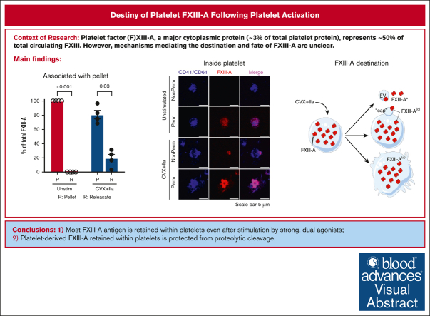
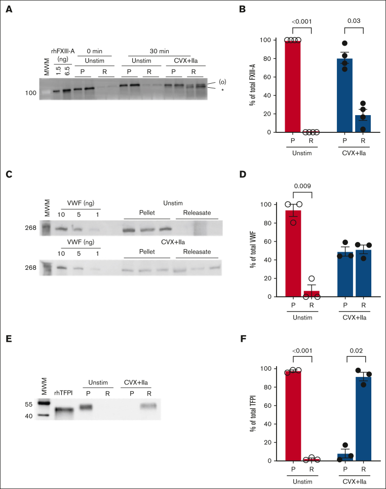
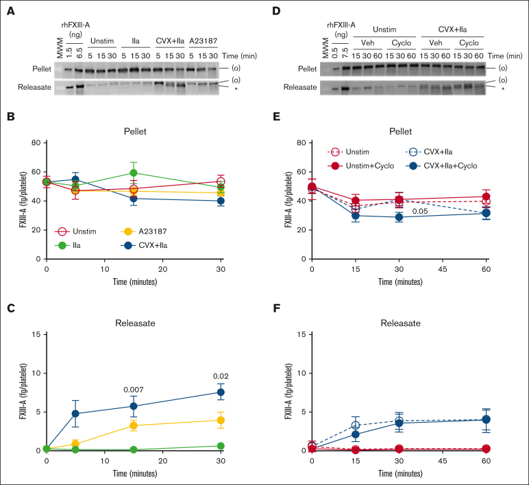
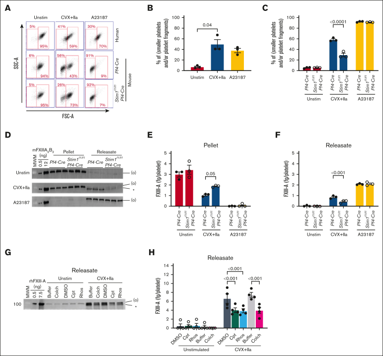
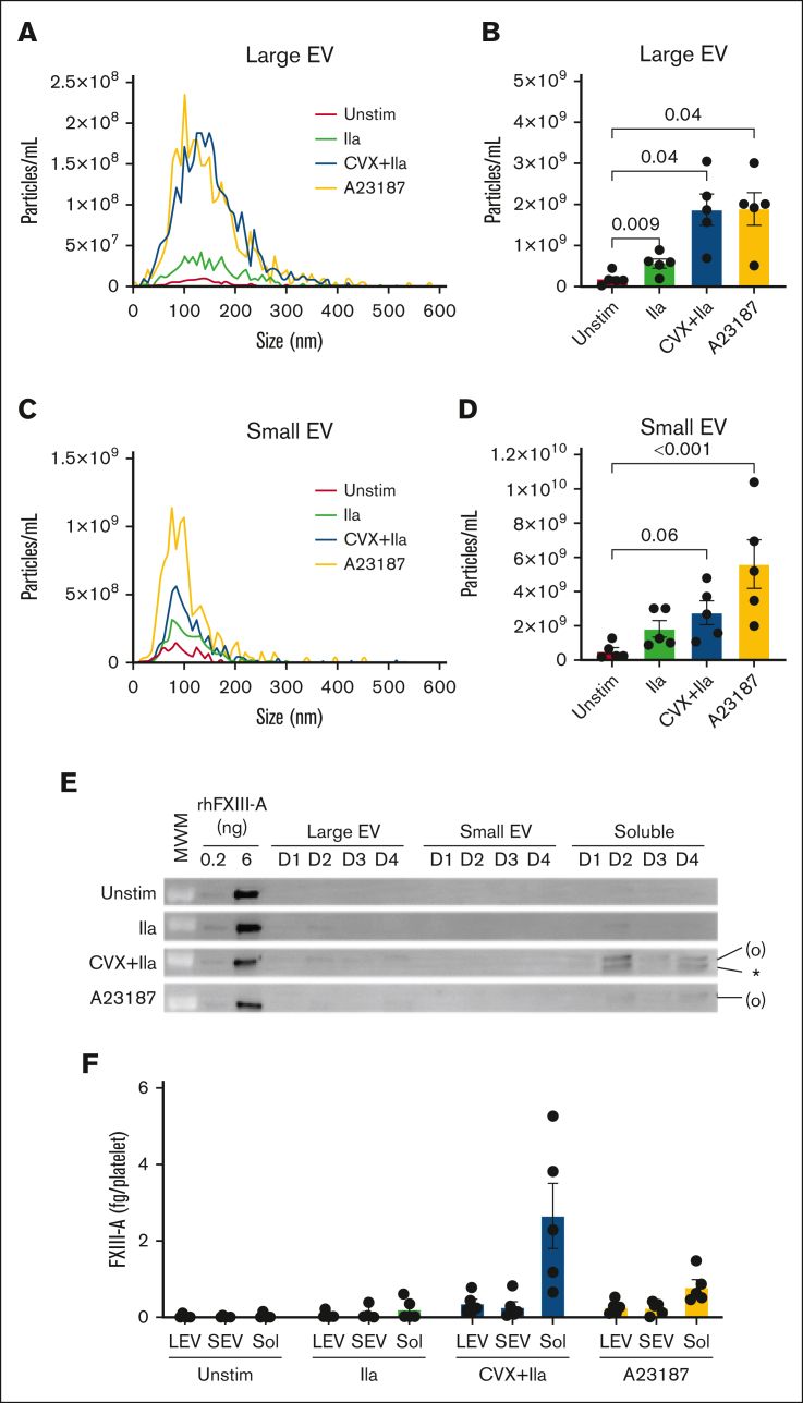
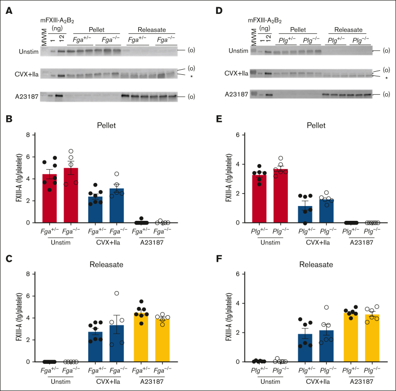
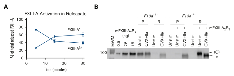
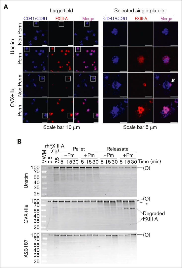
Platelet factor XIII-A (FXIII-A) is a major cytoplasmic protein (∼3% of total), representing ∼50% of total circulating FXIII. However, mobilization of FXIII-A during platelet activation is not well defined. To determine mechanisms mediating the retention vs release of platelet FXIII-A, platelets from healthy humans and mice (F13a1-/-, Fga-/-, Plg-/-, Stim1fl/flPf4-Cre, and respective controls) were stimulated with thrombin, convulxin plus thrombin, or calcium ionophore (A23187), in the absence or presence of inhibitors of transglutaminase activity, messenger RNA (mRNA) translation, microtubule rearrangement, calpain, and Rho GTPase. Platelet releasates and pellets were separated by (ultra)centrifugation. FXIII-A was detected by immunoblotting and immunofluorescence microscopy. Even after strong dual agonist (convulxin plus thrombin) stimulation of human platelets, >80% platelet FXIII-A remained associated with the platelet pellet. In contrast, essentially all tissue factor pathway inhibitor, another cytoplasmic protein in platelets, was released to the supernatant. Pellet-associated FXIII-A was not due to de novo synthesis via platelet F13A1 mRNA. The proportion of platelet FXIII-A retained by vs released from activated platelets was partly dependent on STIM1 signaling, microtubule rearrangement, calpain, and RhoA activation but did not depend on the presence of fibrinogen or plasminogen. Immunofluorescence microscopy confirmed the presence of considerable FXIII-A within the activated platelets. Although released FXIII-A was cleaved to FXIII-A∗ and could be degraded by plasmin, platelet-associated FXIII-A remained uncleaved. Retention of substantial platelet-derived FXIII-A by activated platelets and its reduced susceptibility to thrombin- and plasmin-mediated proteolysis suggest platelet FXIII-A is a protected pool with biological role(s) that differs from plasma FXIII.


Conflict of interest statement

Conflict-of-interest disclosure: The authors declare no competing financial interests.

Figures

None
Graphical abstract
Figure 1.
Figure 1.
Most platelet FXIII-A is retained with the platelet after dual agonist stimulation. Washed human platelets were unstimulated (Unstim) or stimulated with convulxin (CVX) plus thrombin (IIa) for 30 minutes. The platelet pellet (P) and releasate (R) were separated by centrifugation. FXIII-A, TFPI, and von Willebrand factor (VWF) were visualized by immunoblotting and quantified by densitometry. For analysis of pellets, proteins from 1 × 105 and 2 × 105 platelets were loaded in the first and second lanes of each pairing, respectively. For analysis of releasates, supernatants from 0.5 × 106 and 1 × 106 platelets were loaded for the first and second lanes of each pairing, respectively. (A) Representative immunoblot for FXIII-A. Recombinant human FXIII-A loading control is labeled rhFXIII-A. FXIII-A species are labeled on the right of the immunoblot as (O) (representing zymogen FXIII-A or nonproteolytically activated FXIII-A°) and ∗ (representing FXIII-A∗). (B) Quantification of FXIII-A. (C) Representative immunoblot of VWF; each lane represents a separate donor. (D) Quantitation of VWF. (E) Representative immunoblot of TFPI (recombinant human TFPI [rhTFPI] loading control). (F) Quantification of TFPI. For all immunoblots, molecular weight marker (MWM) is indicated on the left. For the bar graphs, the data show mean ± standard error of the mean (SEM); each dot represents a separate donor.
Figure 2.
Figure 2.
Platelet FXIII-A is not synthesized during activation of human platelets. (A-C) Washed human platelets were unstimulated (Unstim) or stimulated with thrombin (IIa), CVX plus IIa, or A23187. The platelet pellet (P) and releasate (R) were separated by centrifugation. FXIII-A was visualized by immunoblotting and quantified by densitometry. (A) Representative immunoblots of FXIII-A from the pellet (1.5 × 105 platelets) or releasate (7.5 × 105 platelets). (B-C) Quantification of total FXIII-A in the pellet and releasate, respectively (mean ± SEM; n = 5 separate donors; same symbols in both panels; P values vs unstimulated platelets at the same time point are indicated). (D-F) Washed human platelets were unstimulated (Unstim) or stimulated with CVX plus IIa in the presence of cycloheximide (cyclo) or vehicle (Veh [ethanol]). Samples were processed as those in panels A-C. (D) Representative immunoblots of FXIII-A. For analysis of the pellet, proteins from ∼2 × 105 platelets were loaded. For analysis of the releasate, supernatant from 1 × 106 platelets were loaded. (E-F) Quantification of total FXIII-A in the pellet and releasate, respectively (mean ± SEM; n = 6 separate donors; same symbols in both panels; P = .05 vs convulxin plus thrombin in the absence of cycloheximide at the same time point; the slight increase in FXIII-A in the pellet in the absence of cycloheximide at 30 minutes is likely an artifact because this is too short of a time frame for significant protein synthesis). For both immunoblots, rhFXIII-A is recombinant human FXIII-A loading control, and the band in the MWM lane indicates 100 kDa. FXIII-A species are labeled on the right of the immunoblot as (O) (representing zymogen FXIII-A or nonproteolytically activated FXIII-A°) and ∗ (representing FXIII-A∗).
Figure 3.
Figure 3.
FXIII-A translocation is partly dependent on STIM1 signaling and cytoskeletal rearrangements. (A-F) Washed human or mouse platelets were unstimulated (Unstim) or stimulated with CVX plus thrombin (IIa) or A23187 for 30 minutes. (A-C) Five-thousand events per sample were analyzed by flow cytometry. (A) Cytograms of human and mouse platelets. Two gates (smaller platelets and/or platelet fragments [left]; intact platelets [right]) were set based on unstimulated platelets. (B) Percentage of smaller platelets and/or platelet fragments for human platelets (n = 3; each dot represents a separate donor). (C) Percentage of smaller platelets and/or platelet fragments for mouse platelets (n = 3; each dot represents a separate mouse). (D-F) Mouse platelet pellet and releasate were separated by centrifugation at 1500g. FXIII-A was visualized by immunoblotting and quantified by densitometry. (D-F) Representative immunoblots (D) and quantitation (E-F) of FXIII-A in the mouse platelet pellet and releasate from 1.5 × 106 platelets. Each lane and dot represents a separate mouse. (G-H) Washed human platelets were unstimulated or stimulated with CVX plus IIa in the absence (buffer, DMSO [vehicle]) or presence of calpeptin (Cpt), rhosin (Rhos), or colchicine (Colch). The platelet pellet and releasate were separated by centrifugation. FXIII-A was quantified by densitometry. (G-H) Representative immunoblot and quantitation of FXIII-A in the human platelet releasate from 1 × 106 platelets (mean ± SEM of 4 separate donors). FXIII-A species are labeled on the right of the immunoblot as (O) (representing zymogen FXIII-A or nonproteolytically activated FXIII-A°) and ∗ (representing FXIII-A∗). Bars show mean ± SEM. DMSO, dimethyl sulfoxide. Statistical comparison in (H) are with appropriate controls (DMSO or buffer [diluted Tyrodes]).
Figure 4.
Figure 4.
Released FXIII-A is mainly free protein. Washed human platelets were unstimulated (Unstim) or stimulated with thrombin (IIa), CVX plus thrombin (IIa), or A23187 for 30 minutes at 37°C. The reaction mixture was then quenched and centrifuged serially to separate the pellet and releasate and then large EVs, small EVs, and remaining soluble fraction (free proteins), as described in the “Methods” and illustrated in supplemental Figure 5. (A-D) Nanoparticle tracking analysis of size distribution and enumeration of large EVs (A-B) and small EVs (C-D). (E) Representative immunoblots of FXIII-A in large EVs (pellet of 20 000g, 15 minutes), small EVs (pellet of 100 000g, 70 minutes), and soluble fraction (supernatant of 100 000g, 70 minutes) from 4 of the donors (D) studied; rhFXIII-A is recombinant human FXIII-A loading control; the band in the MWM lane indicates 100 kDa. FXIII-A species are labeled on the right of the immunoblot as (O) (representing zymogen FXIII-A or nonproteolytically activated FXIII-A°) and ∗ (representing FXIII-A∗). (F) Quantification of FXIII-A in large EVs (LEVs), small EVs (SEVs), and soluble proteins (Sol). The data show mean ± SEM of 5 separate donors; each dot represents a separate donor.
Figure 5.
Figure 5.
Retention and release of FXIII-A from convulxin plus thrombin–stimulated platelets do not depend on endogenous fibrinogen or plasminogen. Washed platelets from fibrinogen-deficient (Fga−/−), plasminogen- deficient (Plg−/−), or littermate control (Fga+/− and Plg+/−, respectively) mice were unstimulated (Unstim) or stimulated with CVX plus thrombin (IIa) or A23187 for 30 minutes. The platelet pellets and releasate were separated by centrifugation. FXIII-A was visualized by immunoblotting and quantified by densitometry. (A-C) Studies with fibrinogen-sufficient and -deficient platelets. (D-F) Studies with plasminogen-sufficient and -deficient platelets. (A,D) Immunoblots of FXIII-A in the pellet or releasate from 1.5 × 106 mouse platelets (mouse [m]FXIII-A2B2 loading control). The band in the MWM lane indicates 100 kDa. FXIII-A species are labeled on the right of the immunoblot as (O) (representing zymogen FXIII-A or nonproteolytically activated FXIII-A°) and ∗ (representing FXIII-A∗). (B,E) Quantification of total FXIII-A in the pellet. (C,F) Quantification of total FXIII-A in the releasate. The data show mean ± SEM; each dot is a separate mouse.
Figure 6.
Figure 6.
Released FXIII-A is cleaved by thrombin, whereas platelet-derived FXIII-A that remains associated with the platelet is protected from thrombin cleavage. (A) Quantification of relative FXIII-A(°) and FXIII-A∗ in the releasate of CVX plus thrombin (IIa)–stimulated platelets as in Figure 2A-C. The data show mean ± SEM of 5 separate donors. (B) Representative immunoblot of FXIII-A in pellets (P) and releasate (R) from experiments with washed platelets from F13a1+/+ or F13a1−/− mice unstimulated or stimulated with CVX plus IIa in the absence (for F13a1+/+ platelets) or presence (for F13a1−/− platelets) of exogenous mouse FXIII (mFXIII-A2B2) for 30 minutes (n = 3). Proteins from ∼1.5 × 106 platelets were loaded. FXIII-A species are labeled on the right of the immunoblot as (O) (representing zymogen FXIII-A or nonproteolytically activated FXIII-A°) and ∗ (representing FXIII-A∗).
Figure 7.
Figure 7.
Activated platelets retain most of their FXIII-A inside the cell, where it is protected from proteolytic degradation. (A) Washed human platelets were unstimulated (Unstim) or stimulated with CVX plus thrombin (IIa) for 30 minutes. Intact (nonpermeabilized [Non-Perm]) or permeabilized (Perm) platelets were labeled with antibodies against CD41/CD61 (surface marker, blue) and FXIII-A (red). A merge of CD41/CD61 and FXIII-A is shown in pink. Platelets were imaged on a Zeiss LSM 900 confocal microscope (UNC Microscopy Service Laboratory) through a Plan-Apochromat 63×/1.40 oil immersion objective. For optimal display, the brightness/contrast was adjusted separately; gray scale images with equivalent settings are shown in supplemental Figure 8. Representative images of experiments performed on platelets from 3 different donors are shown. The white boxes (left images) indicate the selected individual platelets shown on the right images. White arrow points to FXIII-A exposed on the cap-like structure on the platelet surface. (B) Representative immunoblots of FXIII-A from washed human platelets (n = 5 separate donors) that were unstimulated (Unstim) or stimulated with CVX plus thrombin (IIa) in the absence or presence of plasmin (Pm) for up to 30 minutes. The pellet and releasate were separated by centrifugation. For analysis of the pellet, proteins from 2 × 105 platelets were loaded. For analysis of the releasate, supernatant from 1 × 106 platelets were loaded. FXIII-A species are labeled on the right of the immunoblot as (O) (representing zymogen FXIII-A or nonproteolytically activated FXIII-A°) and ∗ (representing FXIII-A∗).

References

    1. Muszbek L, Bereczky Z, Bagoly Z, Komaromi I, Katona E. Factor XIII: a coagulation factor with multiple plasmatic and cellular functions. Physiol Rev. 2011;91(3):931–972. - PubMed
    1. Katona E E, Ajzner E, Toth K, Karpati L, Muszbek L. Enzyme-linked immunosorbent assay for the determination of blood coagulation factor XIII A-subunit in plasma and in cell lysates. J Immunol Methods. 2001;258(1-2):127–135. - PubMed
    1. Mills EW, Green R, Ingolia NT. Slowed decay of mRNAs enhances platelet specific translation. Blood. 2017;129(17):e38–e48. - PMC - PubMed
    1. Mohammed RDS, Ablan FDO, McCann NM, Hindi MM, Maurer MC. Transglutaminase activities of blood coagulant factor XIII are dependent on the activation pathways and on the substrates. Thromb Haemost. 2023;123(4):380–392. - PMC - PubMed
    1. Anokhin BA, Dean WL, Smith KA, et al. Proteolytic and nonproteolytic activation mechanisms result in conformationally and functionally different forms of coagulation factor XIII A. FEBS J. 2020;287(3):452–464. - PMC - PubMed

Publication types

LinkOut - more resources