. 2024 Oct;11(40):e2404159.
doi: 10.1002/advs.202404159. Epub 2024 Aug 8.

Intranasal Multiepitope PD-L1-siRNA-Based Nanovaccine: The Next-Gen COVID-19 Immunotherapy

Affiliations

Intranasal Multiepitope PD-L1-siRNA-Based Nanovaccine: The Next-Gen COVID-19 Immunotherapy

Rita C Acúrcio et al. Adv Sci (Weinh). 2024 Oct.

Abstract

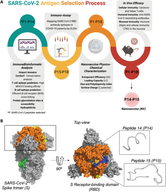
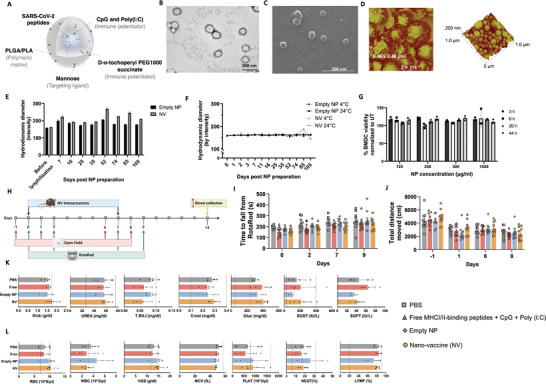
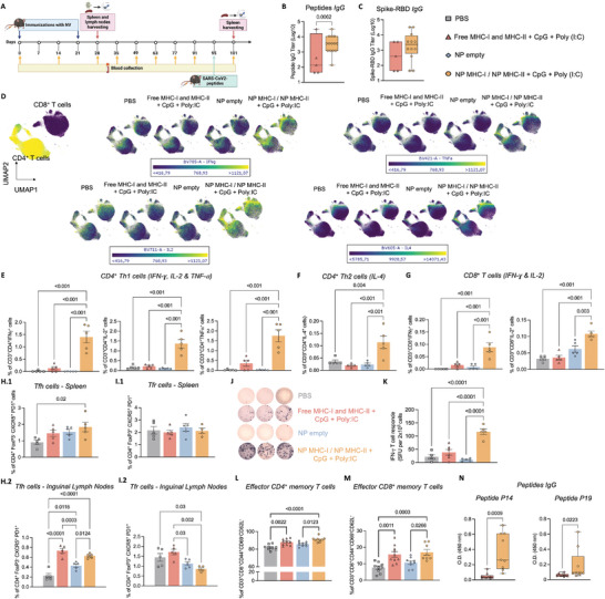
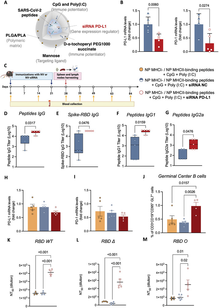
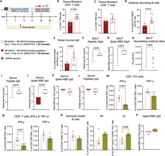
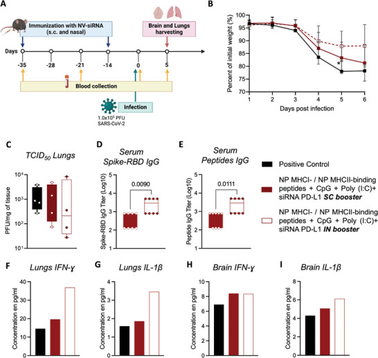
The first approved vaccines for human use against severe acute respiratory syndrome coronavirus 2 (SARS-CoV-2) are nanotechnology-based. Although they are modular, rapidly produced, and can reduce disease severity, the currently available vaccines are restricted in preventing infection, stressing the global demand for novel preventive vaccine technologies. Bearing this in mind, we set out to develop a flexible nanovaccine platform for nasal administration to induce mucosal immunity, which is fundamental for optimal protection against respiratory virus infection. The next-generation multiepitope nanovaccines co-deliver immunogenic peptides, selected by an immunoinformatic workflow, along with adjuvants and regulators of the PD-L1 expression. As a case study, we focused on SARS-CoV-2 peptides as relevant antigens to validate the approach. This platform can evoke both local and systemic cellular- and humoral-specific responses against SARS-CoV-2. This led to the secretion of immunoglobulin A (IgA), capable of neutralizing SARS-CoV-2, including variants of concern, following a heterologous immunization strategy. Considering the limitations of the required cold chain distribution for current nanotechnology-based vaccines, it is shown that the lyophilized nanovaccine is stable for long-term at room temperature and retains its in vivo efficacy upon reconstitution. This makes it particularly relevant for developing countries and offers a modular system adaptable to future viral threats.

Keywords: Dendritic cells; Intranasal; MHC class I and MHC class II peptides; Nanovaccines; PD‐1/PD‐L1 immune checkpoints; SARS‐CoV‐2; siRNA.


Conflict of interest statement

R.S.‐F. is a Board Director at Teva Pharmaceutical Industries, Ltd. All other authors declare that they have no competing interests.

Figures

Figure 1
Figure 1
SARS‐CoV‐2 peptide selection immunoinformatic analysis workflow. A) SARS‐CoV‐2 antigen selection strategy. B) SARS‐CoV‐2 Spike trimer (PDB ID 6VXX) surface representation in gray. The receptor‐binding domain (RBD) from each monomer is highlighted in orange. The top‐ranked epitope sequences of in silico workflow are highlighted in yellow (RBD area) and green (other spike regions). In the top view, selected peptides are highlighted in red (MHC‐I binding peptide) and blue (MHC‐II binding peptide). 3D structure prediction of SARS‐CoV‐2 RBD‐peptide (P) 14 and P15 using PEP‐FOLD server.[ 21 ]
Figure 2
Figure 2
Nanoplatform physicochemical characterization. A) Schematic representation of the nanovaccine (NV). B) Representative image of spherical empty nanoparticle (NP) by transmission electron microscopy (TEM), scale bar = 200 nm. C) Representative image of spherical empty NP by scanning electron microscopy (SEM), scale bar = 1 µm. D) Atomic force microscopy (AFM) images show the spherical shape of NP with a slight roughness surface, scale bar = 200 nm. E,F) Hydrodynamic diameter measurements by dynamic light scattering (DLS) of empty NP (NP w/o immunogens) and NV (NP entrapping one SARS‐CoV‐2‐RBD peptide and the two TLR ligands) over time. E) Lyophilized empty NP and NV stored at 24 °C. F) Empty NP and NV in suspension were stored at 4 and 24 °C. Data represent mean ± s.d. (n =  5). G) Cell viability of bone marrow‐derived dendritic cells (BMDC) after incubation with NP for 44 h by XTT. Data represent mean ± s.d. (a representative graph of 2 independent experiments demonstrating the same trend). All group comparisons were non‐significant (NS) by the One‐way ANOVA test. H) In vivo safety timeline, C57BL/6J mice. I,J) Assessment of motor function. I) RotaRod test. The motor learning of C57BL/6 male mice was analyzed in a five‐lane accelerating RotaRod. J) Open field test. The distance traveled by C57BL/6 male mice during a 15‐min video recording was analyzed using EthoVision 13XT software. Data represent mean ± s.e.m., N = 10 mice. All group comparisons were NS by the One‐way ANOVA test. K,L) Blood test, N = 5 mice. All comparisons between the groups were NS by the One‐way ANOVA test. Data represent mean ± s.e.m. Whisker charts show minimum and maximum values. K) Blood chemistry panel, and L) Blood hematology panel (CBC panel).
Figure 3
Figure 3
Nanovaccine ‐8 (NV‐8) elicited robust receptor‐binding domain (RDB)‐specific T‐ and B‐cell responses. A) Immunization scheme of C57BL/6J mice timeline. B) SARS‐CoV‐2 RBD‐peptide, and C) RBD‐specific IgG antibody titers determined by Enzyme‐Linked Immunosorbent Assay (ELISA) on day 35. Box and whisker plots represent the mean, min, and max, N = 5 (PBS) or 12 (NV), unpaired t‐test. D‐H) Cellular response. D) Uniform manifold approximation and projection (UMAP) plot of lymphocytes (CD45+ CD3+ cells) colored by Interferon gamma (IFN‐γ), Tumor necrosis factor‐alpha (TNF‐α), Interleukin‐2 (IL‐2), and IL‐4 expression. E) Frequency of antigen‐specific CD4+ T cells producing T helper 1 (Th1) cytokines, IFN‐γ, IL‐2, and TNF‐α; F) Th2 cytokine, IL‐4; and G) CD8+ T cells producing IFN‐γ and IL‐2 cytokines were evaluated 1 week after the second vaccination (day 28) in 6 h antigen‐stimulated splenocytes. H) Frequency of Tfh retrieved from (1) spleen and (2) inguinal lymph nodes of mice on day 28. Data represent mean ± s.e.m., N = 5 mice, one‐way ANOVA followed by Tukey's multiple comparisons test. I) Frequency of Tfr cells retrieved from (1) spleen and (2) inguinal lymph nodes of mice on day 28. Data represent mean ± s.e.m., N = 5 mice, one‐way ANOVA followed by Tukey's multiple comparisons test. J) ELISpot representative images of IFN‐γ spot forming cells among splenocytes after ex vivo restimulation with peptides 14 and 15 on day 28. Each condition was repeated five times. K) IFN‐γ spot forming units (SFU). Data represent mean ± s.e.m., N = 5 mice, one‐way ANOVA followed by Tukey's multiple comparisons test. L‐M) Frequency of effector memory T cells (CD4+ and CD8+) evaluated 1 week after IV administration of MHCI/II‐binding peptides and the adjuvants (CpG and Poly(I:C)). N) SARS‐CoV‐2 RBD‐peptides IgG levels were evaluated on day 101 by ELISA (dilution 1:500). The number of mice per group: PBS/Empty NP = 9, Free = 10, and NV‐8 = 8, mean ± s.e.m.
Figure 4
Figure 4
Co‐delivery of receptor‐binding domain (RBD)‐peptides, toll‐like receptor (TLR) ligands, and siRNA against Programmed death‐ligand 1 (PD‐L1) (siPD‐L1 NV‐8) increases NV‐8 neutralizing‐antibody responses. A) Schematic representation of siPD‐L1 NV‐8 B) PD‐L1 and PD‐1 mRNA levels from spleen, 55 h post siNC NV‐8 (negative control of scramble siRNA) or siPD‐L1 NV‐8 immunization. mRNA levels measured by qRT‐PCR. Data represent mean ± s.e.m., N = 5 mice, unpaired t‐test. C) Immunization scheme of C57BL/6J mice timeline. SARS‐CoV‐2 RBD‐peptides, and RBD‐specific IgG D,E), IgG1 F), and IgG2a G) antibody titers determined by ELISA. Box and whiskers represent the mean, min, and max, N = 5 mice per group, unpaired t‐test. H,I) PD‐L1, and PD‐1 mRNA levels, from spleens, on day 28. mRNA levels measured by qRT‐PCR. Data represent mean ± s.e.m., N = 5 mice, one‐way ANOVA followed by Dunnett's multiple comparisons test. J) Frequency of germinal center (GC) B cells detected by flow cytometry on day 28. Data represent mean ± s.e.m., N = 5 mice, one‐way ANOVA followed by Tuke's multiple comparisons test. NT50 in serum against SARS‐CoV‐2 RBD WT K), Delta (Δ) L), and Omicron BA.1.1 (Ο) M) in mice immunized with NV‐8, siNC NV‐8, and siPD‐L1 NV‐8 using surrogate virus neutralization test. Data represent mean ± s.e.m., N = 5 mice, one‐way ANOVA followed by Dunnett's multiple comparisons test.
Figure 5
Figure 5
Intranasal siPD‐L1 NV‐8‐booster elicited robust mucosal immunity. A) Immunization scheme of C57BL/6J mice timeline. B,C) Mucosal‐cellular response determined by flow cytometry on day 28. Frequency of tissue‐resident T cells (TRM) (CD4+ and CD8+) D,C), and antibody‐secreting B cells E) in the nasal mucosa. Data represent mean ± s.e.m., N = 5 mice (subcutaneous (SC) booster), and 10 (intranasal (IN) booster), one‐way ANOVA followed by Tuke's multiple comparisons test. E–K) Mucosal‐humoral response. E) Frequency of immunoglobulin (Ig) IgA‐B cells in nasal mucosa on day 28. F,G) SARS‐CoV‐2 receptor‐binding domain (RBD)‐peptides and RBD‐specific secretory immunoglobulin A (SIgA) titers in bronchoalveolar lavage fluid (BALF) determined by ELISA. Box and whiskers represent the mean, min, and max, N = 5 mice per group, unpaired t‐test. H) Neutralizing antibody titers (NT50) against SARS‐CoV‐2 in BALF determined by surrogate virus neutralization test. Data represent mean ± s.e.m., N = 5 mice, unpaired t‐test. SARS‐CoV‐2 RBD‐peptides and RBD‐specific IgA titers I,J), and IgG in serum K,L) determined by ELISA. Box and whiskers represent the mean, min, and max, N = 5 mice per group, unpaired t‐test. M–P) Comparison between siPD‐L1 NV‐8 versus mRNA vaccine. M,N) Cellular response. The frequency of antigen‐specific CD4+ T cells producing T helper 1 (Th1) cytokines such as interferon‐gamma (IFN‐γ) and tumor necrosis factor‐alpha (TNF‐α) M) was evaluated one week after the intranasal booster (day 28) and after stimulation with RBD‐peptides or spike protein for 6 h. Cytokine production (IFN‐γ and TNF‐α) was also evaluated on CD8+ T cells N). O) Frequency of germinal center B cells, Tfh, and Tfr cells retrieved from mice on day 28. Data represent mean ± s.e.m., N = 5 mice, unpaired t‐test. P) SARS‐CoV‐2 RBD IgG antibody titers determined on day 35 by ELISA. Box and whisker plots represent the mean, min, and max, N = 5, unpaired t‐test.
Figure 6
Figure 6
Intranasal siPD‐L1 NV‐8 booster triggers a protective Th1‐guided immune response in vivo. A) Immunization scheme of K18‐hACE2 mice timeline. K18‐hACE2 mice (N = 10 or 11) received a SC prime immunization with siPD‐L1 NV‐8. Mice received the SC or IN booster three weeks later. Two weeks after the boost immunization, mice were infected intranasally with 1.0 × 105 PFU SARS‐CoV‐2. B) Animals were monitored for body weight. On day 5 post‐infection, viral loads in the lung tissue C) were determined by an assay to quantify PFUs of infectious SARS‐CoV‐2. D,E) The SARS‐CoV‐2 RBD‐ and RBD peptides‐specific IgG titers in mouse serum were determined by ELISA. Box and whiskers represent the mean, min, and max, N = 4 mice per group in duplicate, unpaired t‐test. Mouse cytokine array shows high levels of IFN‐γ and IL‐1β in the lungs F,G) and brain H,I).

Similar articles

Cited by

References

    1. Coronavirus (COVID‐19) Vaccinations, accessed: April, 2024, https://www.frontiersin.org/journals/immunology/articles/10.3389/fimmu.2.... - DOI
    1. Florindo H. F., Kleiner R., Vaskovich‐Koubi D., Acúrcio R. C., Carreira B., Yeini E., Tiram G., Liubomirski Y., Satchi‐Fainaro R., Nat. Nanotechnol. 2020, 15, 630. - PMC - PubMed
    1. Friedrichs S., Bowman D. M., Nat. Nanotechnol. 2021, 16, 362. - PubMed
    1. a) Barda N., Dagan N., Ben‐Shlomo Y., Kepten E., Waxman J., Ohana R., Hernán M. A., Lipsitch M., Kohane I., Netzer D., Reis B. Y., Balicer R. D., N. Engl. J. Med. 2021, 385, 1078; - PMC - PubMed
    2. b) Anderson E. J., Rouphael N. G., Widge A. T., Jackson L. A., Roberts P. C., Makhene M., Chappell J. D., Denison M. R., Stevens L. J., Pruijssers A. J., McDermott A. B., Flach B., Lin B. C., Doria‐Rose N. A., O'Dell S., Schmidt S. D., Corbett K. S., Swanson P. A., Padilla M., Neuzil K. M., Bennett H., Leav B., Makowski M., Albert J., Cross K., Edara V. V., Floyd K., Suthar M. S., Martinez D. R., Baric R., et al., N. Engl. J. Med. 2020, 383, 2427. - PMC - PubMed
    1. Uddin M. N., Roni M. A., Vaccines 2021, 9, 1033. - PubMed

LinkOut - more resources