. 2024 Aug 19;4(8):100835.
doi: 10.1016/j.crmeth.2024.100835. Epub 2024 Aug 7.

Development of a 3-dimensional organotypic model with characteristics of peripheral sensory nerves

Affiliations

Development of a 3-dimensional organotypic model with characteristics of peripheral sensory nerves

Madoka Koyanagi et al. Cell Rep Methods. .

Abstract

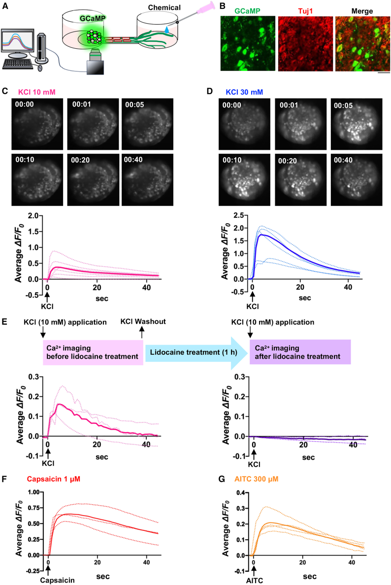
We developed a rat dorsal root ganglion (DRG)-derived sensory nerve organotypic model by culturing DRG explants on an organoid culture device. With this method, a large number of organotypic cultures can be produced simultaneously with high reproducibility simply by seeding DRG explants derived from rat embryos. Unlike previous DRG explant models, this organotypic model consists of a ganglion and an axon bundle with myelinated A fibers, unmyelinated C fibers, and stereo-myelin-forming nodes of Ranvier. The model also exhibits Ca2+ signaling in cell bodies in response to application of chemical stimuli to nerve terminals. Further, axonal transection increases the activating transcription factor 3 mRNA level in ganglia. Axons and myelin are shown to regenerate 14 days following transection. Our sensory organotypic model enables analysis of neuronal excitability in response to pain stimuli and tracking of morphological changes in the axon bundle over weeks.

Keywords: CP: Neuroscience; Schwann cells; ex vivo explant culture; microfluidic device; myelin; nerve regeneration; node of Ranvier; organotypic model; peripheral nervous system; peripheral neuropathy; sensory nerve.


Conflict of interest statement

Declaration of interests The authors declare no competing interests.

Figures

None
Graphical abstract
Figure 1
Figure 1
Generation of the sensory nerve organotypic model (A) Photographs of organoid culture microchambers (left, six-well type; center, three-well type) and schematic of the well within the chambers (right). (B) Schematic of the protocol for seeding DRG explants on the organoid culture microchamber. (C) Bright-field images of organotypic model growth at 2, 11, and 21 days following embryonic rat DRG seeding on the culture microchamber. (D–G) Representative confocal images of an organotypic model 28 days after seeding. (D) Images showing NF200-positive (blue), IB4-positive (green), and CGRP-positive (red) neuronal cell bodies in the ganglion-like structure. (E) Images showing Tuj1-positive (green), NF200-positive (green), IB4-positive (green), and CGRP-positive (green) nerve fibers in organotypic axon fascicles. (F) Images showing NF200-positive nerve fibers (green) and MBP-positive myelin (red) in organotypic axon fascicles. (G) Images showing Ezrin puncta (red) at the node of Ranvier and Caspr puncta (green) at paranodes in organotypic axon fascicles. Scale bars: 300 μm (C), 50 μm (D–F), and 25 μm (G).
Figure 2
Figure 2
Structural characteristics and ganglion expression of representative ion channels, receptors, and sensory neuron markers in the sensory nerve organotypic model (A) Schematic of the organotypic model, showing its structure. (B) Electron micrographs of an axon bundle cross-section containing abundant myelinated nerve fibers 28 days after seeding. (C) Enlarged images of whole-axon-bundle coronal section (dotted red square in B). A fibers (green), myelinating Schwann cells (red), C fibers (orange) and non-myelinating Schwann cells (blue) were present in the axon bundle. The enlarged views of the A fiber (a), multi-layered myelin sheath (b), and C fiber (c) correspond to the dotted squares in (B). (D) Electron micrographs of an axon bundle sagittal section showing a node of Ranvier 28 days after seeding. (E) 3D reconstructed image of the axon bundle sagittal section in (D), showing a node of Ranvier. (F) Schematic showing sample collection of a ganglion-like structure of an organotypic culture for RT-PCR. (G–I) RT-PCR bands showing mRNA levels of (G) cation channels, (H) nerve fiber markers, (I) functional receptors and pro-nociceptive cytokines and chemokines in ganglia of organotypic cultures 28 days after seeding. GAPDH was used as an internal control. n = 3. (J–N) Confocal images showing Nav1.8-positive (J, green), TRPV1-positive (K, green), TRPA1-positive (K, red), TrkA-positive (L, green), CGRP-positive (L, red), TrkB-positive (M, green), NF200-positive (M and N, red), and Npy2r-positive (N, green) neuronal cell bodies in the ganglion-like structure of an organotypic model 28 days after seeding. Scale bars: 10 μm (B), 500 nm (C), 1 μm (D), and 50 μm (J–N).
Figure 3
Figure 3
Chemical stimulus-induced neuronal excitation (A) Schematic showing measurement of GCaMP6m fluorescence in neuronal cell bodies within the ganglion-like structure. (B) Confocal images showing GCaMP6m (green)- and Tuj1 (red)-positive neuronal cell bodies in the ganglion-like structure of an organotypic culture after AAV-hSyn-GCaMP6m infection. See also Figure S3. Scale bar: 50 μm. (C and D) GCaMP6m fluorescence micrographs (top) and traces (bottom) indicating Ca2+ responses in neuronal cell bodies of organotypic cultures upon KCl treatment (C, 10 mM; D, 30 mM). n = 6 (C) and 5 (D). (E) GCaMP6m fluorescence traces showing Ca2+ responses in organotypic neuronal cell bodies upon KCl application (10 mM) before and after lidocaine treatment (3 mM). n = 3. (F and G) GCaMP6m fluorescence traces showing Ca2+ responses in neuronal cell bodies of organotypic cultures upon capsaicin (1 μM, F) and AITC (300 μM, G). n = 3 (F) and 4 (G). Dotted and bold traces show data from individual organotypic cultures and the mean of six (C), five (D), three (E and F), and four (G) organotypic cultures.
Figure 4
Figure 4
Gene expression changes and regeneration of axon bundle nerve fibers and myelin after transection (A) Changes in ganglion-like structure gene expression were analyzed after axon bundle transection in the middle of the microchannel. (B) Fluorescence micrographs of Tuj1 (green) and ATF3 (red) immunoreactivity (left) and percentage of ATF3-positive neurons among Tuj1-positive neurons (right) in ganglion-like structures of organotypic cultures 1 day following axon bundle transection. Data are expressed as means ± SEM. ∗p < 0.05 vs. control (non-transected organoid). n = 4. (C) Quantitative real-time PCR analysis of Atf3 and Il6 mRNA levels in ganglion-like structures of organotypic cultures 1 day after axonal transection. Data are expressed as means ± SEM. ∗∗p < 0.01 vs. control. n = 5. (D) Bright-field images showing axon bundle regeneration 0, 1, 7, and 11 days after transection. Regenerating axons and migrating Schwann cells are indicated by black arrows and arrowheads, respectively. PTD, post-transection day. (E) Confocal images showing NF200 (green) and MBP (red) immunoreactivity in a regenerated axon bundle 14 days after axonal transection. (F) Electron micrographs of a regenerated axon bundle cross-section showing regeneration of stereo-myelin 14 days after axonal transection. Scale bars: 200 μm (B), 100 μm (D), 50 μm (E), and 10 μm (F).

References

    1. Saitoh F., Araki T. Proteasomal degradation of glutamine synthetase regulates Schwann cell differentiation. J. Neurosci. 2010;30:1204–1212. doi: 10.1523/JNEUROSCI.3591-09.2010. - DOI - PMC - PubMed
    1. Malheiro A., Harichandan A., Bernardi J., Seijas-Gamardo A., Konings G.F., Volders P.G.A., Romano A., Mota C., Wieringa P., Moroni L. 3D culture platform of human iPSCs-derived nociceptors for peripheral nerve modeling and tissue innervation. Biofabrication. 2021;14 doi: 10.1088/1758-5090/ac36bf. - DOI - PubMed
    1. Koyanagi M., Imai S., Iwamitsu Y., Matsumoto M., Saigo M., Moriya A., Ogihara T., Nakazato Y., Yonezawa A., Nakagawa S., et al. Cilostazol is an effective causal therapy for preventing paclitaxel-induced peripheral neuropathy by suppression of Schwann cell dedifferentiation. Neuropharmacology. 2021;188 doi: 10.1016/j.neuropharm.2021.108514. - DOI - PubMed
    1. Numata-Uematasu Y., Wakatsuki S., Kobayashi-Ujiie Y., Sakai K., Ichinohe N., Araki T. In vitro myelination using explant culture of dorsal root ganglia: An efficient tool for analyzing peripheral nerve differentiation and disease modeling. PLoS One. 2023;18 doi: 10.1371/journal.pone.0285897. - DOI - PMC - PubMed
    1. Fornaro M., Sharthiya H., Tiwari V. Adult Mouse DRG Explant and Dissociated Cell Models to Investigate Neuroplasticity and Responses to Environmental Insults Including Viral Infection. J. Vis. Exp. 2018;133 doi: 10.3791/56757. - DOI - PMC - PubMed

Substances