. 2024 Nov;1868(11):130690.
doi: 10.1016/j.bbagen.2024.130690. Epub 2024 Aug 6.

Key charged residues influence the amyloidogenic propensity of the helix-1 region of serum amyloid A

Affiliations

Key charged residues influence the amyloidogenic propensity of the helix-1 region of serum amyloid A

Marvin Bilog et al. Biochim Biophys Acta Gen Subj. 2024 Nov.

Abstract

Increased plasma levels of serum amyloid A (SAA), an acute-phase protein that is secreted in response to inflammation, may lead to the accumulation of amyloid in various organs thereby obstructing their functions. Severe cases can lead to a systemic disorder called AA amyloidosis. Previous studies suggest that the N-terminal helix is the most amyloidogenic region of SAA. Moreover, computational studies implicated a significant role for Arg-1 and the residue-specific interactions formed during the fibrillization process. With a focus on the N-terminal region of helix-1, SAA1-13, mutational analysis was employed to interrogate the roles of the amino acid residues, Arg-1, Ser-5, Glu-9, and Asp-12. The truncated SAA1-13 fragment was systematically modified by substituting the key residues with alanine or uncharged but structurally similar amino acids. We monitored the changes in the amyloidogenic propensities, associated conformational markers, and morphology of the amyloids resulting from the mutation of SAA1-13. Mutating out Arg-1 resulted in much reduced aggregation propensity and a lack of detectable β-structures alluding to the importance of salt-bridge interactions involving Arg-1. Our data revealed that by systematically mutating the key amino acid residues, we can modulate the amyloidogenic propensity and alter the time-dependent conformational variation of the peptide. When the behaviors of each mutant peptide were analyzed, they provided evidence consistent with the aggregation pathway predicted by MD simulation studies. Here, we detail the important temporal molecular interactions formed by Arg-1 with Ser-5, Glu-9, and Asp-12 and discuss its mechanistic implications on the self-assembly of the helix-1 region of SAA.

Keywords: AA amyloidosis; Circular dichroism spectroscopy; Fourier transform infrared spectroscopy; Mechanism of aggregation; Serum amyloid a; Thioflavin T fluorescence assay.


Conflict of interest statement

Declaration of competing interest The authors declare that they have no conflicts of interest.

Figures

Figure 1.
Figure 1.
Time course ThT fluorescence assay of wild type SAA1–13 and its mutants in the absence (A) or presence (B) of heparin. Data were obtained at room temperature using 50 µg/mL peptide solutions in 10 mM Tris-HCl buffer (pH 7.4) placed in a 96-well plate and run on a plate reader set to measure ThT fluorescence. Samples were excited at 450 nm, and their ThT fluorescence emission intensities were monitored at 486 nm. Measurements were taken every 15 min for at least 20 h. Whenever needed, heparin was added to a final concentration of 41 µg/mL. The blank in each set of measurements contains all components except the peptide.
Figure 2.
Figure 2.
CD spectra of peptides (A) SAA1–13, (B) SAA1–13R1A, (C) SAA1–13R1Cit, (D) SAA1–13S5A, (G) SAA1–13E9A, (H) SAA1–13E9Q, (I) SAA1–13D12A, and (J) SAA1–13D12N incubated in heparin and 10 mM phosphate buffer (pH 7.4). CD spectra of peptides (E) SAA1–13 and (F) SAA1–13S5A incubated without heparin are also shown. CD measurements from 200 nm to 250 nm were done every hour for 24 hours at room temperature.
Figure 3.
Figure 3.
FTIR spectra, displayed as its second derivate to accentuate the peaks, of the amide I region of (A) aliquots taken from SAA1–13 mutants peptide solutions which exhibited formation of helical structures and (B) washed aggregates collected from the same peptide samples.
Figure 4.
Figure 4.
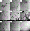
TEM images of the peptides solutions of (A) SAA1–13, (B) SAA1–13S5A, (C) SAA1–13D12A, (D) SAA1–13D12N, (E) SAA1–13E9A, and (F) SAA1–13E9Q, (H) SAA1–13R1A, and (I) SAA1–13R1Cit after incubation with heparin. TEM of (G) SAA1–13S5A fibrils from the solution incubated without heparin is also shown. All samples were incubated for 24 hours in Tris-HCl buffer (pH 7.4) at room temperature. Images were taken from aged samples deposited on 300 mesh copper grids with carbon film negatively stained with 2% uranyl acetate. Scale bars = 1 µm and 2µm in H and I, respectively, and 500 nm in other panels.
Figure 5.
Figure 5.
Rendering of a plausible misfolding mechanism for SAA1–13-based peptides. (A) SAA1–13 helices start to unravel to form (B) a labile helical intermediate that can self-assemble into (C) oligomeric species. Labile helices then transition to (D) bent intermediates. When the critical concentration is reached, the peptide spontaneously folds to a (E) β-hairpin (U-shaped) structure which pile up and grow to (F) mature amyloid fibrils with the characteristic cross-β motif (F).

References

    1. Pettersson T, Konttinen YT, and Maury CP (2008) Treatment Strategies for Amyloid a Amyloidosis, Expert Opin Pharmaco 9, 2117–2128. - PubMed
    1. Li H, Xiang X, Ren H, Xu L, Zhao L, Chen X, Long H, Wang Q, and Wu Q (2020) Serum Amyloid a Is a Biomarker of Severe Coronavirus Disease and Poor Prognosis, J Infect 80, 646–655. - PMC - PubMed
    1. Zinellu A, Paliogiannis P, Carru C, and Mangoni AA (2021) Serum Amyloid a Concentrations, Covid-19 Severity and Mortality: An Updated Systematic Review and Meta-Analysis, Int. J. Infect. Dis 105, 668–674. - PMC - PubMed
    1. Almusalami EM, Lockett A, Ferro A, and Posner J (2023) Serum Amyloid a—a Potential Therapeutic Target for Hyper-Inflammatory Syndrome Associated with Covid-19, Front Med 10. - PMC - PubMed
    1. Gonçalves C-A, Bobermin LD, Sesterheim P, and Netto CA (2022) Sars-Cov-2-Induced Amyloidgenesis: Not One, but Three Hypotheses for Cerebral Covid-19 Outcomes, Metabolites 12, 1099. - PMC - PubMed

Publication types

LinkOut - more resources