. 2024 Aug 8;14(1):18390.
doi: 10.1038/s41598-024-68992-6.

Paeonol alleviates ulcerative colitis by modulating PPAR-γ and nuclear factor-κB activation

Affiliations

Paeonol alleviates ulcerative colitis by modulating PPAR-γ and nuclear factor-κB activation

Shuyu Cheng et al. Sci Rep. .

Abstract

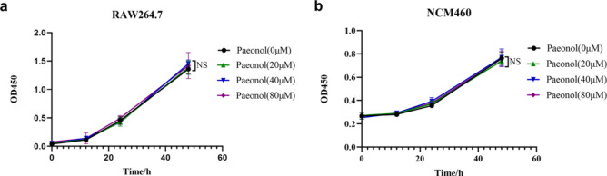
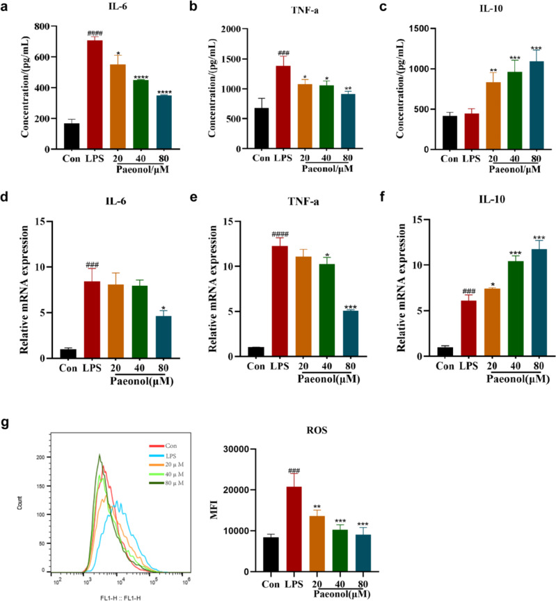
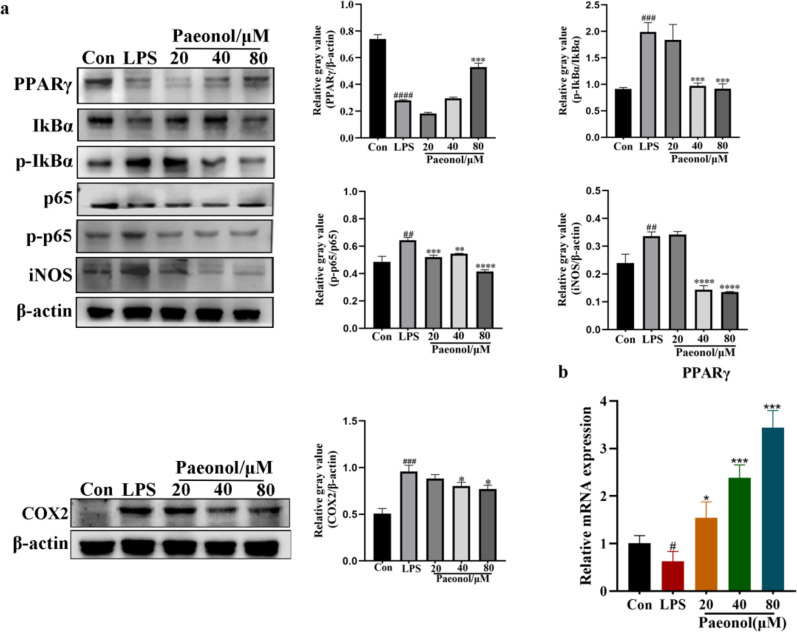
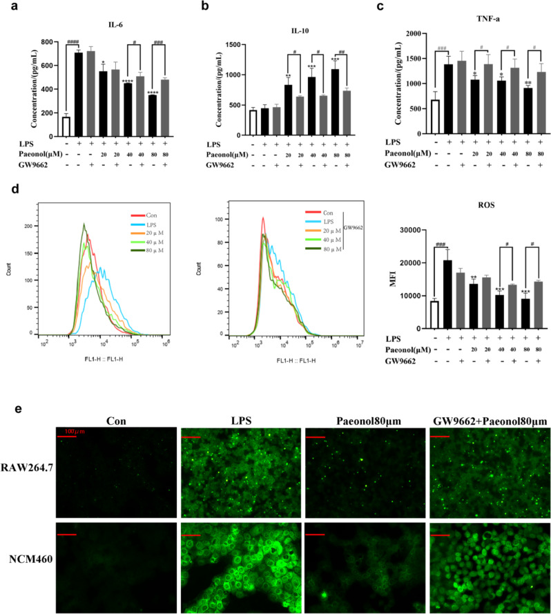
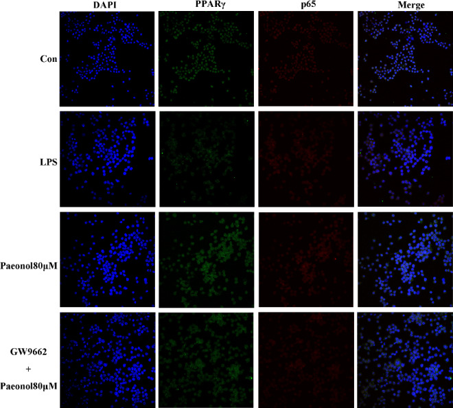
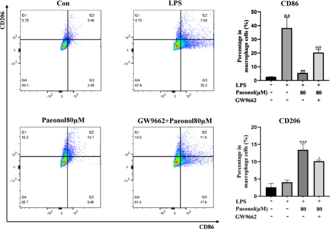
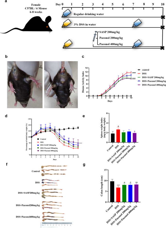
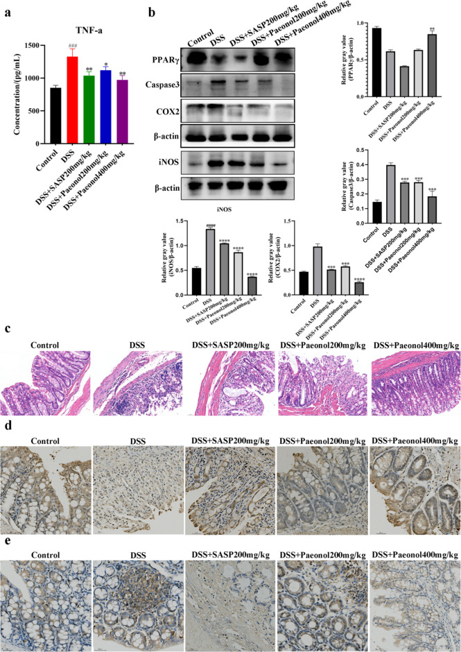
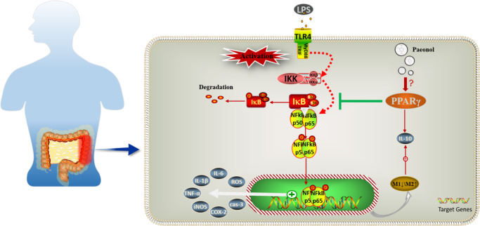
Ulcerative colitis (UC) is a chronic idiopathic inflammatory disease affecting the gastrointestinal tract. Although paeonol has been used for treating UC due to its anti-inflammatory and antioxidant effects, the underlying mechanisms remain unclear. In this study, we investigated the mechanisms of paeonol's action on UC by conducting in-vitro and in-vivo studies using NCM460 cells and RAW264.7 cells, and the DSS-induced mice colitis model. The in vitro studies demonstrate that paeonol exerts inhibitory effects on the activation of the NF-κB signaling pathway through upregulating PPARγ expression, thereby attenuating pro-inflammatory cytokine production, reducing reactive oxygen species levels, and promoting M2 macrophage polarization. These effects are significantly abrogated upon addition of the PPARγ inhibitor GW9662. Moreover, UC mice treated with paeonol showed increased PPARγ expression, which reduced inflammation and apoptosis to maintain intestinal epithelial barrier integrity. In conclusion, our findings suggest that paeonol inhibits the NF-κB signaling pathway by activating PPARγ, reducing inflammation and oxidative stress and improving Dss-induced colitis. This study provides a new insight into the mechanism of treating UC by paeonol.


Conflict of interest statement

The authors declare no competing interests.

Figures

Figure 1
Figure 1
Determination of the administration dosages of paeonol on cell viability. (a) Cell viability of different concentrations of paeonol to RAW264.7 cells. (b) Cell viability of different concentrations of paeonol to RAW264.7 cells.
Figure 2
Figure 2
Effect of paeonol on the inflammatory response of LPS‐stimulated RAW264.7 cells. Expression levels of IL-6 (a), TNF-α (b) and IL-10 (c) were detected by enzyme-linked immunosorbent assay (ELISA). The expression of IL-6 (d), TNF-α (e), and IL-10 (f) was checked by qRT-PCR. (g) ROS was detected by ROS probe DCFH-DA on a flow cytometer. These data represent the mean ± SD (n = 3). *P < 0.5; **P < 0.01; ***P < 0.001; ****P < 0.0001 compared with the LPS group. ###P < 0.001; ####P < 0.0001, compared with the Control group.
Figure 3
Figure 3
Effects of paeonol on PPARγ expression and NF-κB signaling pathway activation. (a) The amount of PPARγ, IκB, p-IκB, p65, p-p65 iNOS and COX2 expression by Western blot analysis. The full blots are shown in Supplementary Information. (b) The expression of PPARγ was checked by qRT-PCR. These data represent the mean ± SD (n = 3). *P < 0.5; **P < 0.01; ***P < 0.001, ****P < 0.0001 compared with the LPS group. #P < 0.5; ##P < 0.01; ###P < 0.001; ####P < 0.0001, compared with the Control group.
Figure 4
Figure 4
Effect of GW9662 on the inhibitory effect of paeonol on the inflammatory response. Expression levels of IL-6 (a), TNF-α (b) and IL-10 (c) were detected by enzyme-linked immunosorbent assay (ELISA). (d) ROS was detected by ROS probe DCFH-DA on a flow cytometer. (e) Then, intracellular ROS of RAW26 4.7 and NCM460 cells was detected by confocal microscopy. These data represent the mean ± SD. *P < 0.5; **P < 0.01; ***P < 0.001, ****P < 0.0001 compared with the LPS group. #P < 0.5; ##P < 0.01; ###P < 0.001; ####P < 0.0001, compared with the Control group.
Figure 5
Figure 5
Effect of paeonol with or without GW9662 on NF-κB nuclear translocation, and PPARγ expression was detected by fluorescence microscopy using an antibody against p65 and PPARγ.
Figure 6
Figure 6
LPS-induced RAW264.7 cells were treated with paeonol and GW9662. Flow cytometric analysis was used to detect M1 and M2 macrophage cells. **P < 0.01; ***P < 0.001 compared with the LPS group. #P < 0.5; ###P < 0.001 compared with the Control group. $P < 0.5; compared with the Control group.
Figure 7
Figure 7
Paeonol attenuates symptoms of DSS-induced colitis in mice. (a) Schematic diagram of the animal experimental design. (b) Normal mice and DSS-induced acute colitis mouse model. (c) Mouse body weights are measured daily. (d) Calculated DAI scores. (e) Spleen body weight ratio. (f, g) Intestine images and statistics for colon length in each group. Data are expressed as mean ± SEM, n = 5–6. #P < 0.5; ###P < 0.001; ####P < 0.0001, compared with the control group, *P < 0.5; ** P < 0.01; ****P < 0.0001, compared with the DSS group.
Figure 8
Figure 8
Paeonol alleviated inflammatory invasion in DSS-induced UC mice. (a) Expression levels of TNF-α in the colon were detected by ELISA. (b) The protein levels of PPARγ, COX2, iNOS and caspase3 in the colon were tested by using Western blotting. The full blots are shown in Supplementary Information. (c) Representative HE-stained images of colon sections (scale bar, 30 µm). Immunohistochemistry staining of PPARγ. (d) and caspase3. (e) (scale bar, 30 µm). Data are expressed as the mean ± SEM, n = 5–6. ### P < 0.001 compared with the control group, *P < 0.5; **P < 0.01; ***P < 0.001, ****P < 0.0001 compared with the DSS group.
Figure 9
Figure 9
Regulation of the PPARγ-NFκB signaling pathway by paeonol.

Similar articles

Cited by

References

    1. Le Berre, C., Ananthakrishnan, A. N., Danese, S., Singh, S. & Peyrin-Biroulet, L. Ulcerative colitis and Crohn’s disease have similar burden and goals for treatment. Clin. Gastroenterol. Hepatol.18, 14–23. 10.1016/j.cgh.2019.07.005 (2020). 10.1016/j.cgh.2019.07.005 - DOI - PubMed
    1. Kobayashi, T. et al. Ulcerative colitis. Nat. Rev. Dis. Primers6, 74. 10.1038/s41572-020-0205-x (2020). 10.1038/s41572-020-0205-x - DOI - PubMed
    1. Olén, O. et al. Colorectal cancer in ulcerative colitis: A Scandinavian population-based cohort study. Lancet395, 123–131. 10.1016/S0140-6736(19)32545-0 (2020). 10.1016/S0140-6736(19)32545-0 - DOI - PubMed
    1. Yao, D., Dong, M., Dai, C. & Wu, S. Inflammation and inflammatory cytokine contribute to the initiation and development of ulcerative colitis and its associated cancer. Inflamm. Bowel Dis.25, 1595–1602. 10.1093/ibd/izz149 (2019). 10.1093/ibd/izz149 - DOI - PubMed
    1. Mitsialis, V. et al. Single-cell analyses of colon and blood reveal distinct immune cell signatures of ulcerative colitis and Crohn’s disease. Gastroenterology10.1053/j.gastro.2020.04.074 (2020). 10.1053/j.gastro.2020.04.074 - DOI - PMC - PubMed

MeSH terms

LinkOut - more resources