. 2024 Aug 9;47(1):414.
doi: 10.1007/s10143-024-02662-z.

Giant unruptured middle cerebral artery aneurysm revealed by intracranial hypertension: is a systematic decompressive hemicraniotomy mandatory?

Affiliations

Giant unruptured middle cerebral artery aneurysm revealed by intracranial hypertension: is a systematic decompressive hemicraniotomy mandatory?

Rabih Aboukais et al. Neurosurg Rev. .

Abstract

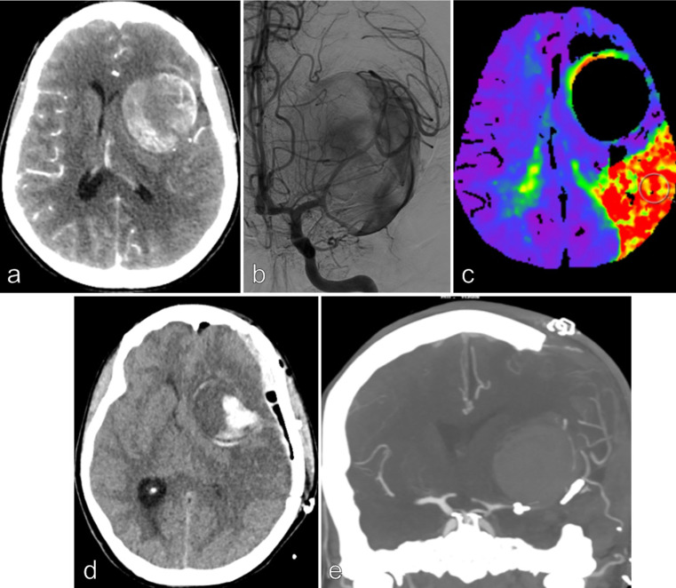
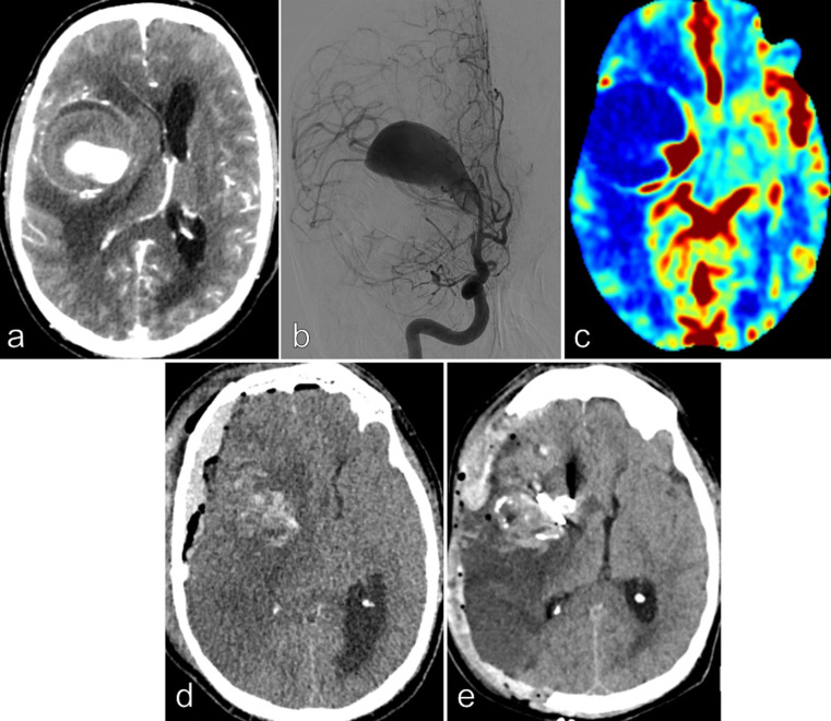
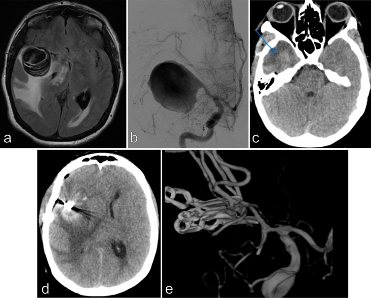
Our study aimed to evaluate the postoperative outcome of patients with unruptured giant middle cerebral artery (MCA) aneurysm revealed by intracranial hypertension associated to midline brain shift. From 2012 to 2022, among the 954 patients treated by a microsurgical procedure for an intracranial aneurysm, our study included 9 consecutive patients with giant MCA aneurysm associated to intracranial hypertension with a midline brain shift. Deep hypothermic circulatory flow reduction (DHCFR) with vascular reconstruction was performed in 4 patients and cerebral revascularization with aneurysm trapping was the therapeutic strategy in 5 patients. Early (< 7 days) and long term clinical and radiological monitoring was done. Good functional outcome was considered as mRS score ≤ 2 at 3 months. The mean age at treatment was 44 yo (ranged from 17 to 70 yo). The mean maximal diameter of the aneurysm was 49 mm (ranged from 33 to 70 mm). The mean midline brain shift was 8.6 mm (ranged from 5 to 13 mm). Distal MCA territory hypoperfusion was noted in 6 patients. Diffuse postoperative cerebral edema occurred in the 9 patients with a mean delay of 59 h and conducted to a postoperative neurological deterioration in 7 of them. Postoperative death was noted in 3 patients. Among the 6 survivors, early postoperative decompressive hemicraniotomy was required in 4 patients. Good functional outcome was noted in 4 patients. Complete aneurysm occlusion was noted in each patient at last follow-up. We suggest to discuss a systematic decompressive hemicraniotomy at the end of the surgical procedure and/or a partial temporal lobe resection at its beginning to reduce the consequences of the edema reaction and to improve the postoperative outcome of this specific subgroup of patients. A better intraoperative assessment of the blood flow might also reduce the occurrence of the reperfusion syndrome.

Keywords: Anastomosis; Bypass; Deep hypothermic circulatory flow arrest; Giant aneurysm; Mca; Microsurgery.


Conflict of interest statement

The authors declare no competing interests.

Figures

Fig. 1
Fig. 1
Patient 1, 41 yo female, smoker with a medical history of migraine, presented with headache and a 1-month history of moderate motor language disorder. CT scan, MRI/MRA and conventional angiography demonstrated a giant left MCA aneurysm injected only by the left temporal M2 segment. No significant edema, intrasaccular thrombus or wall calcifications were noted (a and b). The midline brain shift was 9 mm. A severe hypoperfusion in the left distal MCA territory was noted on the perfusion CT scan (c). A distal STA-MCA bypass was performed, followed by aneurysmal trapping and partial resection of the aneurysm sac. The patient was extubated after 24 h without any neurological deficit. 36 h after treatment, a rapid consciousness disorder with a left mydriasis occurred, related to a left hemispheric diffuse edema (d). A decompressive hemicraniectomy associated to a partial anterior temporal lobe resection was immediately done (e). A severe vasospasm appeared 5 days later and was partially improved by chemical angioplasty. The bypass was always patent. The patient improved, with a moderate language disorder but no motor deficit. The mRS score was 2 after 3 months
Fig. 2
Fig. 2
Patient 2, 66 yo male, smoker with a medical history of chronic bronchitis and peripheral arterial occlusive disease of the lower limbs, presented with headache and a 3 weeks history of left moderate hemiparesis. CT scan, MRI/MRA and conventional angiography demonstrated a right giant MCA aneurysm (maximal diameter = 70 mm) injected only by the M1 segment. A significant peri-aneurysmal edema, wall calcifications and intrasaccular thrombus were noted. The midline brain shift was 13 mm (a and b). A severe hypoperfusion in the left distal MCA territory was noted on the perfusion CT scan (c). Double barrel STA-MCA bypass (on the 2 M2 segments) was done, followed by aneurysmal trapping and a partial resection of the sac and thrombus. Twelve hours after treatment, a rapid consciousness disorder with a right mydriasis occurred, related to a right hemispheric diffuse edema and ischemic lesions (d). Decompressive hemicraniectomy was immediately done (e). Fatal evolution with lung infection and scar necrosis were noted. He died 21 days after the treatment
Fig. 3
Fig. 3
Patient 6, 44 yo female, without any medical history, complained about increasing headache for 1 month. CT scan, MRI/MRA and conventional angiography demonstrated a right giant MCA aneurysm (maximal diameter = 37 mm). A significant peri-aneurysmal edema and intrasaccular thrombus were noted. The midline brain shift was 10 mm (a and b). Deep hypothermic circulatory arrest/extra-corporeal circulation with vascular reconstruction was performed to exclude the aneurysm from the circulation. A deliberate partial temporal lobe resection (blue arrow) was performed at the beginning of the surgical procedure to gain a better exposure of the aneurysm (c and d). The patient was extubated after 48 h without any neurological deficit. Complete occlusion of the aneurysm was noted (e). No neurological sequelae were noted and the mRS score was 1 after 3 months
Fig. 4
Fig. 4
Patient 9, 44 yo female, without any medical history, complained about increasing headache for 3 weeks. CT scan, MRI/MRA and conventional angiography demonstrated a left giant MCA aneurysm (maximal diameter = 50 mm). A moderate peri-aneurysmal edema and intrasaccular thrombus were noted. The midline brain shift was 5 mm (a and b). A severe hypoperfusion in the left distal MCA territory was noted on the perfusion CT scan (c). A distal STA-MCA bypass was performed, followed by aneurysmal trapping and partial resection of the aneurysmal sac. A deliberate decompressive hemicraniectomy was performed at the end of the procedure. The patient was extubated after 24 h without any neurological deficit (d). An improvement of the hypoperfusion was recorded on control Perfusion CT scan (e). A long-term favorable outcome was recoded (mRS score = 1)

References

    1. Aboukais R, Devalckeneer A, Boussemart P, Bourgeois P, Menovsky T, Leclerc X, Vinchon M, Lejeune JP (2022) Is malignant edema and hemorrhage after occlusion of high-flow arteriovenous malformation related to the size of feeding arteries and draining veins? Neurochirurgie 68:e1–e7. 10.1016/j.neuchi.2022.01.008 - PubMed
    1. Amelot A, Smajda S, Terrier LM, Redjem H, Simonneau A, Bekaert O, Piotin M, Lot G, Chauvet D (2017) Posterior Fossa Craniectomy with Endovascular Therapy of Giant Fusiform Basilar Artery Aneurysms: a New Approach to consider? World Neurosurg 98:104–112. 10.1016/j.wneu.2016.10.119 - PubMed
    1. Amili O, Schiavazzi D, Moen S, Jagadeesan B, Van de Moortele PF, Coletti F (2018) Hemodynamics in a giant intracranial aneurysm characterized by in vitro 4D flow MRI. PLoS ONE 13:e0188323. 10.1371/journal.pone.0188323 - PMC - PubMed
    1. Amin-Hanjani S, Charbel FT (2007) Flow-assisted surgical technique in cerebrovascular surgery. Surg Neurol 68(Suppl 1):S4–11. 10.1016/j.surneu.2007.07.051 - PubMed
    1. Cagnazzo F, Mantilla D, Rouchaud A, Brinjikji W, Lefevre PH, Dargazanli C, Gascou G, Riquelme C, Perrini P, di Carlo D, Bonafe A, Costalat V (2018) Endovascular treatment of very large and giant intracranial aneurysms: comparison between reconstructive and deconstructive Techniques-A Meta-analysis. AJNR Am J Neuroradiol 39:852–858. 10.3174/ajnr.A5591 - PMC - PubMed

LinkOut - more resources