. 2024 Sep;129(9):1382-1393.
doi: 10.1007/s11547-024-01862-3. Epub 2024 Aug 8.

ASL, DSC, DCE perfusion MRI and 18F-DOPA PET/CT in differentiating glioma recurrence from post-treatment changes

Affiliations

ASL, DSC, DCE perfusion MRI and 18F-DOPA PET/CT in differentiating glioma recurrence from post-treatment changes

Giulia Moltoni et al. Radiol Med. 2024 Sep.

Abstract

Objectives: To discriminate between post-treatment changes and tumor recurrence in patients affected by glioma undergoing surgery and chemoradiation with a new enhancing lesion is challenging. We aimed to evaluate the role of ASL, DSC, DCE perfusion MRI, and 18F-DOPA PET/CT in distinguishing tumor recurrence from post-treatment changes in patients with glioma.

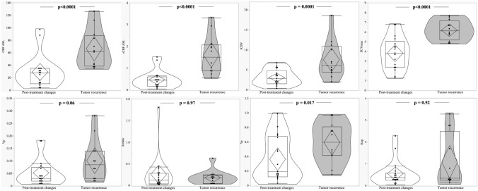
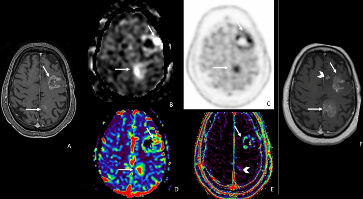
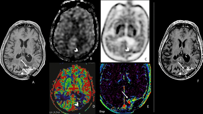
Materials and methods: We prospectively enrolled patients with treated glioma (surgery plus chemoradiation) and a new enhancing lesion doubtful for recurrence or post-treatment changes. Each patient underwent a 1.5T MRI examination, including ASL, DSC, and DCE PWI, and an 18F-DOPA PET/CT examination. For each lesion, we measured ASL-derived CBF and normalized CBF, DSC-derived rCBV, DCE-derived Ktrans, Vp, Ve, Kep, and PET/CT-derived SUV maximum. Clinical and radiological follow-up determined the diagnosis of tumor recurrence or post-treatment changes.

Results: We evaluated 29 lesions (5 low-grade gliomas and 24 high-grade gliomas); 14 were malignancies, and 15 were post-treatment changes. CBF ASL, nCBF ASL, rCBV DSC, and PET SUVmax were associated with tumor recurrence from post-treatment changes in patients with glioma through an univariable logistic regression. Whereas the multivariable logistic regression results showed only nCBF ASL (p = 0.008) was associated with tumor recurrence from post-treatment changes in patients with glioma with OR = 22.85, CI95%: (2.28-228.77).

Conclusion: In our study, ASL was the best technique, among the other two MRI PWI and the 18F-DOPA PET/CT PET, in distinguishing disease recurrence from post-treatment changes in treated glioma.

Keywords: Glioma; MRI perfusion; PET; Post-treatment changes; Tumor recurrence.


Conflict of interest statement

The authors have no relevant financial or non-financial interests to disclose.

Figures

Fig. 1
Fig. 1
Box-plot comparing different parameters at post-treatment changes and tumor recurrence. a CBF_ASL, b nCBF_ASL, c rCBV_DSC, d SUVmax_PET; e Vp_DCE, f Ktrans_DCE, g Ve_DCE, h Kep_DCE). ASL = Arterial Spin Labeling; CBF = Cerebral Blood Flow; DSC = dynamic susceptibility contrast-enhanced perfusion; DCE = Dynamic contrast-enhanced perfusion; rCBV = relative cerebral blood volume; Kep = rate constant; Ktrans = transfer constant; Ve = fractional volume of the extravascular extracellular space; Vp = fractional volume of the plasma space; SUVmax = maximum standardized uptake values
Fig. 2
Fig. 2
A representative case of high-grade glioma with two enhancing lesions in the left frontal lobe (a, arrows) appeared 6 months after radiation therapy. Lesions showed increased ASL_CBF (b, arrows), increased 18F-DOPA uptake (c, arrows), increased DSC_rCBV, particularly evident in the posterior lesion (d, arrows), doubtful DCE_Ve increase in the posterior lesion (e, arrowhead) and increased DCE_Ve values in the anterior lesion (f arrow). f 6-month follow-up MRI showing progression in lesion contrast enhancement and size, and a new lesion (arrowhead) as the expression of the disease recurrence. ASL, PET, and DSC correctly identify the tumoral nature of the lesions; Ve misclassified the posterior lesion. ASL = arterial spin labeling; CBF = cerebral blood flow; DSC = dynamic susceptibility contrast-enhanced perfusion; DCE = dynamic contrast-enhanced perfusion; CBV = cerebral blood volume; Ve = fractional volume of the extravascular extracellular space
Fig. 3
Fig. 3
A representative case of a high-grade glioma with an enhancing lesion in the left parietal lobe (a, arrow) occurring 8 months after radiation therapy. The lesion did not show high values of ASL_CBF (b, arrowhead), DSC_rCBV (d, arrowhead), or a significant increase in 18F-DOPA PET uptake (c, arrowhead). DCE_Ve showed foci of increased values (e, arrow). Nine-month MRI follow-up showed the stability of the lesion with a slight reduction in contrast enhancement (f, arrow). The lesion was classified as post-treatment changes, and ASL, DSC, and PET were concordant and negative, whereas DCE_Ve misclassified the lesion. ASL = arterial spin labeling; CBF = cerebral blood flow; DSC = dynamic susceptibility contrast-enhanced perfusion; DCE = dynamic contrast-enhanced perfusion; CBV = cerebral blood volume; Ve = fractional volume of the extravascular extracellular space
Fig. 4
Fig. 4
A representative case of high-grade glioma with a linear enhancing lesion in the left front-basal region, close to the surgical site, occurred 8 months after radiation therapy (a, arrow). The enhancing lesion did not show an increase in ASL_CBF (b, arrow); DSC-rCBV evaluation was difficult because of the proximity to bone structures (d, circle); and DCE_Ve was slightly increased (e, arrow). The uptake of 18F-DOPA was high (c, arrow). Six months of MRI follow-up showed a massive reduction in contrast enhancement (f, arrow). ASL correctly classified the lesion, whereas the linear 18F-DOPA uptake close to the surgical site could be the expression of macrophages activation. ASL = arterial spin labeling; CBF = cerebral blood flow; DSC = dynamic susceptibility contrast enhanced perfusion; DCE = dynamic contrast enhanced perfusion; CBV = cerebral blood volume; Ve = fractional volume of the extravascular extracellular space
Fig. 5
Fig. 5
A representative case of high-grade glioma with a new enhancing lesion appeared 3 months after radiation therapy (a, arrow); the patient was included in our study, and after 2 months he underwent MRI and PET examination following study protocol (b, squared images). On T1 post-contrast WI, the lesion was increased in size (b, arrow), the ASL_CBF map showed no increment of CBF (b, arrowhead), and 18F-DOPA PET/TC showed an increased uptake (b, circle). T1 post-contrast WI acquired, respectively, after 3 (c) and 6 (d) months, showed a progressive reduction of contrast enhancement and lesion size until, after 9 months (e), the lesion was no longer present. This is a case of pseudoprogression with a false positive PET/CT and a true negative ASL classification. ASL = arterial spin labeling; CBF = cerebral blood flow; DSC = dynamic susceptibility contrast-enhanced perfusion; DCE = dynamic contrast-enhanced perfusion; CBV = cerebral blood volume; Ve = fractional volume of the extravascular extracellular space; WI = weighted imaging

References

    1. Stupp R, Hegi ME, Mason WP et al (2009) Effects of radiotherapy with concomitant and adjuvant temozolomide versus radiotherapy alone on survival in glioblastoma in a randomised phase III study: 5-year analysis of the EORTCNCICtrial. Lancet Oncol 10:459-466 3 10.1016/S1470-2045(09)70025-7 - DOI - PubMed
    1. Stupp R, Mason WP, van den Bent MJ et al (2005) Radiotherapy plus concomitant and adjuvant temozolomide for glioblastoma. N Engl J Med 352(10):987–996 10.1056/NEJMoa043330 - DOI - PubMed
    1. Nael K, Bauer AH, Hormigo A et al (2018) Multiparametric MRI for differentiation of radiation necrosis from recurrent tumor in patients with treated glioblastoma. AJR Am J Roentgenol 210:18–23 10.2214/AJR.17.18003 - DOI - PubMed
    1. Brandsma D, Stalpers L, Taal W, Sminia P, van den Bent MJ (2008) Clinical features, mechanisms, and management of pseudoprogression in malignant gliomas. Lancet Oncol 9:453–461 10.1016/S1470-2045(08)70125-6 - DOI - PubMed
    1. Ellingson BM, Wen PY, Cloughesy TF (2017) Modified criteria for radiographic response assessment in glioblastoma clinical trials. Neurother J Am Soc Exp Neurother 14:307–320 - PMC - PubMed

LinkOut - more resources