. 2024 Aug 8;22(1):100.
doi: 10.1186/s12958-024-01265-z.

Quercetin enhances decidualization through AKT-ERK-p53 signaling and supports a role for senescence in endometriosis

Affiliations

Quercetin enhances decidualization through AKT-ERK-p53 signaling and supports a role for senescence in endometriosis

Julia Delenko et al. Reprod Biol Endocrinol. .

Abstract

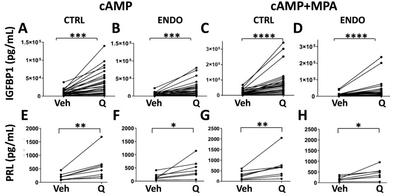
Background: Patients with endometriosis suffer with chronic pelvic pain and infertility, and from the lack of pharmacologic therapies that consistently halt disease progression. Differences in the endometrium of patients with endometriosis vs. unaffected controls are well-documented. Specifically, shed endometrial tissues (delivered to the pelvic cavity via retrograde menstruation) reveal that a subset of stromal cells exhibiting pro-inflammatory, pro-fibrotic, and pro-senescence-like phenotypes is enhanced in endometriosis patients compared to controls. Additionally, cultured biopsy-derived endometrial stromal cells from endometriosis patients exhibit impaired decidualization, a defined differentiation process required for human embryo implantation and pregnancy. Quercetin, a senolytic agent, shows therapeutic potential for pulmonary fibrosis, a disorder attributed to senescent pulmonary fibroblasts. In rodent models of endometriosis, quercetin shows promise, and quercetin improves decidualization in vitro. However, the exact mechanisms are not completely understood. Therefore, we investigated the effects of quercetin on menstrual effluent-derived endometrial stromal cells from endometriosis patients and unaffected controls to define the signaling pathways underlying quercetin's effects on endometrial stromal cells.

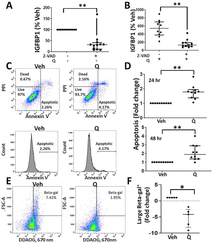
Methods: Menstrual effluent-derived endometrial stromal cells were collected and cultured from unaffected controls and endometriosis patients and then, low passage cells were treated with quercetin (25 µM) under basal or standard decidualization conditions. Decidualization responses were analyzed by measuring the production of IGFBP1 and PRL. Also, the effects of quercetin on intracellular cAMP levels and cellular oxidative stress responses were measured. Phosphokinase arrays, western blotting, and flow cytometry methods were performed to define the effects of quercetin on various signaling pathways and the potential mechanistic roles of quercetin.

Results: Quercetin significantly promotes decidualization of control- and endometriosis-endometrial stromal cells. Quercetin substantially reduces the phosphorylation of multiple signaling molecules in the AKT and ERK1/2 pathways, while enhancing the phosphorylation of p53 and total p53 levels. Furthermore, p53 inhibition blocks decidualization while p53 activation promotes decidualization. Finally, we provide evidence that quercetin increases apoptosis of endometrial stromal cells with a senescent-like phenotype.

Conclusions: These data provide insight into the mechanisms of action of quercetin on endometrial stromal cells and warrant future clinical trials to test quercetin and other senolytics for treating endometriosis.

Keywords: AKT signaling; Apoptosis; Fertility; Menstrual effluent; Senescence; p53 signaling.


Conflict of interest statement

The authors declare no competing interests.

Figures

Fig. 1
Fig. 1
Quercetin inhibits eSC proliferation and enhances decidualization.(A) Control endometrial stromal cells (eSCs) were treated with vehicle (Veh) or quercetin (Q, 6.25-50 µM) for 72 h and the relative cell number was determined. Each point represents data from one individual’s eSCs, with the group mean ± SD for vehicle and each Q dose. (B-C) Control eSCs were treated with vehicle (Veh) or quercetin (1.56-50 µM) for 4 h prior to the addition of cAMP alone (B) or cAMP + MPA (C). After 48 h, decidualization was analyzed by measuring IGFBP1 levels by ELISA. Each point represents data from one individual’s eSCs, as IGFBP1 (percent Veh, where 100% = Veh + cAMP alone (IGFBP1) (B) or Veh + cAMP + MPA (IGFBP1) (C), with the group median ± interquartile range (IQR) for each Q dose. (D) eSCs from two control subjects were treated with vehicle (Veh) or quercetin (Q, 25µM) for 4 h followed by vehicle or cAMP + MPA prior to immunofluorescent staining and confocal imaging; images show IGFBP1 (red), phalloidin (green), and DAPI (blue) staining (at 20x magnification). The scale bars (20 μm) are indicated. IGFBP1 + cells are indicated by yellow arrows. Images showing individual channels are in Supplementary Fig. 2. (EG) The effects of quercetin (Q, 25 µM) on decidualization when added 4 h pre-cAMP ± MPA (E) versus at the same time as (0 h pre-) cAMP ± MPA (F) or 20 h post-cAMP ± MPA stimulation (G). IGFBP1 levels were measured by ELISA 48 h post cAMP ± MPA stimulation. Data are shown as IGFBP1 (% Veh) where 100% = Veh + cAMP alone (IGFBP1). Each point represents data from one individual’s eSCs, with the median ± IQR shown for each group comparing Veh vs. Q. *p < 0.05, **p < 0.01; ***p < 0.001; ****p < 0.0001; ns = non-significant
Fig. 2
Fig. 2
Endometriosis eSCs exhibit impaired decidualization. (A-B) Comparison of decidualization responses of eSCs from controls (CTRL) vs. endometriosis (ENDO) cases induced by cAMP alone (A) or cAMP + MPA (B), as determined by IGFBP1 levels measured 48 h post cAMP ± MPA by ELISA. Each point represents data from one individual’s eSCs, with mean ± SD shown for each group. **p < 0.001 CTRL vs. ENDO (Student’s t test)
Fig. 3
Fig. 3
Quercetin enhances decidualization by control-eSCs and endometriosis-eSCs, as determined by IGFBP1 and PRL protein production. (A-H) Endometrial stromal cells (eSCs) from controls (A, C, E, and G, CTRL) and endometriosis cases (B, D, F, and H, ENDO) were treated with vehicle (Veh) or quercetin (Q, 25µM) for 4 h prior to the addition of cAMP alone (A-B, E-F) or cAMP + MPA (C-D, G-H). After 48 h, decidualization was analyzed by measuring IGFBP1 (A-D) or PRL (E-H) levels by ELISA. Data points connected by a line represent paired data points from one individual’s eSCs (± Q). *p < 0.05 Veh vs. Q-treated; **p < 0.01 Veh vs. Q-treated; ***P < 0.001 Veh vs. Q-treated; ****p < 0.0001 Veh vs. Q-treated
Fig. 4
Fig. 4
Quercetin does not increase [cAMP]i concentrations in eSCs. (A-B) Endometrial stromal cells (eSCs) from controls (CTRL) or endometriosis cases (ENDO) were treated with IBMX (0.1mM, a phosphodiesterase inhibitor that blocks [cAMP]i degradation), followed by addition of either vehicle, forskolin (FOR, 25µM, an activator of adenylyl cyclases and hence, [cAMP]i) or quercetin (Q, 25µM). Lysates were analyzed by ELISA for [cAMP]i concentrations (A-B). For clarity, data without forskolin (FOR) treatment are also shown on a different scale (B). Each point represents data from one individual’s eSCs, with median ± IQR shown for each group. **p < 0.001 vs. vehicle (IBMX alone); ns = not significant
Fig. 5
Fig. 5
Quercetin does not reduce oxidative stress in eSCs. (A-D) Endometrial stromal cells (eSCs) from controls (CTRL, A and C) or endometriosis cases (ENDO, B and D) were treated with either vehicle (Veh), quercetin (Q, 25µM), or N-acetylcysteine (NAC, 10mM) before H2O2 (500µM) (A and B) or after H2O2 (500µM) (C and D), and oxidative stress was measured 3 h later using the DCF-DA assay. Data are shown as % Veh control (oxidative stress) where 100% = Veh-Veh-treated eSCs. Each point represents data from one individual’s eSCs, with median ± IQR shown for each group. *p < 0.05; **p < 0.01; ns = not significant
Fig. 6
Fig. 6
Quercetin inhibits AKT and ERK1/2 phosphorylation and signaling and promotes p53 (Ser46) phosphorylation. (A-B) Control endometrial stromal cells (eSCs) were treated with quercetin (Q, 25µM) for 4 h before analyzing cell lysates using a phospho-kinase array panel. Representative arrays (A) and quantification of array analytes/spots (B) are shown. Data for differentially expressed targets are presented as specific spot density normalized to the reference spot comparing Veh- vs. Q-treated (B). Each point represents the specific density of each duplicate analyte spot as a percentage of Veh-treated, with group median ± IQR shown for control- and endometriosis-eSCs. **p < 0.01. The remaining arrays are in Supplementary Fig. 7. (C-D) Control (CTRL) and endometriosis (ENDO) eSCs were treated with Veh or Q (25µM) for 4 h before western blotting for p-AKT, total AKT, p-ERK1/2, total ERK1/2, p-PRAS40, and total PRAS40. Representative blots and quantification of specific analytes are shown in (C) and (D), respectively. Band densities were normalized to GAPDH and shown as fold-change between Veh- vs. Q-treated eSCs in (D). Each point represents data from one individual’s eSCs, with median ± IQR shown for each group. *p < 0.05; ***p < 0.001; ****p < 0.0001. (E) Control-eSCs were treated with Veh or Q (25µM) for 0.5, 2, and 4 h before western blotting for phospho-AKT, total AKT, phospho-ERK1/2, total ERK1, phospho-PRAS40, and total PRAS40. (F-I) Control-eSCs were treated with vehicle (Veh), AKT inhibitor MK-2206 (MK, 1µM), quercetin (Q, 25µM) or Q + MK for 4 h prior to cAMP (F and H) or cAMP + MPA (G and I) stimulation; decidualization was assessed by measuring IGFBP1 (F and G) or PRL (H and I) by ELISA. Data are presented as fold-change in IGFBP1 or PRL over Veh-treated (Veh-treated = 1). Each symbol represents data from one individual’s eSCs, with median ± IQR shown for each group. *p < 0.05; **p < 0.01 vs. Veh-treated; ***p < 0.001 vs. Veh-treated; ns = non-significant vs. Q-treated
Fig. 7
Fig. 7
Quercetin induces p53, which contributes to decidualization. (A-D) Control (CTRL)- and endometriosis (ENDO)-endometrial stromal cells (eSCs) were treated with vehicle or quercetin (Q, 25 µM) for 4 h (A, B) or 2 days (C, D) before western blot analysis of cell lysates for p53 and GAPDH. Representative blots are shown in (A) and (C). Band densities for control-eSCs were normalized to GAPDH and presented as fold-change after 4 h (B) or 2 days (D) following quercetin treatment, with median ± IQR shown for each group. ***p < 0.001 comparing fold-change in GAPDH vs. p53. (E) Control-eSCs were treated with Veh or Q (25 µM) for 0.5, 2, and 4 h before western blotting for p53 and GAPDH. (F-G) Control-eSCs were treated with vehicle (DMSO diluted 1:250 in media and added as a 10X stock) Pifithrin α (PFT, 40 µM, prepared in DMSO, diluted 1:250 in media and added as a 10X stock) for 4 h prior to cAMP + MPA stimulation (F) or PFT (40 µM) and Q (25µM) for 4 h prior to cAMP + MPA stimulation (G). Decidualization was assessed by measuring IGFBP1 levels 48 h later. (H) Control-eSCs were treated with Veh or nutlin-3a (Nut, 100nM) 24 h after cAMP + MPA stimulation and decidualization was assessed by measuring IGFBP1 production 48 h post-cAMP + MPA. For F-H, data are presented as IGFBP1% vehicle (Veh-treated cAMP = 100%). Each point represents data from one individual’s eSCs, with median ± IQR shown for each group. **p < 0.01 vs. Veh-treatment (F and H) or Q (G)
Fig. 8
Fig. 8
eSCs show evidence of senescence-like phenotype, which is reversed by quercetin. (A) Endometriosis (ENDO)-endometrial stromal cells (eSCs) and control (CTRL)-eSCs were treated with vehicle or quercetin (Q, 25 µM) for 2 days before western blotting for SASPs, IL-6 and MMP3, or GAPDH. A representative blot is shown. (B-C) Quantification of blots (with band densities normalized to GAPDH) from control-eSCs treated with vehicle (Veh) or quercetin (Q, 25 µM) for 2 days for IL-6 and MMP3 are shown in (B) and (C), respectively. Data are presented as IL-6% vehicle or MMP3% vehicle (where Veh-treated = 100%). Each point represents data from one individual’s eSCs, with median ± IQR shown for each group *p < 0.05; **p < 0.01; ns = non-significant. (D) Control-eSCs were treated with vehicle or 250 µM H2O2 for 2 h and then, eSCs were harvested at 2, 4, 6, and 8 days post-H2O2 exposure for western blotting analysis for p21, p16, lamin B1, MMP3, and GAPDH. (E) Control-eSCs were treated with vehicle (Veh) or H2O2 (250 µM) for 2 h and then treated with vehicle (Veh) or quercetin (Q, 25 µM), as indicated, for 4 h prior to cAMP + MPA-induced decidualization. After 48 h, culture supernatants were assessed for IGFBP1 (pg/ml) by ELISA. Data points connected by lines represent paired data points from one individual’s eSCs (comparing Veh vs. H2O2 and H2O2-Q vs. H2O2 + Q). *p < 0.05; **p < 0.01 Veh vs. Q-treated; ns = non-significant
Fig. 9
Fig. 9
Inhibiting apoptosis blocks decidualization and quercetin induces apoptosis in a subset of eSCs. (A-B) Control-eSCs (p3) were treated with vehicle (Veh) or Z-VAD-fmk (0 vs. 40 µM), a pan-caspase inhibitor that blocks apoptosis, in the presence of vehicle (A) or quercetin (Q, 25 µM) (B) prior to inducing decidualization with cAMP + MPA. Decidualization was assessed 48 h later by ELISA for IGFBP1. Each point represents data from one individual’s eSCs, with the median ± IQR shown for each group. **p < 0.01. (C-D) Quercetin induces apoptosis in a subset of control-eSCs (passage 3–4), as determined by flow cytometry. Representative gating (upper panels) and histograms (lower panels) of Annexin V staining comparing vehicle-treated (Veh, left panels) vs. quercetin-treated (Q, 25 µM, right panels) eSCs 24 h post-treatment (C). Quantification of quercetin-induced apoptosis (shown as fold-change) measured 24 h and 48 h post-vehicle (Veh) vs. post-quercetin (Q, 25 µM) treatment (D). Each point represents data from one individual’s eSCs (fold-change in apoptosis), with median ± IQR shown for each group (± Q). **p < 0.01. (E-F) Larger senescence-associated β-galactosidase (SA-βgal) + control-eSCs are reduced following quercetin treatment. SA-βgal was determined using 9 H-(1,3-Dichloro-9,9-Dimethylacridin-2-one) β-D-Galactopyranoside (DDAOG), a substrate of SA-βgal that yields a far-red fluorescent product detected by flow cytometry at 670 nm. Y-axis (FSC-A, size) and X-axis (DDAOG, 670 nm). Representative flow cytometry plots show the gating of larger DDAOG, 670 nm/SA-βgal (Beta-gal) + eSCs comparing vehicle-treated (Veh – left panel) vs. quercetin-treated (Q, 25 µM- right panel) eSCs (p3-4) at 48 h post-treatment (E). The percentage of Beta-gal+/senescent cells (of total viable cells per eSC sample) is indicated on the representative plots in E. The fold-change in the number of larger Beta-gal+/senescent cells comparing vehicle (Veh) vs. quercetin (Q, 25 µM) is shown (F). Each point represents data from one individual’s eSCs (fold-change in larger Beta-gal + cells), with median ± IQR shown for each group (± Q). *p < 0.05 comparing vehicle vs. Q
Fig. 10
Fig. 10
The AKT and ERK1/2 signaling pathways promote cell survival and proliferation and suppress apoptosis. Quercetin (QUE) promotes apoptosis in eSCs by inhibiting the AKT and ERK1/2 pathways and enhancing p53 stability and apoptosis. Green and red solid lines indicate positive and negative effects, respectively, based on our data. Dashed red lines indicate proposed negative effects. Created with biorender.com

References

    1. Nambiar A, Kellogg D 3rd, Justice J, Goros M, Gelfond J, Pascual R, et al. Senolytics dasatinib and quercetin in idiopathic pulmonary fibrosis: results of a phase I, single-blind, single-center, randomized, placebo-controlled pilot trial on feasibility and tolerability. EBioMedicine. 2023;90:104481. 10.1016/j.ebiom.2023.104481 - DOI - PMC - PubMed
    1. Justice JN, Nambiar AM, Tchkonia T, LeBrasseur NK, Pascual R, Hashmi SK, et al. Senolytics in idiopathic pulmonary fibrosis: results from a first-in-human, open-label, pilot study. EBioMedicine. 2019;40:554–63. 10.1016/j.ebiom.2018.12.052 - DOI - PMC - PubMed
    1. Kirkland JL, Tchkonia T. Senolytic drugs: from discovery to translation. J Intern Med. 2020;288(5):518–36. 10.1111/joim.13141 - DOI - PMC - PubMed
    1. Jamali N, Zal F, Mostafavi-Pour Z, Samare-Najaf M, Poordast T, Dehghanian A. Ameliorative effects of Quercetin and Metformin and their combination against experimental endometriosis in rats. Reprod Sci. 2021;28(3):683–92. 10.1007/s43032-020-00377-2 - DOI - PubMed
    1. Park S, Lim W, Bazer FW, Whang KY, Song G. Quercetin inhibits proliferation of endometriosis regulating cyclin D1 and its target microRNAs in vitro and in vivo. J Nutr Biochem. 2019;63:87–100. 10.1016/j.jnutbio.2018.09.024 - DOI - PubMed

MeSH terms