. 2024 Nov;181(22):4610-4627.
doi: 10.1111/bph.16511. Epub 2024 Aug 8.

The protein disulfide isomerase A3 and osteopontin axis promotes influenza-induced lung remodelling

Affiliations

The protein disulfide isomerase A3 and osteopontin axis promotes influenza-induced lung remodelling

Amit Kumar et al. Br J Pharmacol. 2024 Nov.

Abstract

Background and purpose: Fibrotic lung remodelling after a respiratory viral infection represents a debilitating clinical sequela. Studying or managing viral-fibrotic sequela remains challenging, due to limited therapeutic options and lack of understanding of mechanisms. This study determined whether protein disulfide isomerase A3 (PDIA3) and secreted phosphoprotein 1 (SPP1), which are associated with pulmonary fibrosis, can promote influenza-induced lung fibrotic remodelling and whether inhibition of PDIA3 or SPP1 can resolve viral-mediated fibrotic remodelling.

Experimental approach: A retrospective analysis of TriNetX data sets was conducted. Serum from healthy controls and influenza A virus (IAV)-infected patients was analysed. An inhibitor of PDIA3, punicalagin, and a neutralizing antibody for SPP1 were administered in mice. Macrophage cells treated with macrophage colony-stimulating factor (M-CSF) were used as a cell culture model.

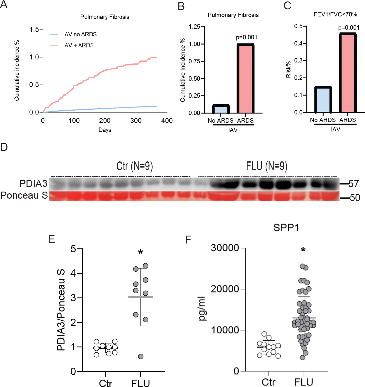
Key results: The TriNetX data set showed an increase in lung fibrosis and decline in lung function in flu-infected acute respiratory distress syndrome (ARDS) patients compared with non-ARDS patients. Serum samples revealed a significant increase in SPP1 and PDIA3 in influenza-infected patients. Lung PDIA3 and SPP1 expression increased following viral infection in mouse models. Punicalagin administration 2 weeks after IAV infection in mice caused a significant decrease in lung fibrosis and improved oxygen saturation. Administration of neutralizing SPP1 antibody decreased lung fibrosis. Inhibition of PDIA3 decreased SPP1secretion from macrophages, in association with diminished disulfide bonds in SPP1.

Conclusion and implications: The PDIA3-SPP1 axis promotes post-influenza lung fibrosis in mice and that pharmacological inhibition of PDIA3 or SPP1 can treat virus-induced lung fibrotic sequela.

Keywords: AHR; M‐CSF; PDIA3; SPP1; influenza; lung fibrosis; lung function.


Conflict of interest statement

COMPETING INTERESTS

Yvonne Janssen-Heininger (YJH), and Vikas Anathy (VA) hold patents: United States Patent No. 8,679,811, “Treatments Involving Glutaredoxins and Similar Agents”, United States Patent No. 8,877,447, “Detection of Glutathionylated Proteins”, United States Patent, 9,907,828 (to YJH), “Treatments of oxidative stress conditions” (YJH and VA). In the past YJH and VA have received consulting fees and laboratory contracts from Celdara Medical LLC, NH. AGJ serves as a member of Scientific Advisory Board of Gen1E Lifesciences, Palo Alto, CA, United States.

Amit Kumar and VA hold patent: United States Patent No. 11883395, “Methods and uses of protein disulfide isomerase inhibitory compounds.”

Figures

FIGURE 1
FIGURE 1. Post-IAV fibrotic sequelae and elevated levels of PDIA3 and SPP1 in patients
(A and B) Cumulative incidence of pulmonary fibrosis within 1 year post-H1N1 infection; (C) FEV1/FVC predicted (≤ 70%) within 1 year post-H1N1 infection. Patient cohort after propensity matching, IAV without ARDS no IPF n=14,936 and IAV with ARDS no IPF n=14,936. (D) Western blot for PDIA3, (E) quantitation of PDIA3 protein expression in western blot, Control (n=9) and FLU (n=9) patients, and (F) ELISA for SPP1, Control (n=12) and FLU (n=50) patients. * Significance was based on unpaired two-tailed non-parametric Mann-Whitney t-test. Error bars represent ± SD.
FIGURE 2
FIGURE 2. IAV-driven temporal changes in lung physiology, fibrosis, and airway hyperresponsiveness (AHR) in mice.
(A) Mock or IAV-PR8 virus challenge and harvest regimen; (B) The mean body weight of mice represented as a percentage of the original weight, and (C) mean oxygen saturation levels (SpO2) as determined by pulse-oximetry. *p<0.05 as compared to mock samples by ANOVA; error bars ±SEM. (n=4–10 mice/group). (D) Time-dependent alterations in collagen content in lungs of mice. (E-H) Measurement of Pdia3, Spp1, Fn-1, and Col1a1 mRNA expression from lung tissue. (I) TCID50 measurement of viral burden in the BAL of MOCK and IAV infected mice at different time points. ND: Not Detected and (J) SPP1 protein concentrartion in lung tissues. (K) dead cell protease in BALF and (L) LDH activity in WLL. *p<0.05 as compared to 3-day mock by ANOVA with Bonnferroni’s multiple comparison test; error bars ±SEM. (n=4–10 mice/group). (M) Western blot showing lung SPP1, PDIA3, and β-Actin levels, and (N) Western blot analysis of immunoprecipitated PDIA3 and SPP1in lung lysates (n=4–8 samples/group). (O and P) Representative histochemical images of Masson’s trichrome (blue) stained lung sections (n=4 mice/group); Scale bar 2mm and 200 μm, respectively.
FIGURE 3
FIGURE 3. IAV infection impaired airway hyperresponsiveness (AHR).
Central airway resistance (Rn), peripheral airway resistance (G), and parenchymal tissue elasticity (H) as measured at (A-C) day 3, (D-F) day 15, (G-I), day 25 and (J-L) day 60 using flexivent. *p<0.05 as compared to saline-treated mock samples by ANOVA with Bonnferroni’s multiple comparison test; error bars ± SEM. (n=4–10 mice/group).
FIGURE 4
FIGURE 4. PDI inhibitor punicalagin decreases lung fibrosis and AHR in mice.
(A) IAV infection, Punicalagin (0.15 mg/kg administered into the lung via oropharyngeal aspiration) treatment, and harvest regimen. (B) The mean body weight of mice represented as percentage of the original weight. (C) The mean oxygen saturation levels (SpO2) as determined by pulse-oximetry. (D) collagen content in the upper right lung lobe, measured by hydroxyproline content. (E and F) Measurement of Col1a1 (Outlier removed in IAV+PUN (n=1)) and Fn-1 mRNA expression. (G and H) Representative histochemical images of Masson’s trichrome (blue) stained lung sections and histological scoring (n=5–10 mice/group); Scale bar 2mm. (I) Measurement of SPP1 by ELISA. (J) Western blot showing SPP1, PDIA3, and β-Actin expression level in mouse lung lysate and (K) central airway resistance (Rn), peripheral airway resistance (G), and parenchymal tissue elasticity (H) as measured using flexivent. *p<0.05 as compared to MOCK+VC group and #p<0.05 as compared to IAV+VC group by ANOVA with Bonnferroni’s multiple comparison test; error bars ±SEM. (n=5–10 mice/group).
FIGURE 5
FIGURE 5. Blocking SPP1 attenuates lung fibrosis and AHR in mice.
(A) IAV infection, IgG/SPP1-Ab treatment, and harvest regimen. (B) Collagen content in the upper right lung lobe, measured by hydroxyproline content. (C and D) Measurement of Col1a1 and Fn-1 mRNA expression in the lungs. (E) Measurement of SPP1 by ELISA. (F and G) Representative histochemical images of Masson’s trichrome (blue) stained lung sections and histological scoring (n=4 mice/group); Scale bar 2mm. and (H) AHR measurement as Rn, G and H using flexivent. *p<0.05 as compared to MOCK/IgG group and #p<0.05 as compared to IAV/IgG group by ANOVA with Bonnferroni’s multiple comparison test; error bars ±SEM. (n=7–10 mice/group).
FIGURE 6
FIGURE 6. The PDI inhibitor punicalagin alters the disulfide bond of SPP1
(A) M-CSF stimulation, Punicalagin treatment, and harvest regimen in RAW 264.7 cells. (B) Measurement of SPP1 24 hr post-M-CSF stimulation and punicalagin treatment in the supernatant, and (C) in whole cell lysate. *p<0.05 compared to PBS treated cells and #p<0.05 compared to M-CSF/PBS group by ANOVA with Bonnferroni’s multiple comparison test; error bars ±SEM. (n=3 samples/group). (D) Schematic showing biotin switch assay and subsequent labeling of reduced sulfhydryl groups by MPB. (E and F) Western blot and densitometry analysis of thiol content of SPP1 following punicalagin treatment and MPB labeling with neutravidin pulldown in whole cell lysate (upper blot) (n=3 samples/group), Western blot showing SPP1, PDIA3, and β-Actin expression level in whole cell lysate (lower blots) (n=3 samples/group) and (G and H) Western blot and densitometry analysis of thiol content of SPP1 following punicalagin treatment and MPB labeling with neutravidin pulldown in mouse lung lysate (upper blot) (n=4 samples/group), Western blot analysis of SPP1, PDIA3 and β-Actin in mouse lung lysate (lower blots) (n=4 samples/group).

References

    1. Aghaei M, Dastghaib S, Aftabi S, Aghanoori MR, Alizadeh J, Mokarram P, Mehrbod P, Ashrafizadeh M, Zarrabi A, McAlinden KD, Eapen MS, Sohal SS, Sharma P, Zeki AA, & Ghavami S (2020). The ER Stress/UPR Axis in Chronic Obstructive Pulmonary Disease and Idiopathic Pulmonary Fibrosis. Life (Basel), 11(1). 10.3390/life11010001 - DOI - PMC - PubMed
    1. Bai L, Gu L, Cao B, Zhai XL, Lu M, Lu Y, Liang LR, Zhang L, Gao ZF, Huang KW, Liu YM, Song SF, Wu L, Yin YD, & Wang C (2011). Clinical features of pneumonia caused by 2009 influenza A(H1N1) virus in Beijing, China. Chest, 139(5), 1156–1164. 10.1378/chest.10-1036 - DOI - PMC - PubMed
    1. Berman JS, Serlin D, Li X, Whitley G, Hayes J, Rishikof DC, Ricupero DA, Liaw L, Goetschkes M, & O’Regan AW (2004). Altered bleomycin-induced lung fibrosis in osteopontin-deficient mice. Am J Physiol Lung Cell Mol Physiol, 286(6), L1311–1318. 10.1152/ajplung.00394.2003 - DOI - PubMed
    1. Bocci V, Zanardi I, & Travagli V (2011). Ozone: a new therapeutic agent in vascular diseases. Am J Cardiovasc Drugs, 11(2), 73–82. 10.2165/11539890-000000000-00000 - DOI - PubMed
    1. Bozanich EM, Gualano RC, Zosky GR, Larcombe AN, Turner DJ, Hantos Z, & Sly PD (2008). Acute Influenza A infection induces bronchial hyper-responsiveness in mice. Respir Physiol Neurobiol, 162(3), 190–196. 10.1016/j.resp.2008.06.011 - DOI - PubMed

MeSH terms

LinkOut - more resources