. 2024 Aug 7;57(3):365-377.
doi: 10.3724/abbs.2024103.

Endogenous RBM4 prevents Ang II-induced cardiomyocyte hypertrophy via downregulating the expression of PTBP1

Endogenous RBM4 prevents Ang II-induced cardiomyocyte hypertrophy via downregulating the expression of PTBP1

Weihan Sun et al. Acta Biochim Biophys Sin (Shanghai). .

Abstract

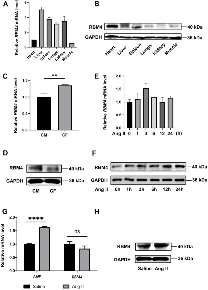
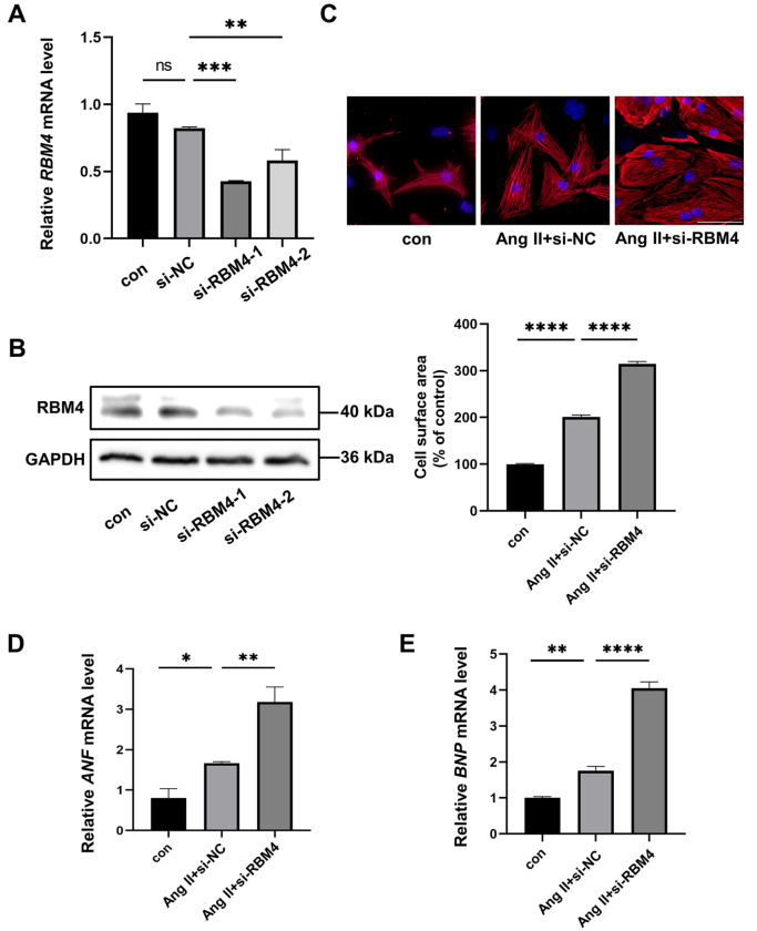
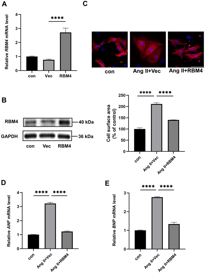
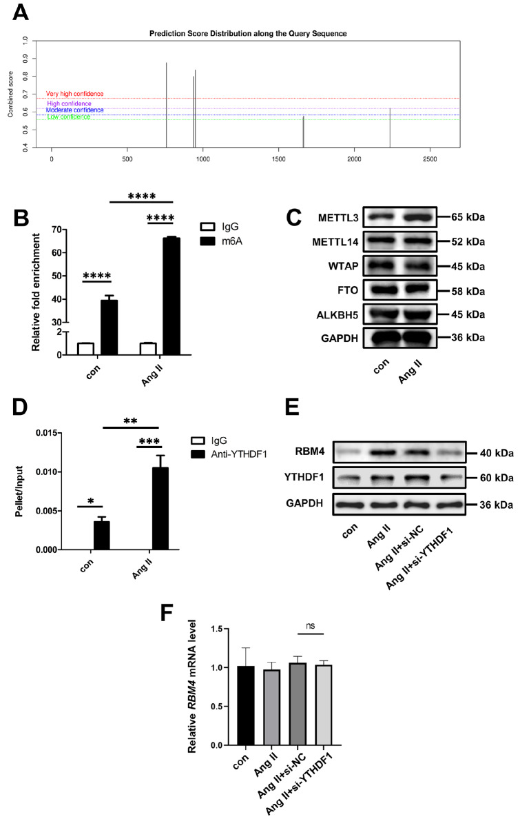
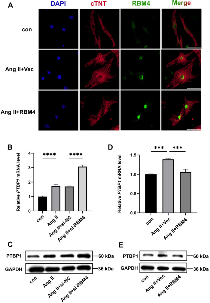
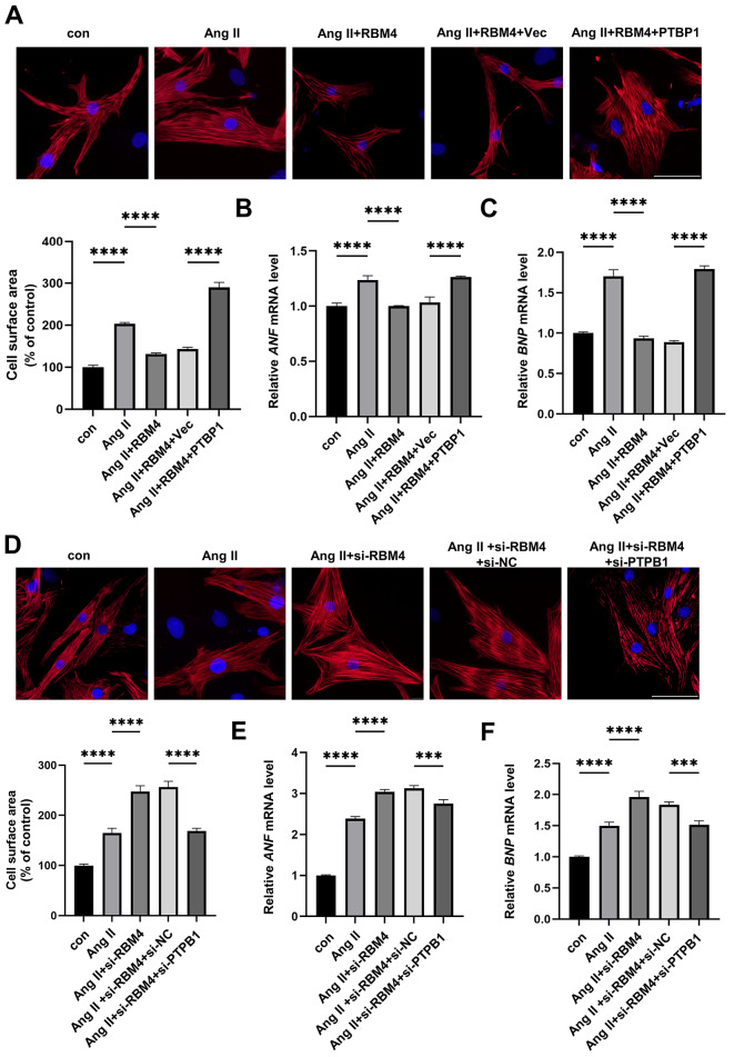
Aberrant gene expression in cardiomyocyte has been revealed to be the fundamental essence of pathological cardiac hypertrophy. However, the detailed mechanisms are not fully understood. The underlying regulators of gene expression involved in cardiac hypertrophy remain to be further identified. Here, we report that the RNA-binding protein RNA-binding motif protein 4 (RBM4) functions as an endogenic protector that is able to fight against cardiomyocyte hypertrophy in vitro. Under pro-hypertrophic stimulation of angiotensin II (Ang II), the protein level of RBM4 in cardiomyocyte and myocardium is elevated. Knockdown of RBM4 can further aggravate cardiomyocyte hypertrophy, while over-expression of RBM4 represses cardiomyocyte hypertrophy. Mechanistically, RBM4 is localized in the nucleus and down-regulates the expression of polypyrimidine tract-binding protein 1 (PTBP1), which has been shown to aggravate cardiomyocyte hypertrophy. In addition, we suggest that the up-regulation of RBM4 in cardiomyocyte hypertrophy is caused by N6-methyladenosine (m6A). Ang II induces m6A methylation of RBM4 mRNA, which further enhances the YTH domain-containing family protein 1 (YTHDF1)-mediated translation of RBM4. Thus, our results reveal a novel pathway consisting of m6A, RBM4 and PTBP1, which is involved in cardiomyocyte hypertrophy.

Keywords: PTBP1; RBM4; cardiomyocyte hypertrophy; m6A.


Conflict of interest statement

The authors declare that they have no conflict of interest.

Figures

None
Figure 1
The expression pattern of RBM4 (A,B) The expression of RBM4 in mouse tissue at the mRNA level analyzed by qPCR (A), and the protein level analyzed by immunoblotting (B). n = 3. (C,D) The expression of RBM4 in rat cardiomyocyte (CM) and cardiac fibroblast (CF) at the mRNA level analyzed by qPCR (**P < 0.01) and the protein level analyzed by immunoblotting (D). n = 3. (E,F) The expression of RBM4 in rat cardiomyocyte treated with Ang II. (E) The mRNA level was analyzed by qPCR. (F) The protein level was analyzed by immunoblotting. n = 3. (G) The mRNA levels of ANF and RBM4 in mouse hearts were analyzed by qPCR. ****P < 0.0001, ns, P ≥ 0.05. n = 3. (H) The RBM4 protein level in mouse hearts was analyzed by immunoblotting. n = 3.
None
Figure 2
Knockdown of RBM4 aggravates Ang II-induced cardiomyocyte hypertrophy (A,B) The expression of endogenous RBM4 was interfered by transfection with siRNA. si-NC, negative control siRNA. (A) The RBM4 mRNA level was analyzed by qPCR. ** P < 0.01, ***P < 0.001, ns, P ≥ 0.05. (B) The RBM4 protein level was analyzed by immunoblotting. si-RBM4-1 was selected for use in the subsequent assays. n = 3. (C‒E) Knockdown of RBM4 aggravated Ang II-induced cardiomyocyte hypertrophy. Cardiomyocytes were transfected with control siRNA or RBM4 siRNA and then treated with Ang II. (C) Representative images of cardiomyocytes stained for cTnT (red). The nuclei were stained with DAPI (blue). Scale bar: 50 μm. The cardiomyocyte cell surface area was quantified based on the pixel size using ImageJ software (below). ****P < 0.0001. (D) The ANF mRNA level was analyzed by qPCR. *P < 0.05, ** P < 0.01. (E) The BNP mRNA level was analyzed by qPCR. **P < 0.01, ****P < 0.0001. n = 3.
None
Figure 3
RBM4 overexpression attenuates Ang II-induced cardiomyocyte hypertrophy (A,B) Cardiomyocytes were transfected with empty vector (Vec) or RBM4 expression vector. (A) The RBM4 mRNA level was analyzed by qPCR. ****P < 0.0001. (B) The RBM4 protein level was analyzed by immunoblotting. n = 3. (C‒E) Over-expression of RBM4 attenuates Ang II-induced cardiomyocyte hypertrophy. Cardiomyocytes were transfected with an empty vector or an RBM4 vector and then treated with Ang II. (C) Representative images of cardiomyocytes stained for cTnT (red). The nuclei were stained with DAPI (blue). Scale bar: 50 μm. The cardiomyocyte cell surface area was quantified based on the pixel size using ImageJ software (below). ****P < 0.0001. (D) The ANF mRNA level was analyzed by qPCR. ****P < 0.0001. (E) The BNP mRNA level was analyzed by qPCR. **** P < 0.0001. n = 3.
None
Figure 4
The expression of RBM4 in cardiomyocyte is mediated by m6A-YTHDF1 (A) The potential m6A sites in RBM4 mRNA predicted by SRAMP. (B) The m6A methylation of RBM4 mRNA in cardiomyocyte is increased by Ang II stimulation. The N6-methylated RNAs captured from cardiomyocytes treated with Ang II or saline (con) by m6A antibody or control IgG, were purified with TRIzol. The RBM4 mRNA level was analyzed by qPCR. ****P < 0.0001. n = 3. (C) The levels of m6A writer or eraser proteins in cardiomyocytes were analyzed by immunoblotting. n = 3. (D) The interaction between RBM4 mRNA and YTHDF1 was increased by Ang II stimulation. The RNA captured from the lysates of cardiomyocytes treated with Ang II or saline (con) by YTHDF1 antibody or control IgG was purified with TRIzol. The RBM4 mRNA level was analyzed by qPCR. *P < 0.05, **P < 0.01, ***P < 0.001. n = 3. (E,F) YTHDF1 enhances RBM4 translation during cardiomyocyte hypertrophy. Cardiomyocytes were transfected with control siRNA or YTHDF1 siRNA and then treated with Ang II. (E) The protein levels of RBM4 and YTHDF1 were analyzed by immunoblotting. (F) The RBM4 mRNA level was analyzed by qPCR. ns, P ≥ 0.05. n = 3.
None
Figure 5
RBM4 down-regulates PTBP1 expression during cardiomyocyte hypertrophy (A) RBM4 is localized in the nucleus of cardiomyocyte. Represent images of cardiomyocytes stained for cTnT (red) and RBM4 (green). The nuclei were stained with DAPI (blue). Scale bar: 50 μm. (B,C) Knockdown of RBM4 increases the expression of PTBP1 during cardiomyocyte hypertrophy. Cardiomyocytes were transfected with control siRNA or RBM4 siRNA and then treated with Ang II. (B) The PTBP1 mRNA level was analyzed by qPCR. ****P < 0.0001. (C) PTBP1 protein levels were analyzed by immunoblotting. n = 3. (D,E) Over-expression of RBM4 decreases the expression of PTBP1 during cardiomyocyte hypertrophy. (D) The PTBP1 mRNA level was analyzed by qPCR. ***P < 0.001. (E) PTBP1 protein levels were analyzed by immunoblotting. n = 3.
None
Figure 6
PTBP1 induces cardiomyocyte hypertrophy (A,B) Cardiomyocytes were transfected with an empty vector or a PTPB1 vector. (A) The PTBP1 mRNA level was analyzed by qPCR. ****P < 0.0001. (B) PTBP1 protein levels were analyzed by immunoblotting. n = 3. (C‒E) Over-expression of PTBP1 induces cardiomyocyte hypertrophy. (C) Representative images of cardiomyocytes stained for cTnT (red). The nuclei were stained with DAPI (blue). Scale bar: 50 μm. The cardiomyocyte cell surface area was quantified based on the pixel size using ImageJ software (below). ***P < 0.001. n = 3. (D) The ANF mRNA level was analyzed by qPCR. ****P < 0.0001. (E) The BNP mRNA level was analyzed by qPCR. ****P < 0.0001. n = 3.
None
Figure 7
RBM4 inhibits cardiomyocyte hypertrophy by regulating PTBP1 (A‒C) PTBP1 abolishes the protective effect of RBM4 on cardiomyocyte hypertrophy. Cardiomyocytes were co-transfected with RBM4 vector and PTBP1 vector and then treated with Ang II. (A) Representative images of cardiomyocytes stained for cTnT (red). The nuclei were stained with DAPI (blue). Scale bar: 50 μm. The cardiomyocyte cell surface area was quantified based on the pixel size using ImageJ software (below). ****P < 0.0001. n = 3. (B) The ANF mRNA level was analyzed by qPCR. ****P < 0.0001. n = 3. (C) The BNP mRNA level was analyzed by qPCR. ****P < 0.0001. n = 3. (D‒F) Knockdown of PTBP1 counteracts excessive cardiomyocyte hypertrophy caused by RBM4 deficiency. Cardiomyocytes were cotransfected with RBM4 siRNA and PTBP1 siRNA and then treated with Ang II. (D) Representative images of cardiomyocytes stained for cTnT (red). The nuclei were stained with DAPI (blue). Scale bar: 50 μm. The cardiomyocyte cell surface area was quantified based on the pixel size using ImageJ software (below). ****P < 0.0001. n = 3. (E) The ANF mRNA level was analyzed by qPCR. ***P < 0.001, ****P < 0.0001. n = 3. (F) The BNP mRNA level was analyzed by qPCR. ***P < 0.001, ****P < 0.0001. n = 3.

Similar articles

References

    1. Nakamura M, Sadoshima J. Mechanisms of physiological and pathological cardiac hypertrophy. Nat Rev Cardiol. . 2018;15:387–407. doi: 10.1038/s41569-018-0007-y. - DOI - PubMed
    1. Shimizu I, Minamino T. Physiological and pathological cardiac hypertrophy. J Mol Cell Cardiol. . 2016;97:245–262. doi: 10.1016/j.yjmcc.2016.06.001. - DOI - PubMed
    1. Maris C, Dominguez C, Allain FH- The RNA recognition motif, a plastic RNA-binding platform to regulate post-transcriptional gene expression. FEBS J. . 2005;272:2118–2131. doi: 10.1111/j.1742-4658.2005.04653.x. - DOI - PubMed
    1. Li Z, Guo Q, Zhang J, Fu Z, Wang Y, Wang T, Tang J. The RNA-binding motif protein family in cancer: friend or foe? Front Oncol. . 2021;11:757135. doi: 10.3389/fonc.2021.757135. - DOI - PMC - PubMed
    1. van den Hoogenhof MMG, Beqqali A, Amin AS, van der Made I, Aufiero S, Khan MAF, Schumacher CA, et al. RBM20 mutations induce an arrhythmogenic dilated cardiomyopathy related to disturbed calcium handling. Circulation. . 2018;138:1330–1342. doi: 10.1161/CIRCULATIONAHA.117.031947. - DOI - PubMed

MeSH terms