. 2024 Jul 25:6:1412864.
doi: 10.3389/ftox.2024.1412864. eCollection 2024.

Diesel exhaust particles alter mitochondrial bioenergetics and cAMP producing capacity in human bronchial epithelial cells

Affiliations

Diesel exhaust particles alter mitochondrial bioenergetics and cAMP producing capacity in human bronchial epithelial cells

Isabella Cattani-Cavalieri et al. Front Toxicol. .

Abstract

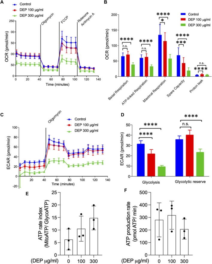
Introduction: Air pollution from diesel combustion is linked in part to the generation of diesel exhaust particles (DEP). DEP exposure induces various processes, including inflammation and oxidative stress, which ultimately contribute to a decline in lung function. Cyclic AMP (cAMP) signaling is critical for lung homeostasis. The impact of DEP on cAMP signaling is largely unknown. Methods: We exposed human bronchial epithelial (BEAS-2B) cells to DEP for 24-72 h and evaluated mitochondrial bioenergetics, markers of oxidative stress and inflammation and the components of cAMP signaling. Mitochondrial bioenergetics was measured at 72 h to capture the potential and accumulative effects of prolonged DEP exposure on mitochondrial function. Results: DEP profoundly altered mitochondrial morphology and network integrity, reduced both basal and ATP-linked respiration as well as the glycolytic capacity of mitochondria. DEP exposure increased gene expression of oxidative stress and inflammation markers such as interleukin-8 and interleukin-6. DEP significantly affected mRNA levels of exchange protein directly activated by cAMP-1 and -2 (Epac1, Epac2), appeared to increase Epac1 protein, but left phospho-PKA levels unhanged. DEP exposure increased A-kinase anchoring protein 1, β2-adrenoceptor and prostanoid E receptor subtype 4 mRNA levels. Interestingly, DEP decreased mRNA levels of adenylyl cyclase 9 and reduced cAMP levels stimulated by forskolin (AC activator), fenoterol (β2-AR agonist) or PGE2 (EPR agonist). Discussion: Our findings suggest that DEP induces mitochondrial dysfunction, a process accompanied by oxidative stress and inflammation, and broadly dampens cAMP signaling. These epithelial responses may contribute to lung dysfunction induced by air pollution exposure.

Keywords: air pollution; cAMP; diesel exhaust particles; lung; mitochondria; oxidative stress.


Conflict of interest statement

The authors declare that the research was conducted in the absence of any commercial or financial relationships that could be construed as a potential conflict of interest.

Figures

FIGURE 1
FIGURE 1
Exposure of BEAS-2B cells to DEP alters mitochondrial morphology. (A) Representative MitoTracker images are shown. Scale bar of 20 μm. (B) Relative quantification of mitochondria in categories I-III. (C) Mitochondrial footprint (µm2), (D) branches (counts) and (E) branch length was analyzed using the ImageJ MINA-Macro. BEAS-2B cells were exposed to 100 μg/mL DEP or 0.5% DMSO (control). Data represent 4 independent experiments (15–20 technical replicates per group) and are expressed as mean ± SD; *p < 0.05 and ****p < 0.0001 significant difference between indicated groups.
FIGURE 2
FIGURE 2
Exposure of BEAS-2B cells to DEP negatively affects mitochondrial respiration and glycolysis. The injection of oligomycin was used to inhibit ATP synthase (complex V), leading to a reduction in mitochondrial respiration. The parameters measured after oligomycin injection are indicated as ATP-linked respiration (ATP produced by the mitochondria) and proton leak (remining basal respiration not coupled to ATP production). FCCP was then used as an uncoupling agent, which stimulates the respiratory chain to operate at maximum capacity, revealing the maximum rate of respiration of the cell. Spare capacity shows the ability of the cell to respond to an energetic demand. Rotenone and actinomycin were then used to inhibit complex I and III, respectively, shutting down mitochondrial respiration (Divakaruni et al., 2014) (A,B) Basal respiration, ATP-linked respiration, maximal respiration and spare capacity were analyzed using Seahorse analyzer. (C) Glycolysis, (D) glycolytic reserve, (E) ATP rate index and (F) ATP production rate was determined based on the ECAR profile under different conditions. BEAS-2B cells were exposed to DEP (100 or 300 μg/mL) or 0.5% DMSO (control) for 72 h. Data represent 3–5 independent experiments and are expressed as mean ± SD; *p < 0.05, ***p < 0.001, and ****p < 0.0001 significant difference between indicated groups.
FIGURE 3
FIGURE 3
Exposure of BEAS-2B cells to DEP did not alter cell viability and induces expression of inflammation markers. (A) xCELLigence measurement in BEAS-2B cells exposed to DEP (100 or 300 μg/mL) or 5% DMSO (control) for 24 h. (B–D) MTT measurement of BEAS-2B cells exposed to the indicated concentrations of DEP for 24, 48 and 72 h. Exposure of BEAS-2B cells to DEP induces expression of inflammation markers. (E) IL-8 and IL-6 gene expression and (F,G) secreted protein was determined by real-time quantitative PCR and ELISA, respectively, in BEAS-2B cells exposed to 100 μg/mL DEP for 24 h. Real-time quantitative PCR data is expressed as fold over the control (vehicle treated) condition using the ΔΔCt method. Data are representative of 3–5 independent experiments and expressed as mean ± SD; *p < 0.05, significant difference between indicated groups.
FIGURE 4
FIGURE 4
Exposure of BEAS-2B cells to DEP alters Epac mRNA content but does not affect expression of phospho-PKA substrates and PDE4 isoforms mRNA. (A) Gene expression levels of Epac1 and Epac2 were assessed using real-time quantitative PCR. Data is expressed as fold over the control (vehicle treated) condition using the ΔΔCt method. (B,C,E) Protein expression of Epac1, Epac2 and phospho-PKA substrates were analyzed using Western blotting. (D) Representative immunofluorescence images are shown. Scale bar of 200 μm. (F–I) Gene expression levels of PDE4A, PDE4B, PDE4D and AKAP1 were assessed using real-time quantitative PCR. BEAS-2B were exposed to 100 μg/mL DEP or 0.5% DMSO (control) (A–D), unless indicated otherwise (E–I), for 24 h. Data represent 3–4 independent experiments and are expressed as mean ± SD; *p < 0.05, significant difference between indicated groups.
FIGURE 5
FIGURE 5
Exposure of BEAS-2B cells to DEP alters β2-AR, EP4 and cAMP kinetics. BEAS-2B cells were exposed to DEP (100 or 300 μg/mL) or 0.5% DMSO (control) for 24 h. (A,B) Gene expressions of β2-AR and EP4 were measured using real-time quantitative PCR. Data is expressed as fold over the control (vehicle treated) condition using the ΔΔCt method. (C–F) cAMP was measured in real-time using the GloSenor cAMP assay in the absence and presence of either forskolin, fenoterol (each 10 µM) or a PGE2 analogue (3 µM). The increase in luminescence signal was expressed as RLU; relative luminescence units. Data represent 2–6 independent experiments and are expressed as mean ± SD; *p < 0.05, significant difference between indicated groups.
FIGURE 6
FIGURE 6
Exposure of BEAS-2B cells to DEP alters specific AC mRNA content. BEAS-2B cells were exposed to DEP (100 or 300 μg/mL) or 0.5% DMSO (control) for 24 h. (A–E) Gene expressions of AC1, AC3, AC6, AC7, AC9 were measured using real-time quantitative PCR. Data is expressed as fold over the control (vehicle treated) condition using the ΔΔCt method. Data represent 4–6 independent experiments and are expressed as mean ± SD; *p < 0.05, significant difference between indicated groups.
FIGURE 7
FIGURE 7
Proposed molecular mechanism showing the effect of DEP in the cAMP production and mitochondrial bioenergetics in human bronchial epithelial cells. (A) Unknown effects of DEP on inflammation and oxidative stress, GPCRs and cAMP signaling, and mitochondrial function. (B) DEP induces oxidative stress and inflammation; and induced cAMP generating receptor (β2-AR and EP4). However, the reduction of AC9 affected the cAMP production levels. AKAP1 and SOD2 elevation show the potential alteration in the mitochondrial function.

Similar articles

Cited by

References

    1. Acin-Perez R., Salazar E., Kamenetsky M., Buck J., Levin L. R., Manfredi G. (2009). Cyclic AMP produced inside mitochondria regulates oxidative phosphorylation. Cell. Metab. 9 (3), 265–276. 10.1016/j.cmet.2009.01.012 - DOI - PMC - PubMed
    1. Bach N. S., Lag M., Ovrevik J. (2014). Toll like receptor-3 priming alters diesel exhaust particle-induced cytokine responses in human bronchial epithelial cells. Toxicol. Lett. 228 (1), 42–47. 10.1016/j.toxlet.2014.03.021 - DOI - PubMed
    1. Baldwin T. A., Li Y., Brand C. S., Watts V. J., Dessauer C. W. (2019). Insights into the regulatory properties of human adenylyl cyclase type 9. Mol. Pharmacol. 95 (4), 349–360. 10.1124/mol.118.114595 - DOI - PMC - PubMed
    1. Cao X., Fu M., Bi R., Zheng X., Fu B., Tian S., et al. (2021). Cadmium induced BEAS-2B cells apoptosis and mitochondria damage via MAPK signaling pathway. Chemosphere 263, 128346. 10.1016/j.chemosphere.2020.128346 - DOI - PubMed
    1. Cattani-Cavalieri I., Valenca S. D. S., Schmidt M. (2020). Nanodomains in cardiopulmonary disorders and the impact of air pollution. Biochem. Soc. Trans. 48, 799–811. 10.1042/BST20190250 - DOI - PMC - PubMed