Review
. 2024 Aug 1;12(4):75.
doi: 10.21037/atm-23-1430. Epub 2023 Aug 11.

Review on endobronchial therapies-current status and future

Affiliations
Review

Review on endobronchial therapies-current status and future

Joyce W Y Chan et al. Ann Transl Med. .

Abstract

There is a growing demand for lung parenchymal-sparing localized therapies due to the rising incidence of multifocal lung cancers and the growing number of patients who cannot undergo surgery. Lung cancer screening has led to the discovery of more pre-malignant or early-stage lung cancers, and the focus has shifted from treatment to prevention. Transbronchial therapy is an important tool in the local treatment of lung cancers, with microwave ablation showing promise based on early and mid-term results. To improve the precision and efficiency of transbronchial ablation, adjuncts such as mobile C-arm platforms, software to correct for computed tomography (CT)-to-body divergence, metal-containing nanoparticles, and robotic bronchoscopy are useful. Other forms of energy such as steam vapor therapy, pulsed electric field, and photodynamic therapy are being intensively investigated. In addition, the future of transbronchial therapies may involve the intratumoral injection of novel agents such as immunomodulating agents, gene therapies, and chimeric antigen receptor T cells. Extensive pre-clinical and some clinical research has shown the synergistic abscopal effect of combination of these agents with ablation. This article aims to provide the latest updates on these technologies and explore their most likely future applications.

Keywords: Transbronchial ablation; microwave ablation; photo dynamic therapy; pulsed electric field; robotic bronchoscopy.


Conflict of interest statement

Conflicts of Interest: All authors have completed the ICMJE uniform disclosure form (available at https://atm.amegroups.com/article/view/10.21037/atm-23-1430/coif). The series “Lung Cancer Management—The Next Decade” was commissioned by the editorial office without any funding or sponsorship. C.S.H.N. served as the unpaid Guest Editor of the series and serves as the Editor-in-Chief of Annals of Translational Medicine from January 2022 to December 2023. He is also a consultant for Johnson and Johnson, Medtronic USA and Siemens Healthineer. R.W.H.L. is a consultant for Medtronic USA and Siemens Healthineer. The authors have no other conflicts of interest to declare.

Figures
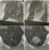
Figure 1
Figure 1
Pre- and post-ablation cone-beam CT images of a right lower lobe nodule. Column (A) is a pre-ablation cone-beam CT in two axes showing a 7 mm right lower lobe solid colorectal lung metastasis in a patient who had undergone right upper and middle lobectomy. Column (B) shows the post-ablation CT after microwave ablation (100W × 10 minutes × 2), the lesion (yellow outline) is covered adequately by surrounding ground glass opacity which represents the ablation zone, with minimal margin of 8 mm. CT, computed tomography.
Figure 2
Figure 2
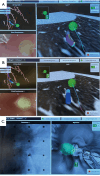
Images during ENB navigation using the IllumisiteTM platform. Figure (A) shows the tip of locatable guide pointing directly at the lesion (represented by green ball) after peripheral navigation. In Figure (B), the locatable guide is removed, but there are positional sensors embedded in the extended working channel, showing its tip pointing also at the lesion. In Figure (C), the cross hair is shown pointing slightly off center at the lesion which would require further manipulation to ensure target view, while the tip of catheter is shown to be 2 cm from the target lesion. ENB, electromagnetic navigation bronchoscopy.
Figure 3
Figure 3
This figure shows the operating room setup during Cios Spin® mobile C-arm image acquisition.
Figure 4
Figure 4
During robotic bronchoscopy by Auris MonarchTM, the left hand panel shows the real-time bronchoscopic view, middle panels show bull’s eye view with cross hair in centre and lesion represented by yellow ball, while the right hand panels show the pre-operative CT scan images in 3 perpendicular axes. CT, computed tomography.
Figure 5
Figure 5
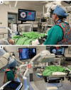
Intraoperative layout during robotic bronchoscopy. Figure (A) shows the operator standing on the right side of patient manipulating the robotic bronchoscope by Auris MonarchTM (which has been inserted through the endotracheal tube) using a controller. The screen (*) shows real time bronchoscopic view during the initial registration process. Figure (B) shows robotic bronchoscopy used together with cone-beam CT in hybrid operating room to further improve the accuracy of navigation, biopsy or treatment. A screen shows robotic navigation (*) and another screen shows real time fluoroscopy (#), while another operator advances biopsy tools through the robotic bronchoscope (@). CT, computed tomography.
Figure 6
Figure 6
Operative setup when performing robotic ENB microwave ablation is similar to that during robotic ENB biopsy using Auris MonarchTM, except that the treatment catheter is connected to the microwave generator on the left. ENB, electromagnetic navigation bronchoscopy.
Figure 7
Figure 7
This shows the overlay of two pre-ablation CT scans with ablation catheter positioned on each side of nodule (target 2) to perform bracket ablation. The catheter was put in position 1 with ablation performed, then renavigated to position 2 to deliver a second ablation, in order to ensure adequate margin on both sides of nodule. CT, computed tomography.
Figure 8
Figure 8
Operative set up during transbronchial pulsed electric field ablation. The endobronchial applicator is inserted into the bronchoscope on the right side of photo, and is connected to appliances on the left side which includes the electrical signal generator, and impedance and ECG monitors. ECG, electrocardiography.

Similar articles

Cited by

  • Robotic-assisted bronchoscopy-advancing lung cancer management.
    Chang ATC, Chan JWY, Siu ICH, Liu W, Lau RWH, Ng CSH. Chang ATC, et al. Front Surg. 2025 Jun 3;12:1566902. doi: 10.3389/fsurg.2025.1566902. eCollection 2025. Front Surg. 2025. PMID: 40529888 Free PMC article. Review.
  • Chinese expert consensus on shape-sensing robotic-assisted bronchoscopy (ssRAB) in the management of peripheral pulmonary lesions.
    Xie F, Zhang C, Li C, Liu D, Zhang Q, Mu C, Hou G, Li M, Tang C, Xu F, Chen H, Chen Z, Gu Y, Guan Z, Ji C, Li H, Li W, Li X, Li Y, Lian H, Liao J, Luo Z, Ouyang H, Shen Y, Shi Y, Wan N, Wang T, Wang H, Wang H, Wang J, Wu J, Wu X, Xia Y, Xiao K, Xu W, Yang H, Yang J, Ye T, Ye X, Yu P, Zhang N, Zhang P, Zhao Q, Zheng X, Zou J, Chen E, Zhang H, Sun J; Professional Committee on Interventional Pulmonology of China Association for Promotion of Health Science and Technology; Expert Group on Technology of Shape-sensing Robotic-assisted Bronchoscopy. Xie F, et al. Transl Lung Cancer Res. 2025 May 30;14(5):1500-1515. doi: 10.21037/tlcr-2025-400. Epub 2025 May 28. Transl Lung Cancer Res. 2025. PMID: 40535069 Free PMC article. Review.
  • Two-phase radial endobronchial ultrasound bronchoscopy registration.
    Zhao W, Kuhlengel T, Chang Q, Daneshpajooh V, He Y, Kao A, Bascom R, Ahmad D, Htwe YM, Toth J, Schaer T, Brewer L, Hilliard R, Higgins WE. Zhao W, et al. J Med Imaging (Bellingham). 2025 Mar;12(2):025001. doi: 10.1117/1.JMI.12.2.025001. Epub 2025 Mar 7. J Med Imaging (Bellingham). 2025. PMID: 40061857

References

    1. Leventakos K, Peikert T, Midthun DE, et al. Management of Multifocal Lung Cancer: Results of a Survey. J Thorac Oncol 2017;12:1398-402. 10.1016/j.jtho.2017.05.013 - DOI - PMC - PubMed
    1. Chan JWY, Lau R, Chang A, et al. 96P Transbronchial microwave ablation: Important role in the battle of lung preservation for multifocal lung primaries or metastases. Ann Oncol 2022;33:S76-7.
    1. Jung W, Cho S, Yum S, et al. Stepwise Disease Progression Model of Subsolid Lung Adenocarcinoma with Cystic Airspaces. Ann Surg Oncol 2020;27:4394-403. 10.1245/s10434-020-08508-4 - DOI - PubMed
    1. Inamura K. Clinicopathological Characteristics and Mutations Driving Development of Early Lung Adenocarcinoma: Tumor Initiation and Progression. Int J Mol Sci 2018;19:1259. 10.3390/ijms19041259 - DOI - PMC - PubMed
    1. Suzuki K, Kusumoto M, Watanabe S, et al. Radiologic classification of small adenocarcinoma of the lung: radiologic-pathologic correlation and its prognostic impact. Ann Thorac Surg 2006;81:413-9. 10.1016/j.athoracsur.2005.07.058 - DOI - PubMed

LinkOut - more resources