. 2024:4:1257880.
doi: 10.3389/fnume.2024.1257880. Epub 2024 Feb 7.

Motion-correction strategies for enhancing whole-body PET imaging

Affiliations

Motion-correction strategies for enhancing whole-body PET imaging

James Wang et al. Front Nucl Med. 2024.

Abstract

Positron Emission Tomography (PET) is a powerful medical imaging technique widely used for detection and monitoring of disease. However, PET imaging can be adversely affected by patient motion, leading to degraded image quality and diagnostic capability. Hence, motion gating schemes have been developed to monitor various motion sources including head motion, respiratory motion, and cardiac motion. The approaches for these techniques have commonly come in the form of hardware-driven gating and data-driven gating, where the distinguishing aspect is the use of external hardware to make motion measurements vs. deriving these measures from the data itself. The implementation of these techniques helps correct for motion artifacts and improves tracer uptake measurements. With the great impact that these methods have on the diagnostic and quantitative quality of PET images, much research has been performed in this area, and this paper outlines the various approaches that have been developed as applied to whole-body PET imaging.

Keywords: cardiac gating; data-driven gating; hardware-driven gating; motion correction; positron emission tomography (PET); respiratory gating.


Conflict of interest statement

Conflict of interest AM is a consultant for Weinberg Medical Physics. The remaining authors declare that the research was conducted in the absence of any commercial or financial relationships that could be construed as a potential conflict of interest.

Figures
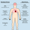
Figure 1
Figure 1
Diagram of techniques used to correct for head motion, respiratory motion, and cardiac motion for PET imaging. Multiple techniques have been developed based on the specific application when imaging certain anatomical areas, thus these techniques can be categorized based on the motion source that they are addressing. The left column shows a list of hardware driven techniques, and the right column shows data driven techniques.
Figure 2
Figure 2
Implementing hardware-driven gating can provide drastic improvement to PET images by reducing motion artifacts. It helps improve SNR and remove motion artifacts. Abdominal PET images of the liver and kidneys are shown with (A) no gating and (B) respiratory belt gating. The red arrow shows a distinct motion artifact adjacent to an FDG-avid lesion in the ungated image.
Figure 3
Figure 3
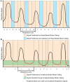
Phase-based and amplitude-based gating are two ways to process motion signals for motion gating. The red components represent regions used for image reconstruction. (A) Phase-based gating divides the motion signal into gates. Specific gates can be chosen for the corresponding PET data to be used for image reconstruction. Using this method, multiple images can be generated based on each phase gate. (B) Amplitude-based gating divides the motion signal based on the amplitude of motion. The PET data with motion that is within the specified amplitude band is kept for image reconstruction. Data with motion that is outside of the amplitude band is discarded.
Figure 4
Figure 4
The application of data-driven gating can vastly improve image quality of PET images. (A) Uncorrected images can show lots of blurring and other motion artifacts. (B) Data-driven motion correction using an image registration approach was applied to this brain image and shows much greater clarity than the uncorrected version.
Figure 5
Figure 5
Cardiac gating must take into account the different phases of the cardiac cycle. (A) This outline shows the different sections of the cardiac cycle with the PQRST peaks and how different stages could affect cardiac outlines. (B) Sample images shown here are non-gated, EKG-gated, cardiac-gated, and dual-gated from left to right.
Figure 6
Figure 6
Processing flowchart for hardware-driven and data-driven gating. The red pathway represents processing pipelines of motion gating stemming from multi-modality PET imaging which can include images or data acquired from other sources such as MRI, CT, or ultrasound. The blue pathway represents the general processing pipeline for hardware-driven gating.

References

    1. Shukla AK, Kumar U. Positron emission tomography: an overview. J Med Phys. (2006) 31(1):13–21. 10.4103/0971-6203.25665 - DOI - PMC - PubMed
    1. Anand SS, Singh H, Dash AK. Clinical applications of PET and PET-CT. Med J Armed Forces India. (2009) 65(4):353–8. 10.1016/S0377-1237(09)80099-3 - DOI - PMC - PubMed
    1. Wardak M, Wong KP, Shao W, Dahlbom M, Kepe V, Satyamurthy N, et al. Movement correction method for human brain PET images: application to quantitative analysis of dynamic 18F-FDDNP scans. J Nucl Med. (2010) 51(2):210. 10.2967/jnumed.109.063701 - DOI - PMC - PubMed
    1. Rahmim A, Lodge MA, Karakatsanis NA, Panin VY, Zhou Y, McMillan A, et al. Dynamic whole-body PET imaging: principles, potentials and applications. Eur J Nucl Med Mol Imaging. (2019) 46(2):501–18. 10.1007/s00259-018-4153-6 - DOI - PubMed
    1. Kyme AZ, Fulton RR. Motion estimation and correction in SPECT, PET and CT. Phys Med Biol. (2021) 66(18):18TR02. 10.1088/1361-6560/ac093b - DOI - PubMed

LinkOut - more resources