. 2024 Oct;11(38):e2404693.
doi: 10.1002/advs.202404693. Epub 2024 Aug 9.

Methuosis Inducer SGI-1027 Cooperates with Everolimus to Promote Apoptosis and Pyroptosis by Triggering Lysosomal Membrane Permeability in Renal Cancer

Affiliations

Methuosis Inducer SGI-1027 Cooperates with Everolimus to Promote Apoptosis and Pyroptosis by Triggering Lysosomal Membrane Permeability in Renal Cancer

Yu Luo et al. Adv Sci (Weinh). 2024 Oct.

Abstract

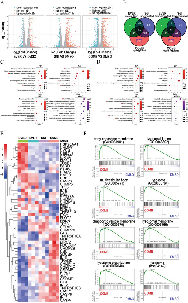
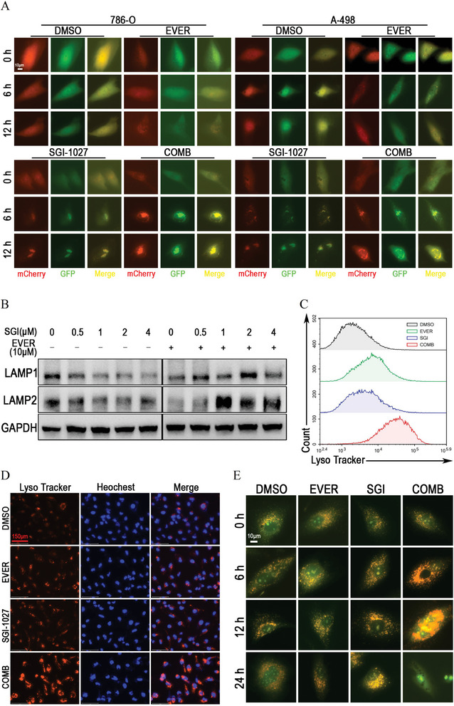
The mTOR inhibitor everolimus has been approved as a sequential or second-line therapy for renal cell carcinoma (RCC). However, the development of drug resistance limits its clinical applications. This study aims to address the challenge of everolimus resistance and provide new insights into the treatment of advanced RCC. Here, the cytotoxicity of the DNA methyltransferase 1 (DNMT1) inhibitor SGI-1027 in inducing cell vacuolation and methuosis is discovered and demonstrated for the first time. Additionally, SGI-1027 exerts synergistic effects with everolimus, as their combination suppresses the growth, migration, and invasion of renal cancer cells. Mechanistically, apoptosis and GSDME-dependent pyroptosis triggered by lysosomal membrane permeability (LMP) are observed. The upregulation of GSDME expression and increased lysosomal activity in renal cancer cells provide a therapeutic window for the combination of these two drugs to treat renal cancer. The combination treatment exhibits effective anti-tumor activity and is well tolerated in a subcutaneous tumor model. Overall, this study validates and reveals the specific cytotoxicity property of SGI-1027 and its potent synergistic effect with everolimus, offering new insights into advanced RCC therapy and everolimus-resistance overcoming.

Keywords: SGI‐1027; everolimus; lysosome; methuosis; pyroptosis; renal cell carcinoma.


Conflict of interest statement

The authors declare no conflict of interest.

Figures

Figure 1
Figure 1
SGI‐1027 induced vacuoles via macropinocytosis in renal cancer cells. A–D) The half maximal inhibitory concentration (IC50) of SGI‐1027 in 786‐O, A‐498, Caki‐1 and HK‐2 cells calculated by nonlinear regression. 786‐O, A‐498, Caki‐1 and HK‐2 cells were treated with SGI‐1027 at indicated concentrations for 48 h. E) HK‐2, 786‐O, A‐498, and Caki‐1 cells were treated with 1 × 10−6 m or 2 × 10−6 m SGI‐1027 for 24 h and living cell images were captured under microscope. F) Lucifer yellow staining of 786‐O and A‐498 cells. 786‐O and A‐498 cells were treated with 1.5 × 10⁻⁶ m SGI‐1027 and 2 mg mL−1 Lucifer yellow for 24 h. G) 786‐O and A‐498 cells were treated with 1.5 × 10⁻⁶ m SGI‐1027 for 24 h and then incubated with Lyso‐Tracker, Mito‐Tracker and ER‐Tracker for 30 min. Hoechst was used for specifically staining of the nucleus. H) Immunofluorescent staining of Rab7, LAMP1 and LAMP2 in 786‐O and A‐498 cells treated with 1.5 × 10−6 m SGI‐1027 for 24 h.
Figure 2
Figure 2
SGI‐1027‐induced cytotoxicity involved methuosis in renal cancer cells. A) 786‐O cells were treated with 1.5 × 10⁻⁶ m SGI‐1027 for 24 h and then observed under transmission electron microscopy after the preparation of cell sections. The green and purple arrows indicated the endoplasmic reticulum and mitochondria, respectively. B) Living cell images illustrating the vacuolation induced by SGI‐1027 independently on apoptosis, autophagy or necrosis. 786‐O cells were treated with1.5 × 10−6 m SGI‐1027, 50 × 10−6 m Z‐VAD‐FMK, 1 × 10−3 m 3‐MA, 50 × 10−6 m necrostatin‐1 or their combinations as indicated for 24 h. C) Typical Western blot showing the expression of PARP and cleaved PARP (Cl‐PARP). 786‐O and A‐498 cells were treated with DMSO (control), SGI‐1027 at indicated concentrations, or 10 × 10−6 m cis‐diaminodichloroplatinum (CDDP, positive control) for 48 h. PARP and cleaved PARP (Cl‐PARP) were detected by Western blot, and β‐actin was used as loading control. D) For validating the relationship between SGI‐1027 and apoptosis, 786‐O cells were pretreated with 50 × 10−6 m Z‐VAD‐FMK for 2 h and then treated with 2 × 10⁻⁶ m SGI‐1027 or 10 × 10−6 m cis‐diaminodichloroplatinum (CDDP, positive control) for 48 h. For validating the relationship between SGI‐1027 and autophagy or necrosis, 786‐O cells were treated with 2 × 10−6 m SGI‐1027, 1 × 10−3 m 3‐MA, 50 × 10−6 m necrostatin‐1 or their combinations as indicated for 48 h. The cell viability was detected by CCK‐8 assay. NS, not significant; **P < 0.01; ****P < 0.0001. E,F) 786‐O cells were treated with 2 × 10⁻⁶ m SGI‐1027 for the indicated time or treated with SGI‐1027 for 24 h at the indicated concentrations. LC3B was detected by Western blot, and GAPDH was used as loading control.
Figure 3
Figure 3
SGI‐1027 and everolimus synergically suppress the proliferation of renal cancer cells. A) Everolimus promoted SGI‐1027‐induced vacuolation in renal cancer cells. 786‐O and A‐498 cells were treated with DMSO (control), 5 × 10⁻⁶ m everolimus, 1.5 × 10⁻⁶ m SGI‐1027, and their combination for 12 h. B) Synergy scoring models demonstrating the synergistic effect of SGI‐1027 and everolimus. 786‐O and A‐498 cells were treated with different concentrations of SGI‐1027 and everolimus, or their combination as indicated for 24 h. Subsequently, the synergy scoring models were constructed by Synergyfinder software according to the cell viability. C) CCK‐8 assay for detecting the cell proliferation of 786‐O and A‐498 cells treated with DMSO (control), 5 × 10⁻⁶ m everolimus, 1.5 × 10⁻⁶ m SGI‐1027, or their combination for indicated time. D) EdU assay for labeling the cells in the state of DNA synthesis. 786‐O and A‐498 cells were treated with DMSO (control), 5 × 10⁻⁶ m everolimus, 1.5 × 10⁻⁶ m SGI‐1027, or their combination for 24 h before EdU staining. E) The synergistic effect of SGI‐1027 and everolimus on the cell cycle of renal cancer cells. 786‐O and A‐498 cells were treated with DMSO (control), 5 × 10⁻⁶ m everolimus, 1.5 × 10⁻⁶ m SGI‐1027, or their combination for 24 h before propidium iodide (PI) staining and flow cytometry analysis. NS, not significant; *P < 0.05; **P < 0.01; ***P < 0.001; ****P < 0.0001. EVER, everolimus; SGI, SGI‐1027; COMB, SGI‐1027 combined with everolimus.
Figure 4
Figure 4
SGI‐1027 and everolimus synergically inhibited the colony formation, migration and invasion in renal cancer cells. A) Colony formation assay detecting the proliferation ability of renal cancer cells. 786‐O and A‐498 cells were stained with crystal violet after incubation with DMSO (control), 1 × 10⁻⁶ m everolimus, 1 × 10⁻⁶ m SGI‐1027, or their combination for 14 d. B) Wound‐healing assay and C) transwell migration assay detecting the migration of renal cancer cells. 786‐O and A‐498 cells were treated with 5 × 10⁻⁶ m everolimus, 1.5 × 10⁻⁶ m SGI‐1027, or their combination for 24 h in each assay. D) Transwell invasion assay detecting the invasion of renal cancer cells. 786‐O and A‐498 cells were treated with DMSO (control), 5 × 10⁻⁶ m everolimus, 1.5 × 10⁻⁶ m SGI‐1027, or their combination for 48 h. NS, not significant; **P < 0.01; ***P < 0.001; ****P < 0.0001. EVER, everolimus; SGI, SGI‐1027; COMB, SGI‐1027 combined with everolimus.
Figure 5
Figure 5
SGI‐1027 cooperated with everolimus to induce apoptosis and GSDME‐dependent pyroptosis. A) The transmission electron microscopy imaging of 786‐O cells treated with DMSO (control), 5 × 10⁻⁶ m everolimus, 1.5 × 10⁻⁶ m SGI‐1027, or their combination for 24 h. B) Annexin V and propidium iodide (PI) staining for 786‐O and A‐498 cells treated with DMSO (control), 10 × 10−6 m everolimus, different concentrations of SGI‐1027 and their combinations as indicated. C) LDH release assay detecting the LDH releasing of renal cancer cells treated with DMSO (control), 5 × 10⁻⁶ m everolimus, 1.5 × 10⁻⁶ m SGI‐1027, or their combination for indicated time. D) A‐498 cells were treated with DMSO (control), 5 × 10⁻⁶ m everolimus, 2 × 10⁻⁶ m SGI‐1027, or their combination for 24 h. PARP, cleaved PARP (Cl‐PARP), GSDME, GSDMD were detected by Western blot, and GAPDH was used as loading control. E) A‐498 cells were treated with the combination of 5 × 10⁻⁶ m everolimus and 2 × 10⁻⁶ m SGI‐1027 for indicated time. PARP, cleaved PARP, GSDME, pro‐Caspase 3, cleaved Caspase 3 (Cl‐Caspase 3) were detected by Western blot. GAPDH was used as loading control. ***P < 0.001; ****P < 0.0001. EVER, everolimus; SGI, SGI‐1027; COMB, SGI‐1027 combined with everolimus.
Figure 6
Figure 6
RNA sequencing analysis unveiled the association between the synergistic effect and lysosome. A) The volcano plots for everolimus group, SGI‐1027 group, and SGI‐1027 combined with everolimus group. B) Venn plots of upregulated or downregulated DEGs in everolimus group, SGI‐1027 group and SGI‐1027 combined with everolimus group. C,D) GO and KEGG enrichment analyses for up‐regulated DEGs C) and down‐regulated DEGs D) unique to the combination group. BP, biological process; CC, cellular component; MF, molecular function; KEGG, Kyoto Encyclopedia of Genes and Genomes. E) Heatmap of the expression of pyroptosis‐related genes in the control group (DMSO), everolimus, SGI‐1027, and combination treatment group. F) GSEA enrichment analyses for gene sets related to cytophagy and lysosome in combination group. ES, enrichment score; NES, normalized enrichment score. EVER, everolimus; SGI, SGI‐1027; COMB, SGI‐1027 combined with everolimus.
Figure 7
Figure 7
SGI‐1027 cooperated with everolimus to induce lysosomal membrane permeability (LMP) in renal cancer cells. A) Detection of autophagy flux in renal cancer cells. 786‐O and A‐498 cells were infected by Ad‐mCherry‐GFP‐LC3B, after which cells were treated with DMSO (control), 5 × 10⁻⁶ m everolimus, 2 × 10⁻⁶ m SGI‐1027, or their combination for indicated time. B) A‐498 cells were treated with 10 µM everolimus, different concentrations of SGI‐1027, or their combination as indicated for 24 h. LAMP1 and LAMP2 were detected by Western blot. GAPDH was used as loading control. C) Flow cytometry analysis and D) Lyso‐Traker staining for A‐498 cells treated with DMSO (control), 5 × 10⁻⁶ m everolimus, 2 × 10⁻⁶ m SGI‐1027, or their combination for 12 h. E) Acridine orange staining for A‐498 cells treated with DMSO (control), 5 × 10⁻⁶ m everolimus, 2 × 10⁻⁶ m SGI‐1027, or their combination for indicated time. EVER, everolimus; SGI, SGI‐1027; COMB, SGI‐1027 combined with everolimus.
Figure 8
Figure 8
Targeting LMP and GSDME‐dependent pyroptosis manifests feasibility for RCC therapy. A) Detection of the endogenous expression of GSDME in HK‐2 and renal cancer cells as indicated by Western blot. GAPDH was used as loading control. B) GSDME protein expression was examined utilizing a ccRCC tissue array consisting of 30 normal kidney tissues and 150 tumor tissues. GSDME protein level was significantly higher in ccRCC tissues compared to normal kidney tissues. **P < 0.01. C) The differential expression of GSDME protein between clear cell renal cell carcinoma (ccRCC) and normal kidney tissues in CPTAC database. D,E) The expression of GSDME protein in different stage and grade of ccRCC in CPTAC database. F,G) Lyso‐Tracker staining and flow cytometry analysis for HK‐2, 786‐O, A‐498, Caki‐1, and ACHN cells.
Figure 9
Figure 9
SGI‐1027 combined with everolimus exerted synergetic anti‐tumor effects in vivo. A) Tumor volume of subcutaneous xenograft models during treatment. Carboxymethyl cellulose (vehicle), SGI‐1027 (SGI, 30 mg kg−1 per day), everolimus (EVER, 2 mg kg−1 per day) and their combination (COMB) were administered once daily by oral gavage. B) Image and tumor weight of the subcutaneous xenograft tumors harvested from nude mice models. TGI, tumor growth inhibition value. C) Body weight of subcutaneous xenograft models during 16 d of treatment. COMB, SGI‐1027 combined with everolimus. D) IHC staining analysis of Ki‐67 and PCNA for the harvested subcutaneous tumors. EVER, everolimus; SGI, SGI‐1027; COMB, SGI‐1027 combined with everolimus. E) Western blot analysis to detect the cleavage state of GSDME in subcutaneous tumors. NS, not significant; *P < 0.05; **P < 0.01; ***P < 0.001; ****P < 0.0001. F) A possible schematic model for cell death induced by SGI‐1027 in combination of everolimus suggested by the present study: SGI‐1027 induces vacuolation by macropinocytosis and inhibits the fusion of vacuoles and lysosomes. Everolimus enhances the process of macropinocytosis and cooperates with SGI‐1027 to induce LMP, subsequently leading to apoptosis and GSDME dependent pyroptosis.

References

    1. Bukavina L., Bensalah K., Bray F., Carlo M., Challacombe B., Karam J. A., Kassouf W., Mitchell T., Montironi R., O'Brien T., Panebianco V., Scelo G., Shuch B., van Poppel H., Blosser C. D., Psutka S. P., Eur. Urol. 2022, 82, 529. - PubMed
    1. Klatte T., Rossi S. H., Stewart G. D., World J. Urol. 2018, 36, 1943. - PubMed
    1. Chow W. H., Dong L. M., Devesa S. S., Nat. Rev. Urol. 2010, 7, 245. - PMC - PubMed
    1. Dudani S., de Velasco G., Wells J. C., Gan C. L., Donskov F., Porta C., Fraccon A., Pasini F., Lee J. L., Hansen A., Bjarnason G. A., Beuselinck B., Pal S. K., Yuasa T., Kroeger N., Kanesvaran R., Reaume M. N., Canil C., Choueiri T. K., Heng D. Y. C., JAMA Network Open 2021, 4, e2021869. - PMC - PubMed
    1. Lam J. S., Shvarts O., Leppert J. T., Pantuck A. J., Figlin R. A., Belldegrun A. S., J. Urol. 2005, 174, 466. - PubMed