. 2024 Sep 11;15(9):e0185624.
doi: 10.1128/mbio.01856-24. Epub 2024 Aug 9.

Blocking HXA3-mediated neutrophil elastase release during S. pneumoniae lung infection limits pulmonary epithelial barrier disruption and bacteremia

Affiliations

Blocking HXA3-mediated neutrophil elastase release during S. pneumoniae lung infection limits pulmonary epithelial barrier disruption and bacteremia

Shuying Xu et al. mBio. .

Abstract

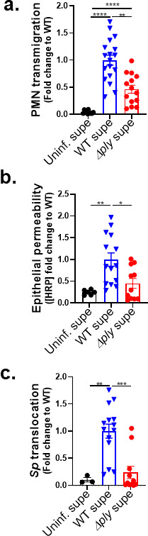
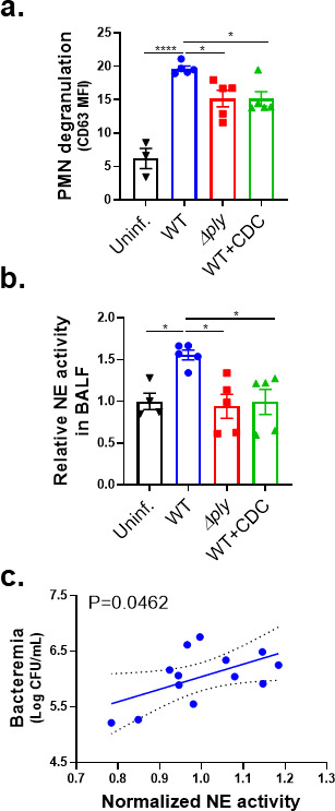
Streptococcus pneumoniae (Sp), a leading cause of community-acquired pneumonia, can spread from the lung into the bloodstream to cause septicemia and meningitis, with a concomitant threefold increase in mortality. Limitations in vaccine efficacy and a rise in antimicrobial resistance have spurred searches for host-directed therapies that target pathogenic immune processes. Polymorphonuclear leukocytes (PMNs) are essential for infection control but can also promote tissue damage and pathogen spread. The major Sp virulence factor, pneumolysin, triggers acute inflammation by stimulating the 12-lipoxygenase (12-LOX) eicosanoid synthesis pathway in epithelial cells. This pathway is required for systemic spread in a mouse pneumonia model and produces a number of bioactive lipids, including hepoxilin A3 (HXA3), a hydroxy epoxide PMN chemoattractant that has been hypothesized to facilitate breach of mucosal barriers. To understand how 12-LOX-dependent inflammation promotes dissemination during Sp lung infection and dissemination, we utilized bronchial stem cell-derived air-liquid interface cultures that lack this enzyme to show that HXA3 methyl ester (HXA3-ME) is sufficient to promote basolateral-to-apical PMN transmigration, monolayer disruption, and concomitant Sp barrier breach. In contrast, PMN transmigration in response to the non-eicosanoid chemoattractant N-formyl-L-methionyl-L-leucyl-phenylalanine (fMLP) did not lead to epithelial disruption or bacterial translocation. Correspondingly, HXA3-ME but not fMLP increased the release of neutrophil elastase (NE) from Sp-infected PMNs. Pharmacologic blockade of NE secretion or activity diminished epithelial barrier disruption and bacteremia after pulmonary challenge of mice. Thus, HXA3 promotes barrier-disrupting PMN transmigration and NE release, pathological events that can be targeted to curtail systemic disease following pneumococcal pneumonia.IMPORTANCEStreptococcus pneumoniae (Sp), a leading cause of pneumonia, can spread from the lung into the bloodstream to cause systemic disease. Limitations in vaccine efficacy and a rise in antimicrobial resistance have spurred searches for host-directed therapies that limit pathologic host immune responses to Sp. Excessive polymorphonuclear leukocyte (PMN) infiltration into Sp-infected airways promotes systemic disease. Using stem cell-derived respiratory cultures that reflect bona fide lung epithelium, we identified eicosanoid hepoxilin A3 as a critical pulmonary PMN chemoattractant that is sufficient to drive PMN-mediated epithelial damage by inducing the release of neutrophil elastase. Inhibition of the release or activity of this protease in mice limited epithelial barrier disruption and bacterial dissemination, suggesting a new host-directed treatment for Sp lung infection.

Keywords: 12-lipoxygenase; Streptococcus pneumoniae; airway mucosal barrier; neutrophil elastase; neutrophil transmigration.


Conflict of interest statement

The authors declare no conflict of interest.

Figures

Fig 1
Fig 1
The 12-LOX pathway, stimulated by PLY-producing Sp, promotes PMN infiltration, lung permeability, and bacteremia following Sp lung infection in mice. BALB/c mice were infected i.t. with 1 × 10⁷ CFU WT or PLY-deficient mutant (Δply) TIGR4 Sp for 18 h, with or without intraperitoneal (i.p.) injection of 8 mg/kg of the 12-LOX inhibitor CDC. (a) Bacterial lung burden determined by measuring CFU in lung homogenates. (b) PMN infiltration determined by flow cytometric enumeration of Ly6G+. (c) Lung permeability quantitated by measuring the concentration of 70 kDa FITC-dextran in the lung relative to serum after i.v. administration. (d) Bacteremia measured by enumerating CFU in serum. Each panel is representative of three independent experiments, or pooled data from three independent experiments. Error bars represent the mean ± SEM. Statistical analysis was performed using ordinary one-way ANOVA: *P-value < 0.05, **P-value < 0.01, ***P-value < 0.001, and ****P-value  < 0.0001.
Fig 2
Fig 2
The 12-LOX pathway promotes PMN transmigration and epithelial barrier breach upon apical infection of ALI monolayers by PLY-producing Sp. Human BSC-derived ALI monolayers (left column) or WT B6 and 12-LOX-deficient Alox15−/− mouse BSC-derived ALI monolayers (right column) were apically infected with 1 × 10⁷ WT or Δply Sp in the presence of basolateral PMNs. (a) After 2 hours of PMN migration, the degree of transmigration as determined by myeloperoxidase (MPO) activity in the apical chamber. (b) PMN infiltration and monolayer integrity assessed by fluorescence confocal microscopy after staining nuclei with 4′,6-diamidino-2-phenylindole (DAPI) and F-actin with fluorescent phalloidin. For clarity, the images shown are of extended projections (all z-sections collapsed into one plane). Arrows indicate examples of PMN nuclei. Scale bar = 40 µm for all images. Quantitation of epithelial retention is shown in the graph below the images, performed by enumerating epithelial cell nuclei relative to uninfected ALI in five images per experiment. (c) Epithelial permeability measured by horseradish peroxidase (HRP) flux relative to monolayers infected with WT Sp. (d) Sp translocation quantitated by measuring basolateral CFU. Each panel is representative of three independent experiments or pooled data from three independent experiments. Error bars represent the mean ± SEM. Statistical analysis was performed using ordinary one-way ANOVA: *P-value < 0.05, **P-value < 0.01, ***P-value < 0.001, and ****P-value < 0.0001.
Fig 3
Fig 3
A soluble factor produced by ALI monolayers via the 12-LOX pathway upon apical Sp infection promotes both PMN migration and barrier disruption. Alox15−/− mouse BSC-derived ALI monolayers were apically infected with 1 × 10⁷ WT Sp and transferred into apical chambers containing supernatant generated from WT Sp infection (WT supe) or Δply infection (Δply supe) of B6 mouse BSC-derived ALI monolayers. (a) After 2 hours of PMN migration, the degree of transmigration as determined by MPO activity in the apical chamber. (b) Epithelial permeability measured by HRP flux relative to monolayers infected with WT Sp. (c) Sp translocation quantitated by measuring basolateral CFU. Each panel is representative of three independent experiments or pooled data from three independent experiments. Error bars represent the mean ± SEM. Statistical analysis was performed using ordinary one-way ANOVA: *P-value < 0.05, **P-value < 0.01, and ***P-value < 0.001.
Fig 4
Fig 4
Upon Sp infection of ALI monolayers, PMN transmigration induced by HXA3 but not fMLP promotes barrier breach. Alox15−/− mouse BSC-derived ALI monolayers were apically infected with 1 × 10⁷ WT Sp and transferred into apical chambers containing 10 nM HXA3 methyl ester (“HXA3”), or 10 µM fMLP, in the presence of basolateral PMNs. (a) After 2 hours of PMN migration, the degree of transmigration as determined by MPO activity in the apical chamber. (b) Monolayer integrity assessed by fluorescence confocal microscopy after staining nuclei with DAPI and F-actin with fluorescent phalloidin. For clarity, the images shown are of extended projections (all z-sections collapsed into one plane). Scale bar = 40 µm for all images. Shown below the images is epithelial retention quantitated by enumerating epithelial cell nuclei relative to uninfected monolayers in five images per experiment. (c) Epithelial permeability measured by HRP flux relative to monolayers infected with WT Sp. (d) Sp translocation quantitated by measuring basolateral CFU. Each panel is representative of three independent experiments or pooled data from three independent experiments. Error bars represent the mean ± SEM. Statistical analysis was performed using ordinary one-way ANOVA: *P-value < 0.05, **P-value < 0.01, ***P-value < 0.001, and ****P-value < 0.0001.
Fig 5
Fig 5
HXA3 enhances NE secretion by Sp-infected PMNs. 1 × 10⁶ PMNs were infected with 1 × 10⁷ Sp after treatment with control HBSS, 10 µM fMLP, or 10 nM HXA3 methyl ester (“HXA3”) and evaluated for functional performance via (a) PMN membrane permeability determined by propidium iodide staining (PI+), (b) opsonophagocytic killing quantitated by plating for CFU, (c) NETosis determined by Sytox and anti-MPO staining (Sytox+ MPO+), (D) released MMP activity by substrate conversion and expressed relative to uninfected PMNs, (e) apoptosis determined by lack of straining by propidium iodide and positive staining of annexin V (PI annexin V+), (f) ROS production by intracellular oxidation of substrate (DCF+), and (g) released NE activity by substrate conversion and expressed relative to uninfected PMNs. (h) Sp-infected PMNs were treated with HXA3 methyl ester in the presence or absence of 50 µM Nexinhib20 (Nex.) and relative NE activity in supernatant quantitated by substrate conversion as in panel (g). (I) Radar plot summary of log fold change in PMN activities in (a–g). Each panel shown is representative of three independent experiments. Error bars represent the mean ± SEM. Statistical analysis was performed using ordinary one-way ANOVA: *P-value < 0.05, **P-value < 0.01, ***P-value < 0.001, and ****P-value < 0.0001.
Fig 6
Fig 6
PLY-producing Sp promotes the release of NE and primary granules in a 12-LOX-dependent manner during experimental lung infection. BALB/c mice were infected i.t. with 1 × 10⁷ CFU WT or Δply Sp for 18 hours, with or without i.p. injection of 8 mg/kg of the 12-LOX inhibitor CDC. (a) NE activity in cell-free bronchial alveolar lavage fluid (BALF) determined by substrate conversion, expressed relative to the NE activity in cell-free BALF from uninfected mice. (b) FACS analysis of degranulation determined by CD63 expression on Ly6G+ lung-infiltrating PMNs. (c) Correlation between normalized NE activity in panel a and bacteremia determined by enumerating CFU in serum. Each panel shown is representative of three independent experiments or pooled data from three independent experiments. Error bars represent the mean ± SEM. Statistical analysis was performed using ordinary one-way ANOVA: *P-value < 0.05, ***P-value < 0.001, and ****P-value < 0.0001.
Fig 7
Fig 7
Inhibition of NE release mitigates disruption of the lung epithelial barrier and bacteremia following Sp lung infection. BALB/c mice were infected i.t. with 1 × 10⁷ CFU WT Sp for 18 hours, with or without i.p. injection of 30 mg/kg Nexinhib20 (Nex) or 30 mg/kg Sivelestat (Siv) 1 hour prior to infection. (a) Bacterial lung burden determined by measuring CFU in lung homogenates; (b) PMN infiltration determined by flow cytometric enumeration of Ly6G+; (C) degranulation determined by CD63 expression on Ly6G+ lung-infiltrating PMNs by FACS; (d) relative NE activity in BALF determined by substrate conversion; (e) lung permeability determined by measuring the concentration of 70 kD FITC-dextran in lung relative to serum after i.v. administration; and (f) bacteremia determined by enumerating CFU in serum. Each panel is representative of three independent experiments or pooled data from three independent experiments. Error bars represent the mean ± SEM. Statistical analysis was performed using ordinary one-way ANOVA: *P-value < 0.05, **P-value < 0.01, and ****P-value < 0.0001.

Update of

References

    1. Henriques-Normark B, Tuomanen EI. 2013. The pneumococcus: epidemiology, microbiology, and pathogenesis. Cold Spring Harb Perspect Med 3:a010215. doi:10.1101/cshperspect.a010215 - DOI - PMC - PubMed
    1. Chen H, Matsumoto H, Horita N, Hara Y, Kobayashi N, Kaneko T. 2021. Prognostic factors for mortality in invasive pneumococcal disease in adult: a system review and meta-analysis. Sci Rep 11:11865. doi:10.1038/s41598-021-91234-y - DOI - PMC - PubMed
    1. Backhaus E, Berg S, Andersson R, Ockborn G, Malmström P, Dahl M, Nasic S, Trollfors B. 2016. Epidemiology of invasive pneumococcal infections: manifestations, incidence and case fatality rate correlated to age, gender and risk factors. BMC Infect Dis 16:367. doi:10.1186/s12879-016-1648-2 - DOI - PMC - PubMed
    1. Zumla A, Rao M, Wallis RS, Kaufmann SHE, Rustomjee R, Mwaba P, Vilaplana C, Yeboah-Manu D, Chakaya J, Ippolito G, Azhar E, Hoelscher M, Maeurer M, Host-Directed Therapies Network consortium . 2016. Host-directed therapies for infectious diseases: current status, recent progress, and future prospects. Lancet Infect Dis 16:e47–63. doi:10.1016/S1473-3099(16)00078-5 - DOI - PMC - PubMed
    1. Cremers AJH, Mobegi FM, de Jonge MI, van Hijum SAFT, Meis JF, Hermans PWM, Ferwerda G, Bentley SD, Zomer AL. 2015. The post-vaccine microevolution of invasive Streptococcus pneumoniae. Sci Rep 5:14952. doi:10.1038/srep14952 - DOI - PMC - PubMed

MeSH terms

Substances

LinkOut - more resources